Why Do My Knees Pop? Medical Causes, Diagnosis, and Evidence-Based Relief

Have you ever stood up from a chair, climbed a flight of stairs, or simply shifted your weight and heard a sudden pop or crack echoing from your knee joint? You are far from alone. Knee popping, medically referred to as crepitus, stands as one of the most frequently reported musculoskeletal sensations across all age demographics. While the auditory feedback can be startling or even unsettling, the underlying physiological reasons range from entirely harmless joint processes to early warning signs of mechanical stress. Understanding why do my knees pop during everyday movements is essential for distinguishing between normal joint function and potential orthopedic issues that warrant clinical attention. In this comprehensive guide, we will explore the intricate biomechanical foundations of knee joint acoustics, examine the most common benign and pathological triggers, and provide actionable, evidence-based strategies to maintain optimal joint resilience. Whether you are a dedicated endurance athlete, a weekend hiker navigating uneven terrain, or someone navigating a predominantly sedentary office lifestyle, learning how to accurately interpret your body’s mechanical signals will empower you to protect your lower extremity mobility and sustain pain-free movement for decades to come.
Understanding Joint Mechanics and Knee Anatomy
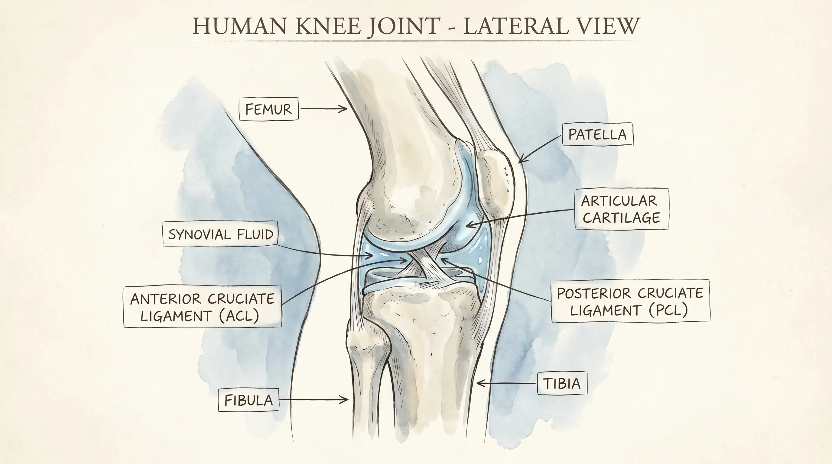
The human knee is a marvel of biological engineering, functioning as a complex, modified hinge joint that bears the majority of your body’s weight while facilitating fluid, multi-planar movement. Structurally, the knee articulates three primary skeletal components: the distal femur, the proximal tibia, and the patella. These osseous structures are shielded by articular cartilage, a smooth, viscoelastic tissue meticulously evolved to minimize friction during dynamic weight-bearing. Encasing the joint is a robust fibrous capsule containing synovial fluid, a highly specialized viscous secretion that functions simultaneously as a mechanical lubricant and a critical nutrient transport medium for the avascular cartilage matrix. During flexion and extension, this fluid redistributes across articular surfaces, ensuring seamless gliding mechanics and shock absorption. Surrounding this intricate system, a network of tendons, ligaments, and fascial tissues—including the anterior cruciate ligament, posterior cruciate ligament, medial and lateral collateral ligaments, and the iliotibial band—collaborate dynamically to stabilize joint alignment and guide the patella through its anatomical trochlear groove. Any disruption to this highly coordinated physiological ecosystem, whether stemming from muscular tension, fluid viscosity changes, or subtle tracking deviations, can generate the audible or palpable sensations that prompt many individuals to ask why do my knees pop during routine activities. Mastering these foundational anatomical principles is the first step toward implementing targeted, scientifically validated interventions that restore harmonious joint function.
Free calculatorBMI CalculatorCalculate Body Mass IndexOpen the calculatorThe Role of Synovial Fluid and Cavitation
One of the most rigorously documented explanations for transient joint acoustics centers around a well-understood physiological phenomenon known as tribonucleation or cavitation. Synovial fluid naturally contains dissolved atmospheric gases, predominantly nitrogen, alongside smaller concentrations of oxygen and carbon dioxide. When a joint undergoes rapid traction or sudden positional shifts, intra-articular pressure temporarily drops below ambient atmospheric levels, creating a localized vacuum effect. This abrupt pressure differential forces dissolved gases to rapidly coalesce into a gaseous cavity, which subsequently collapses within milliseconds, producing a sharp, audible pop. This entire process is entirely physiological, highly predictable, and fundamentally unrelated to tissue degradation. Orthopedic research utilizing high-speed dynamic magnetic resonance imaging has definitively captured the exact moment cavity formation and collapse occurs, confirming its benign nature. Notably, following a cavitation event, it typically requires twenty to thirty minutes for atmospheric gases to fully re-dissolve into the synovial matrix before another acoustic event can manifest in the exact same joint compartment, which perfectly explains why individuals cannot consecutively crack the same knee without allowing adequate recovery time.
Tendons, Ligaments, and the Patellar Tracking
Beyond intra-articular gas dynamics, extracapsular soft tissue interactions play an equally vital role in joint acoustics. The knee’s stability relies heavily on tendons and ligaments that glide over prominent bony landmarks during daily flexion and extension cycles. When these connective tissues experience increased tensile stress, fatigue accumulation, or minor positional misalignment, they can abruptly snap over the femoral condyles, tibial plateau, or fibular head, generating a distinct popping sensation that is frequently mistaken for internal joint pathology. The patella demonstrates particular vulnerability to tracking irregularities under repetitive loading conditions. Under ideal biomechanical circumstances, it travels smoothly within the femoral trochlear groove, maintaining consistent contact with underlying cartilage. However, persistent muscular imbalances—such as disproportionately weak vastus medialis obliquus fibers operating in tandem with excessively tight lateral quadriceps, lateral retinaculum, or iliotibial band fibers—can gradually alter patellar kinematics. Even subtle deviations of two to three millimeters from optimal tracking pathways generate measurable increases in patellofemoral contact pressure, resulting in rhythmic clicking, grinding, or popping during repetitive bending motions. Understanding these precise biomechanical relationships clarifies why do my knees pop during activities demanding sustained knee flexion, such as cycling, deep squatting, or prolonged stair negotiation.
Common Benign Causes of Knee Popping
While it remains psychologically tempting to immediately associate joint noises with degeneration or injury, clinical data consistently demonstrates that the overwhelming majority of knee popping falls securely within the benign spectrum. Physiological crepitus, frequently described as a mild grinding, crunching, or rhythmic popping sensation, appears with remarkable frequency across active populations and older adults alike. It characteristically intensifies during prolonged sedentary periods, following significant hydration fluctuations, or after intense physical exertion without adequate cool-down. Developing the discernment to separate harmless acoustic phenomena from genuine pathological indicators represents a foundational pillar of orthopedic self-management and long-term joint preservation. Environmental variables, systemic hydration status, genetic collagen variations, and daily ergonomic habits collectively influence baseline joint acoustics. By thoroughly examining these common, non-threatening triggers, individuals can confidently implement preventative lifestyle modifications and targeted movement protocols before minor physiological sensations gradually escalate into functionally limiting discomfort.
Crepitus: When It’s Just Air Bubbles and Minor Surface Irregularities
As thoroughly established, transient cavitation explains the sudden, singular pops many experience during rapid joint positioning. However, crepitus can equally manifest as a continuous, granular, or sandpaper-like grinding sensation throughout full range of motion. This variation typically stems from microscopic surface irregularities along the articular cartilage or subtle textural variations within the synovial lining. In the complete absence of localized tenderness, measurable effusion, or functional impairment, this acoustic presentation remains widely classified as a normal physiological variant rather than a disease state. The chondral surface naturally undergoes gradual micro-textural evolution across decades of weight-bearing activity. While the acoustic feedback may sound clinically concerning, it rarely progresses into structural compromise provided the joint maintains adequate muscular support and consistent low-impact movement. Regular, controlled articulation actively promotes synovial fluid circulation, delivering essential nutrients to avascular tissues and naturally preventing the accumulation of inflammatory mediators or the development of more severe friction-based symptoms.
Muscle Imbalances and Tight IT Bands
The kinetic chain governing the lower extremity depends fundamentally upon precise muscular harmony to preserve optimal skeletal alignment throughout dynamic movement. When specific muscle groups develop disproportionately elevated strength or experience chronic adaptive shortening, they fundamentally alter vector forces acting upon the patella and adjacent stabilizing structures. A hypertensive or fibrotic iliotibial band represents one of the most prevalent culprits responsible for lateral knee snapping, particularly among endurance runners and competitive cyclists whose training volumes demand repetitive flexion-extension cycles paired with insufficient posterior chain stretching. Similarly, underactive gluteus medius and maximus muscles frequently force compensatory valgus collapse at the knee joint during weight-bearing transitions, generating abnormal shear forces and subsequent acoustic feedback. Systematically correcting these kinetic chain discrepancies through progressive loading protocols and targeted tissue mobilization not only diminishes joint noises but simultaneously elevates movement efficiency, reduces injury susceptibility, and extends overall athletic longevity.
Age-Related Changes in Cartilage and Collagen Elasticity
As individuals progress into their fourth, fifth, and sixth decades, the resident chondrocytes responsible for maintaining cartilage extracellular matrix integrity gradually experience diminished metabolic activity and reduced proteoglycan synthesis. Simultaneously, the intracellular water content within chondrocyte networks slowly decreases while collagen cross-linking patterns undergo natural stiffening adaptations. These cumulative physiological transitions frequently produce subtle increases in articular friction during dynamic loading, commonly manifesting as mild popping, creaking, or rhythmic clicking during transitional movements. Importantly, age-associated crepitus does not automatically equate to clinical pathology or progressive degeneration. Countless individuals maintaining active lifestyles well into their later decades preserve pain-free mobility despite frequent joint sounds. The defining factor lies not in the acoustic presence but in functional preservation—sustaining lean muscle mass, maintaining dynamic flexibility, and consistently supporting connective tissue through targeted nutrition and evidence-based movement programming.
When Knee Popping Signals a Medical Concern
Not every joint sound represents harmless physiological noise. When popping consistently coincides with acute or chronic pain, visible swelling, prolonged morning stiffness, or subjective feelings of mechanical instability, it frequently signals underlying structural compromise that warrants systematic clinical evaluation. Dismissing these symptomatic clusters can inadvertently accelerate joint deterioration, prolong tissue inflammation, and necessitate increasingly invasive therapeutic interventions over time. Differentiating between benign joint acoustics and genuine orthopedic pathology requires a thorough understanding of symptom correlation, biomechanical stress patterns, and established clinical warning signs. By proactively recognizing these indicators, individuals can pursue timely diagnostic assessment, secure appropriate therapeutic referrals, and effectively prevent manageable conditions from evolving into chronic, functionally limiting disorders.
Meniscus Tears and Ligament Injuries
The medial and lateral menisci function as semi-lunar fibrocartilaginous shock absorbers, distributing compressive loads and enhancing joint congruency between the femoral condyles and tibial plateau. Acute rotational trauma, heavy axial loading, or cumulative degenerative micro-trauma can precipitate partial or complete meniscal disruption. A displaced meniscal fragment frequently catches, shifts, or catches during knee articulation, producing a loud, distinctly painful pop that rarely resolves without intervention. This mechanical event is typically accompanied by rapid effusion, restricted terminal flexion, and a subjective sensation of joint locking or catching. Analogously, acute ligamentous injuries—most notably complete anterior cruciate ligament ruptures or severe medial collateral ligament sprains—are classically heralded by a sudden, explosive popping sound at the precise moment of tissue failure, immediately followed by pronounced instability, significant hemarthrosis, and profound weight-bearing intolerance. These acute structural injuries mandate prompt orthopedic evaluation to determine whether conservative rehabilitation or surgical reconstruction offers the most favorable functional prognosis.
Osteoarthritis and Progressive Cartilage Degeneration
Osteoarthritis remains the most prevalent degenerative joint disorder worldwide and disproportionately targets weight-bearing articulations like the tibiofemoral compartment. As hyaline cartilage gradually erodes, the joint permanently loses its friction-reducing interface, resulting in direct bone-on-bone contact during routine weight-bearing. This irreversible degenerative cascade consistently manifests as persistent grinding, irregular popping, and profound stiffness, particularly noticeable following prolonged inactivity. Clinical markers include morning stiffness persisting beyond thirty minutes, recurrent activity-related swelling, crepitus that intensifies with weight-bearing, and progressive range-of-motion restriction. While osteoarthritis cannot be pharmacologically reversed, comprehensive early intervention incorporating targeted weight optimization, structured neuromuscular re-education, joint-sparing exercise modalities, and strategic anti-inflammatory management can dramatically decelerate disease progression and preserve independent mobility.
Patellofemoral Pain Syndrome and Chondromalacia
Widely recognized within sports medicine as runner’s knee, patellofemoral pain syndrome (PFPS) presents as localized discomfort and audible clicking situated directly anterior or immediately posterior to the patella. It typically emerges secondary to overuse patterns, repetitive micro-trauma, or fundamental lower limb malalignment. Movements demanding deep knee flexion under load—such as descending stairs, performing weighted squats, or maintaining prolonged seated positions with flexed knees—consistently exacerbate symptom intensity. The characteristic popping in PFPS originates from aberrant patellar tracking against the femoral trochlear groove, generating focal cartilage irritation, localized subchondral bone stress, and progressive chondral softening. Conservative rehabilitation emphasizing comprehensive quadriceps recruitment, proximal hip stabilization, and strategic activity modification consistently yields high-resolution rates and effectively restores pain-free articulation.
| Symptom Cluster | Typical Benign Presentation | Potential Pathological Indicator |
|---|---|---|
| Auditory Quality | Sharp, singular pop or mild, consistent grinding | Loud, painful pop followed by catching or locking |
| Pain Association | Completely painless or mild discomfort that resolves quickly | Moderate to severe pain during or immediately after movement |
| Swelling Presence | None or minimal transient fluid after extreme exertion | Rapid onset effusion or persistent swelling beyond 48 hours |
| Mechanical Stability | Full range of motion maintained without buckling | Sensations of joint giving way, instability, or restricted flexion/extension |
| Response to Rest | Resolves completely with 24-48 hours of normal activity | Persists or worsens despite adequate rest and conservative care |
Diagnostic Approaches: How Professionals Evaluate Popping Knees
When self-directed management strategies yield insufficient symptomatic relief or clinical warning signs progressively intensify, consulting a licensed healthcare professional becomes medically imperative. Board-certified orthopedic specialists, licensed physical therapists, and sports medicine physicians implement highly structured, stepwise diagnostic frameworks designed to isolate the precise origin of abnormal knee acoustics. Achieving accurate diagnostic clarity depends entirely upon systematic clinical history acquisition, specialized physical stress testing, and judicious utilization of advanced imaging modalities. Familiarizing yourself with this standardized evaluation process effectively demystifies clinical workflows, reduces patient anxiety, and empowers you to advocate proactively for targeted, personalized joint care.
Physical Examination and Specialized Range of Motion Tests
A thorough clinical evaluation invariably commences with comprehensive patient history documentation, meticulously tracking symptom onset, frequency, aggravating factors, prior lower extremity injuries, and current activity patterns. The examining clinician subsequently conducts meticulous joint inspection, palpates for localized tenderness or warmth, measures active versus passive range of motion, and assesses baseline muscular activation patterns. Specialized orthopedic provocation maneuvers—including the Lachman and pivot shift tests for cruciate ligament integrity, the McMurray and Apley compression tests for meniscal pathology, the Clarke sign for chondromalacia patellae, and functional step-down assessments—serve as highly reliable clinical indicators for isolating compromised anatomical structures. Directly observing joint mechanics during simulated daily tasks like squatting, lunging, and single-leg stance provides invaluable real-time insights into dynamic tracking deviations, compensatory movement patterns, and underlying kinetic chain inefficiencies.
Imaging Techniques: Radiographs, Advanced MRI, and Dynamic Ultrasound
When structural pathology becomes clinically suspected, diagnostic imaging transitions from supportive tool to definitive diagnostic cornerstone. Standard weight-bearing anteroposterior and lateral radiographs typically represent the initial imaging tier, providing exceptional visualization of bone alignment, joint space narrowing, osteophyte development, and subchondral sclerosis—classic radiographic hallmarks of degenerative joint disease. However, conventional X-ray technology fundamentally cannot visualize intra-articular soft tissue structures including cruciate ligaments, meniscal fibrocartilage, tendinous attachments, or cartilage surface topography. High-resolution magnetic resonance imaging (MRI) comprehensively addresses this limitation, delivering multi-planar, high-contrast cross-sectional imaging that precisely identifies ligamentous tears, meniscal disruptions, bone marrow edema, and varying degrees of chondral degradation. Dynamic musculoskeletal ultrasound continues gaining substantial clinical traction for real-time evaluation of tendon and ligament mobility during active flexion, rendering it exceptionally valuable for diagnosing snapping syndromes, tendinopathies, and dynamic impingement phenomena.
Proven Strategies to Manage and Prevent Knee Popping
Whether your primary objective involves eliminating benign acoustic sensations or successfully rehabilitating a structurally compromised articulation, a proactive, multidisciplinary therapeutic approach consistently delivers optimal clinical outcomes. Peer-reviewed sports medicine literature and long-term orthopedic cohort studies unequivocally demonstrate that targeted neuromuscular training, strategic nutritional optimization, and precise biomechanical refinement collectively yield profound improvements in joint resilience and acoustic reduction. By systematically integrating these scientifically validated interventions into your daily movement routines, you can substantially enhance connective tissue durability, normalize joint tracking, and significantly diminish the frequency of both benign and symptomatic popping events.
Targeted Strengthening and Progressive Mobility Protocols
Developing comprehensive muscular support around the knee joint remains the single most impactful intervention for stabilizing joint mechanics and reducing abnormal tracking patterns. Clinical guidelines emphasize balanced recruitment of the quadriceps, hamstrings, gluteal complex, and gastrocnemius-soleus musculature to establish a functional muscular corset that dynamically stabilizes the articulation under varying load conditions. Foundational strengthening exercises including terminal knee extensions, straight-leg raises with external rotation cues, Romanian deadlifts, lateral band walks, and controlled eccentric step-downs effectively build load tolerance without imposing excessive compressive stress on vulnerable articular surfaces. Complementary mobility programming featuring dynamic warm-up sequences like lateral lunges, hip CARs (controlled articular rotations), and ankle dorsiflexion drills preserves optimal fascial elasticity. Incorporating systematic myofascial release using high-density foam rollers targeting the IT band, tensor fasciae latae, vastus lateralis, and gastrocnemius dramatically reduces resting soft tissue tension that directly contributes to abnormal patellar tracking. Maintaining strict adherence to this progressive loading protocol three to four times weekly ensures sustained muscular equilibrium, optimal synovial circulation, and long-term joint preservation.
Nutritional Support and Systemic Inflammation Management
Dietary composition exerts profound influence over baseline systemic inflammation levels, connective tissue regeneration capacity, and synovial fluid viscosity. Long-chain omega-3 polyunsaturated fatty acids (EPA and DHA), abundantly present in wild-caught salmon, sardines, mackerel, and high-quality algal supplements, demonstrate well-documented clinical efficacy in downregulating pro-inflammatory cytokine production and supporting joint comfort during active movement. Hydrolyzed collagen peptides have accumulated substantial clinical evidence supporting their role in improving cartilage matrix composition, with randomized controlled trials consistently reporting meaningful reductions in exercise-induced joint discomfort and improved functional mobility scores in mild-to-moderate osteoarthritis populations. Equally critical is maintaining robust micronutrient availability: vitamin C serves as an indispensable cofactor for collagen cross-linking, zinc facilitates chondrocyte proliferation and extracellular matrix synthesis, while manganese activates essential metalloenzymes responsible for proteoglycan assembly. Hydration status cannot be overstated; since synovial fluid comprises approximately 85 percent water, chronic systemic dehydration directly compromises joint lubrication efficiency, elevates articular friction, and significantly amplifies benign crepitus. Aim for consistent daily fluid intake paired with electrolyte balance during periods of elevated sweat loss.
Footwear Modifications and Gait Biomechanical Adjustments
The kinetic chain operates as an integrated mechanical system wherein distal structural alterations directly impact proximal joint loading patterns. Chronically worn athletic shoes, improper arch support, or unsuitable footwear choices fundamentally alter ground reaction force vectors, dramatically increasing rotational shear stress transmitted directly to the knee articulation during ambulation and impact activities. Investing in purpose-engineered, structurally supportive athletic footwear specifically matched to your foot strike pattern, medial longitudinal arch morphology, and intended activity surface can dramatically normalize load distribution and minimize abnormal joint torque. For individuals presenting with significant biomechanical deviations, pronounced overpronation, or rigid cavus foot structures, custom-molded or prescription-grade foot orthotics frequently become necessary interventions to systematically correct lower extremity alignment and protect vulnerable cartilage surfaces. Additionally, strategically modifying training terrain—prioritizing shock-absorbing rubberized tracks, well-maintained dirt trails, or synthetic turf over unyielding concrete surfaces—naturally dissipates impact energy before it reaches the tibiofemoral joint, substantially reducing cumulative tissue microtrauma over extended training cycles.
Lifestyle Adjustments and Long-Term Joint Care
Preserving optimal knee function extends far beyond isolated exercise sessions and targeted supplementation protocols; it remains deeply interwoven with consistent daily habits, ergonomic awareness, and sustainable lifestyle architecture. Implementing incremental, highly manageable modifications to your daily routines consistently yields profound protective effects that compound significantly over months and years of dedicated practice. By prioritizing joint preservation as an integral component of your long-term wellness framework, you effectively maintain pain-free articulation well into advanced age while dramatically reducing future reliance on surgical interventions or chronic pain management protocols.
Maintaining Healthy Body Composition to Reduce Mechanical Load
Excess adipose tissue places profoundly disproportionate mechanical stress upon lower extremity weight-bearing joints. Biomechanical research consistently demonstrates that each additional pound of systemic body weight translates to approximately four to six pounds of additional compressive force transmitted through the knee joint during level-ground walking, escalating to nearly eight to ten times body weight during stair negotiation or rapid directional changes. Strategically achieving and maintaining an optimal body weight through balanced macronutrient intake, mindful portion regulation, and consistent cardiovascular conditioning represents one of the most powerful, evidence-backed interventions available for minimizing joint loading, decelerating cartilage wear rates, and reducing inflammatory adipokine production. Remarkably, clinical studies confirm that even a modest 5 to 10 percent reduction in total body weight yields dramatic improvements in pain scores, functional mobility, and overall joint comfort in individuals experiencing symptomatic crepitus or early degenerative changes.
Strategic Activity Pacing and Systematic Recovery Protocols
High-intensity resistance training, plyometric development, and repetitive impact activities are not inherently destructive to healthy knee structures when implemented intelligently. However, excessively rapid training volume progressions, inadequate recovery intervals, and chronic neglect of tissue adaptation timelines consistently precipitate overuse syndromes and inflammatory flare-ups. Adopting the established 10 percent progressive loading rule for weekly volume increases, systematically alternating high-impact training days with low-impact cross-training modalities like aquatic therapy, stationary cycling, or elliptical conditioning, and rigorously scheduling mandatory central nervous system and tissue recovery days allows musculoskeletal structures adequate time to remodel, strengthen, and adapt to imposed stressors. Integrating active recovery modalities—including parasympathetic breathing exercises, contrast water therapy, targeted mobility flows, and strategic nutrient timing post-exertion—accelerates metabolic waste clearance, enhances local circulation, and substantially reduces residual joint stiffness while preparing tissues for subsequent loading cycles.
Recognizing Critical Thresholds for Professional Intervention
While numerous popping sensations represent completely harmless physiological noise, specific clinical presentations demand immediate professional medical assessment. You should seek prompt orthopedic evaluation if you encounter persistent, activity-limiting pain that significantly disrupts daily functional tasks, noticeable joint effusion failing to subside after standard forty-eight-hour conservative management, recurrent sensations of mechanical instability or unexpected buckling during weight-bearing, physical locking phenomena that completely prevent full knee extension or deep flexion, or an acute, explosive pop immediately following traumatic impact followed by rapid, visible swelling within the first few hours. Early diagnostic intervention remains absolutely critical for preserving structural joint integrity, preventing secondary compensatory damage, and eliminating the progression toward chronic musculoskeletal disability. Securing appropriate physical therapy referrals, obtaining comprehensive orthopedic consultations, and utilizing advanced diagnostic imaging ensures you receive precise clinical diagnostics and fully personalized therapeutic pathways perfectly aligned with your unique anatomical and functional requirements. Prioritizing proactive joint stewardship today guarantees your knees will reliably support your active lifestyle, recreational pursuits, and natural aging processes for decades to come.
Frequently Asked Questions
Why does my knee make a popping sound without pain?
Painless knee popping is typically caused by benign physiological processes like cavitation, where dissolved gases in synovial fluid form and collapse, or soft tissue moving over bony prominences. Unless accompanied by swelling, instability, or sharp pain, it is generally not a cause for concern and reflects normal joint mechanics rather than structural damage.
Can knee popping lead to arthritis over time?
Current medical research does not support the claim that painless joint popping or cracking causes osteoarthritis. Arthritis develops from a complex interplay of genetic predisposition, cartilage degeneration, chronic inflammation, and mechanical overload. While persistent, painful crepitus may sometimes coexist with early osteoarthritis, the sound itself does not initiate or accelerate the disease process.
What are the best exercises to reduce knee popping?
Focusing on muscular balance is key. Strengthen the quadriceps, hamstrings, and glutes with exercises like squats, lunges, straight-leg raises, and clamshells. Incorporate hip mobility drills and foam rolling for the IT band to ensure proper patellar tracking. Low-impact cardio like swimming or cycling can also lubricate the joint and improve tissue elasticity without excessive wear.
Should I stop working out if my knees pop during exercise?
Not necessarily. If the popping is painless and consistent throughout your routine, it is likely harmless. However, if it occurs suddenly alongside pain, swelling, or a feeling of the knee giving way, pause the activity immediately. Modify your training intensity, focus on proper form, and consider consulting a physical therapist to address potential biomechanical imbalances before they progress.
How long should knee swelling last after a painful pop?
Acute swelling following a painful pop or injury should be evaluated promptly. Inflammatory joint swelling typically peaks within the first 24 to 48 hours. If it does not begin to subside with rest, ice, compression, and elevation, or if it persists beyond three to five days, it may indicate structural damage like a meniscus tear or ligament sprain requiring professional imaging and treatment.
Conclusion
Understanding why do my knees pop requires a nuanced, scientifically grounded appreciation of joint biomechanics, connective tissue physiology, and individual lifestyle variables. While clinical evidence overwhelmingly confirms that the vast majority of popping sensations represent entirely benign byproducts of intra-articular cavitation or physiological soft tissue gliding, maintaining acute awareness of accompanying clinical symptoms—including localized pain, progressive swelling, mechanical instability, or sudden functional impairment—remains absolutely crucial for early pathological detection and timely intervention. By systematically implementing evidence-based joint preservation strategies encompassing targeted neuromuscular strengthening, strategic nutritional optimization, sustainable weight management, intelligent activity pacing, and mindful biomechanical refinement, you can dramatically enhance connective tissue resilience, normalize patellar tracking mechanics, and confidently preserve long-term, pain-free mobility. Remember that your joints were biologically designed for consistent, purposeful movement rather than prolonged static postures. With proactive, consistent joint stewardship and a commitment to professional medical guidance whenever clinical warning signs emerge, you can successfully navigate the natural aging process while maintaining an active, independent lifestyle. Take control of your joint health today, and your knees will reliably support your goals for countless active years ahead.
How we work
Researched and drafted with AI assistance, then reviewed by a human editor against the cited sources.
This article is general health information, not medical advice. Consult a qualified healthcare professional about your own situation.