Understanding Recovery Time Broken Jaw: A Complete Healing Guide

Sustaining a mandibular fracture is a profoundly disruptive injury that extends far beyond immediate pain and discomfort. It fundamentally alters your ability to eat, speak, and even breathe normally, creating a cascade of physical and emotional challenges that demand careful, structured management. Understanding the recovery time broken jaw involves navigating requires patience, medical expertise, and proactive self-care is the first critical step toward restoring full facial function. While the initial trauma can feel overwhelming, modern oral and maxillofacial surgery has refined treatment protocols to optimize bone union, minimize complications, and accelerate functional restoration. Healing is not a linear process; it follows a complex biological timeline influenced by cellular repair mechanisms, nutritional status, and adherence to clinical guidelines. Whether your treatment involves conservative wiring or advanced titanium plating, knowing what to expect at each stage empowers you to collaborate effectively with your healthcare team. This comprehensive guide breaks down the physiological healing phases, evidence-based recovery strategies, and practical daily management techniques to help you transition safely from injury to complete rehabilitation. By integrating clinical protocols with lifestyle adjustments, you can confidently navigate the healing journey while minimizing risks and maximizing long-term outcomes.
Understanding Jaw Fractures: Anatomy and Mechanisms of Injury
The mandible is the largest, strongest, and only mobile bone in the facial skeleton, serving as the structural foundation for mastication, speech, and facial aesthetics. It forms a complex U-shaped ring composed of the symphysis (chin), body, angle, and two ascending rami that culminate in the condylar and coronoid processes. This unique architecture allows it to absorb and distribute significant mechanical forces, but it also contains several anatomical weak points where trauma frequently results in fractures. The mandibular condyles, in particular, are highly susceptible to impact due to their articulation with the temporal bone at the temporomandibular joint (TMJ). When sufficient blunt or penetrating force exceeds the bone’s tensile strength, microtrauma progresses to macroscopic disruption, initiating an immediate inflammatory cascade that marks the beginning of the biological repair sequence.
Free calculatorHeadache Location ChartIdentify headache types by locationOpen the calculatorCommon Causes and High-Risk Demographics
Jaw fractures predominantly affect young adult males due to higher participation in contact sports, motor vehicle collisions, and physical altercations. However, shifting epidemiological trends show rising incidence among older adults, largely attributed to falls, osteoporosis-related bone demineralization, and increased mobility in aging populations. High-impact motor vehicle accidents remain a leading cause of bilateral or comminuted fractures, while sports-related trauma typically produces unilateral or condylar injuries. Occupational hazards in construction, manufacturing, and military environments also contribute significantly to the injury burden. Regardless of etiology, the mechanism of force directly correlates with fracture complexity: direct anterior blows often fracture the symphysis or body, while lateral impacts compromise the angle or ramus. Understanding the biomechanics of your specific injury helps clinicians select the most appropriate stabilization method and provides realistic expectations for your recovery trajectory.
Classification of Mandibular Fractures
Medical professionals classify mandibular injuries based on location, displacement, communication with the oral cavity, and fracture pattern. Simple fractures maintain bone alignment with a single break line, while displaced fractures exhibit significant gap formation or overlapping fragments. Comminuted fractures involve multiple bone fragments, often requiring surgical reconstruction. Open fractures breach the oral mucosa, dramatically increasing infection risk and necessitating immediate antibiotic prophylaxis. Condylar fractures, which affect the TMJ articulation, present unique rehabilitation challenges due to potential joint ankylosis or chronic malocclusion. The fracture classification directly dictates the recovery time broken jaw patients will experience, as complex or intra-articular injuries demand extended immobilization and more intensive physical therapy. Mayo Clinic provides comprehensive clinical overviews of these classifications and their treatment implications.
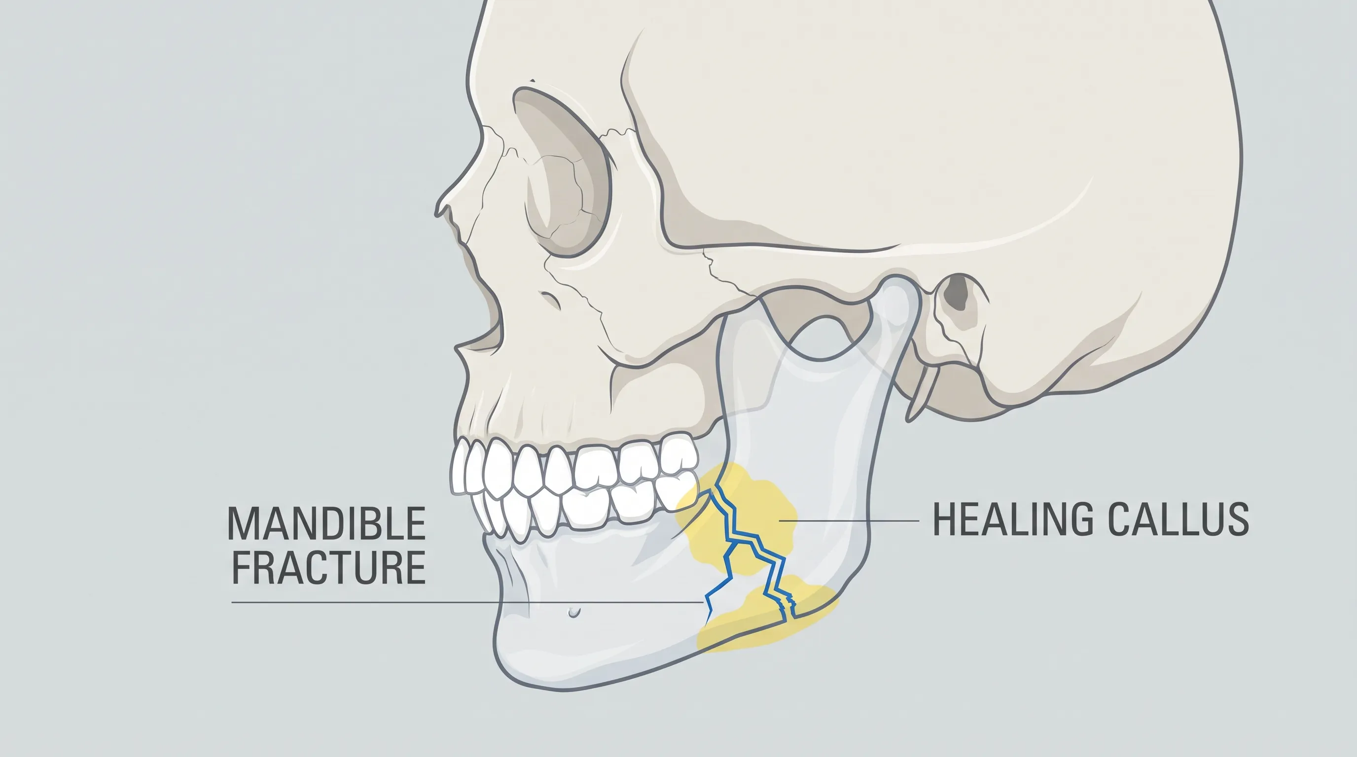
The Healing Timeline: What to Expect Stage by Stage
Bone regeneration follows a highly orchestrated biological sequence divided into inflammatory, reparative, and remodeling phases. While individual timelines vary based on age, genetics, and treatment modality, the physiological milestones remain remarkably consistent across patients. Recognizing each phase helps differentiate normal healing from concerning complications and guides clinical interventions at the appropriate time.
Phase 1: Immediate Post-Injury and Inflammatory Response (Days 1-10)
Within hours of trauma, vascular disruption triggers a localized inflammatory response characterized by hematoma formation, cytokine release, and macrophage recruitment. This acute phase manifests as pronounced swelling, ecchymosis, trismus, and moderate-to-severe pain. Pain management during this window is critical, as uncontrolled inflammation can delay cellular migration and compromise initial stability. Patients typically undergo diagnostic imaging, including panoramic radiographs, cone-beam CT, or conventional CT scans to assess displacement and plan stabilization. Whether treated with closed reduction or open surgery, the first week focuses on edema control through ice application, head elevation, and short-course corticosteroids when indicated. Antibiotic therapy is routinely prescribed for open fractures to prevent osteomyelitis or soft tissue infection. During this phase, strict dietary modification begins immediately, with patients restricted to full liquids or blenderized nutrition depending on immobilization status.
Phase 2: Soft Callus Formation and Early Stabilization (Weeks 2-6)
By the second week, fibroblasts and osteoprogenitor cells migrate into the fracture gap, depositing collagen and initiating woven bone formation. The resulting soft callus provides provisional stability but cannot withstand masticatory forces. If maxillomandibular fixation (MMF) is employed, the jaw remains immobilized for 3 to 6 weeks to prevent micromotion that could disrupt early mineralization. Open reduction internal fixation (ORIF) allows earlier guided mobility, as titanium plates and screws provide rigid mechanical stability that bypasses the need for prolonged wiring. During this period, patients often report reduced pain, improved swallowing tolerance, and gradual resolution of acute swelling. Clinical follow-ups typically occur at weeks 2 and 6 to assess occlusion, hardware integrity, and soft tissue healing. Nutritional intake must remain calorie-dense and protein-optimized, as the body’s metabolic demand increases by 20-30% during active tissue regeneration.
Phase 3: Hard Callus Maturation and Functional Remodeling (Weeks 7-24+)
The reparative phase transitions into remodeling around week 6 to 8, where woven bone is progressively replaced by lamellar bone through coupled osteoblastic and osteoclastic activity. Wolff’s Law governs this process: mechanical stress from gradual chewing stimulates trabecular reorganization along functional load lines. Recovery time broken jaw reaches its most variable window during this stage, as full biomechanical restoration depends on rehabilitation compliance, bite alignment, and joint mobility progression. Patients begin transitioning from pureed to soft diets, incorporating foods that require minimal shearing force. Physical therapy focuses on restoring mandibular excursion, lateral movements, and muscular endurance. Imaging at 12 weeks typically confirms radiographic union, though complete cortical thickening and medullary canal reformation may continue for 9-12 months. Long-term monitoring addresses TMJ function, dental occlusion, and potential hardware sensitivity.
Factors That Influence Your Recovery Time Broken Jaw
Individual healing trajectories are shaped by a complex interplay of intrinsic and extrinsic variables. While biological bone repair follows predictable cellular pathways, modifying factors can significantly accelerate or delay the timeline. Understanding these determinants enables proactive optimization and realistic expectation setting.
Age, Bone Quality, and Systemic Health
Pediatric patients exhibit remarkably rapid healing due to high periosteal vascularity and robust osteogenic potential, often achieving union within 4-6 weeks. Young adults typically follow the 6-8 week standard, while elderly patients experience prolonged timelines due to diminished osteoblast activity, reduced growth factor secretion, and comorbid conditions like diabetes or hypertension. Diabetes mellitus, particularly when poorly controlled, impairs angiogenesis and collagen synthesis, increasing infection risk and delaying callus maturation by 20-40%. Osteoporosis or vitamin D deficiency compromises bone mineral density, requiring extended immobilization and aggressive nutritional supplementation. Immunocompromised states, autoimmune disorders, or chronic kidney disease similarly disrupt the inflammatory-to-remodeling transition, necessitating closer clinical surveillance.
Lifestyle Impacts: Smoking, Alcohol, and Medication Use
Nicotine exerts potent vasoconstrictive effects that reduce peripheral blood flow by up to 40%, severely compromising oxygen and nutrient delivery to the fracture site. Carbon monoxide further displaces oxygen from hemoglobin, while free radicals generated by smoking increase oxidative stress and apoptosis in osteoblasts. Clinical evidence from the National Institutes of Health (NIH) demonstrates that active smokers face a 2-3 times higher risk of delayed union or nonunion, extending the average recovery time broken jaw by 3-5 weeks. Alcohol consumption interferes with calcium homeostasis, inhibits osteocalcin production, and disrupts sleep architecture, which is essential for growth hormone release. Certain medications, including long-term corticosteroids, bisphosphonates, NSAIDs (when used chronically in the remodeling phase), and anticoagulants, require careful management to avoid compromising bone metabolism or increasing bleeding risks.

Medical Treatments and Their Role in Accelerating Healing
Treatment selection depends on fracture displacement, patient health, occupational demands, and dental occlusion requirements. Modern maxillofacial surgery emphasizes anatomical reduction, rigid fixation, and early mobilization to restore function while minimizing complications.
Maxillomandibular Fixation (MMF) and Conservative Management
Closed reduction with intermaxillary fixation remains the standard for non-displaced, minimally mobile, or pediatric fractures. Arch bars or skeletal screws secure elastic bands that maintain dental occlusion, preventing fragment displacement during callus formation. While effective, MMF restricts oral intake, complicates oral hygiene, and increases aspiration risk. The typical recovery time broken jaw with MMF spans 4-6 weeks of strict immobilization, followed by gradual elastic weaning and mobility exercises. Patients must carry wire cutters at all times due to potential airway obstruction from vomiting or trauma. Despite these challenges, MMF offers a non-surgical alternative with comparable union rates when patient selection criteria are strictly met. American Association of Oral and Maxillofacial Surgeons (AAOMS) outlines current clinical indications for closed versus open management.
Open Reduction Internal Fixation (ORIF) and Surgical Protocols
Surgical intervention is indicated for displaced fractures, comminution, intra-articular involvement, or when MMF fails to maintain occlusion. ORIF utilizes titanium miniplates and monocortical screws to achieve absolute stability, enabling early functional movement and reducing nonunion rates. Advantages include improved oral hygiene access, earlier dietary progression, and lower aspiration risk. Post-operative care involves 1-2 weeks of edema management, followed by guided jaw exercises starting at week 3. Hardware removal is rarely necessary unless symptomatic, with titanium remaining biocompatible indefinitely. Surgical recovery typically shortens the immobilization phase by 2-4 weeks compared to MMF, though the overall biological healing timeline remains unchanged.
| Treatment Modality | Immobilization Duration | Diet Progression | Typical Union Time | Major Advantages | Primary Considerations |
|---|---|---|---|---|---|
| Maxillomandibular Fixation (MMF) | 3-6 weeks | Liquids only initially | 6-8 weeks | Non-invasive, low cost, high union rate | Airway risk, difficult hygiene, muscle atrophy |
| Open Reduction Internal Fixation (ORIF) | 1-3 weeks (guidance only) | Puree to soft by week 4 | 6-10 weeks | Early mobility, improved hygiene, faster rehab | Surgical risks, hardware sensitivity, higher cost |
Daily Management and Self-Care Strategies
Successful rehabilitation extends beyond surgical intervention. Patient adherence to structured home care protocols directly correlates with complication reduction, nutritional adequacy, and functional restoration speed.
Pain Management and Inflammation Control
Acute pain peaks within 48-72 hours and gradually declines over two weeks. A multimodal approach combining scheduled acetaminophen, short-course NSAIDs (if not contraindicated), and targeted opioids for breakthrough pain optimizes comfort while minimizing side effects. Ice packs applied for 15-20 minutes every 2 hours during days 1-3 reduce vascular permeability and edema. Transitioning to moist heat after day 5 promotes circulation and muscle relaxation. Sleep positioning with 30-degree head elevation prevents nocturnal fluid accumulation and reduces morning stiffness. Avoiding excessive NSAID use beyond week 2 is critical, as chronic prostaglandin inhibition can impair late-stage bone remodeling.
Nutritional Guidelines: Liquid, Soft, and Gradual Diet Transitions
Malnutrition is a leading preventable cause of delayed healing. Following Cleveland Clinic nutritional guidelines for fracture recovery, daily caloric intake should meet or exceed 1,800-2,200 kcal, with protein targets of 1.2-1.5g/kg body weight. Micronutrients like vitamin C (500-1000mg/day), vitamin D3 (2000-5000 IU/day), calcium (1000-1500mg/day), zinc (15-30mg/day), and magnesium are essential cofactors for collagen synthesis and hydroxyapatite formation. Commercial oral nutritional supplements, fortified blenderized meals, and smoothies with Greek yogurt, spinach, and protein powder provide balanced macronutrient profiles. When transitioning to soft foods, prioritize options that dissolve easily with minimal mastication, such as scrambled eggs, flaky fish, well-cooked pasta, and ripe avocados. Chew exclusively on the unaffected side when permitted, and avoid hard, sticky, or crunchy items until cleared by your surgeon.
Oral Hygiene and Infection Prevention
Maintaining asepsis in the oral cavity is non-negotiable during fracture recovery. Patients with MMF cannot brush normally, requiring chlorhexidine gluconate rinses (0.12%) twice daily, water flossing with reduced pressure, and syringe irrigation with sterile saline to flush food debris. Soft-bristle toothbrushes can clean accessible surfaces, while cotton-tipped applicators clean gum lines. Foul odor, yellow exudate, or worsening erythema warrants immediate clinical evaluation for gingival infection or hardware exposure. Regular professional dental cleanings resume once immobilization ends and occlusion stabilizes.
Physical Therapy and Jaw Mobility Exercises
Post-immobilization stiffness is universal and requires structured rehabilitation. Gentle stretching begins at week 4-6, progressing through five phases: passive range of motion (manual assistance), active assisted movement, resistance training with bite blocks, dynamic stabilization, and functional integration. Exercises include controlled mouth opening (target: 35-45mm lateral excursion), protrusion/retrusion drills, and lateral glide movements. Ultrasound therapy, myofascial release, and neuromuscular electrical stimulation may be prescribed for severe trismus. Consistency prevents fibrous ankylosis, restores muscular strength, and reduces chronic TMJ strain.
Navigating Complications and When to Seek Emergency Care
Despite optimal care, approximately 10-15% of mandibular fractures develop secondary complications requiring prompt intervention. Early recognition prevents irreversible damage and short-term morbidity.
Signs of Malunion, Infection, or Nerve Damage
Malocclusion, bite shifting, or palpable step-offs indicate fragment displacement or premature mobilization. Infection presents as escalating pain after initial improvement, purulent drainage, fever >100.4°F, and localized warmth. Inferior alveolar nerve involvement causes persistent chin or lower lip paresthesia, typically resolving within 3-6 months but occasionally requiring microsurgical exploration if permanent deficits persist. Hardware failure, though rare, manifests as sudden mobility, clicking, or palpable plate edges. Immediate radiographic evaluation and possible revision surgery are indicated.
Addressing TMJ Dysfunction and Chronic Pain
Post-traumatic TMJ arthritis or internal derangement affects up to 20% of condylar fractures. Symptoms include joint crepitus, limited opening, radiating otalgia, and muscle fatigue. Conservative management includes anti-inflammatories, occlusal splints, and physical therapy. Refractory cases may require arthrocentesis or arthroscopic debridement. Central sensitization can transform acute pain into chronic myofascial syndrome, necessitating neuropathic pain modulators, cognitive behavioral therapy, and multidisciplinary pain clinic referral.
Psychological Support During Recovery
Facial trauma disrupts self-image, communication ability, and social participation. Anxiety, depression, and social withdrawal are common during prolonged immobilization. Counseling, peer support groups, and teletherapy sessions improve coping strategies and treatment adherence. Recognizing mental health impacts as integral to physical recovery promotes holistic healing and prevents long-term psychological sequelae.
Returning to Normalcy: Work, Exercise, and Lifestyle Adjustments
Functional restoration extends beyond clinical discharge. Strategic reintegration ensures sustainable recovery and prevents reinjury.
Gradual Reintegration into Professional Duties
Desk workers typically return within 2-3 weeks post-ORIF or after MMF release, while physically demanding jobs require 8-12 weeks. Employers should accommodate modified schedules, speech therapy referrals, and flexible communication methods during the transition period. Driving resumes only when opioid medications cease and head rotation tolerance meets safety standards.
Safe Return to Physical Activity and Contact Sports
Cardiovascular exercise begins at week 2 (walking, stationary cycling), progressing to moderate intensity by week 6. Heavy lifting and high-impact activities remain restricted until radiographic union confirms structural integrity. Contact sports mandate custom-fitted protective mouthguards and clearance from maxillofacial and sports medicine specialists. Premature return risks refracture, hardware displacement, and extended recovery time broken jaw timelines by several months.
Long-Term Dental and Orthodontic Follow-Up
Occlusal monitoring continues for 12 months, with periodic adjustments for minor shifts. Endodontic evaluation of traumatized teeth prevents delayed pulp necrosis. Implants or prosthetics deferred until bone maturation completes ensure optimal osseointegration and functional longevity.
Frequently Asked Questions
What is the average recovery time broken jaw patients experience?
The average recovery time broken jaw patients experience typically spans 6 to 12 weeks for initial bone union, with complete structural remodeling and full functional restoration taking 6 to 12 months. Individual timelines depend on fracture severity, treatment type, age, nutritional status, and adherence to rehabilitation protocols.
How long will my jaw be wired shut after a fracture?
Maxillomandibular fixation generally remains in place for 3 to 6 weeks, depending on fracture location and healing progress. During this period, elastics secure the upper and lower dentition to maintain occlusion while the initial bone callus forms. Your surgeon will determine weaning schedules based on clinical and radiographic assessment.
Can a broken jaw heal without surgery?
Non-displaced or minimally displaced fractures frequently heal successfully with conservative management using closed reduction and intermaxillary fixation alone. However, significantly displaced, comminuted, intra-articular, or infected fractures usually require open reduction internal fixation (ORIF) surgery to restore proper alignment, prevent malunion, and preserve temporomandibular joint function.
What are the best foods to eat during jaw fracture recovery?
Optimal recovery nutrition includes high-calorie, high-protein liquids and pureed foods. Prioritize Greek yogurt, protein shakes, blended bean soups, mashed avocados, scrambled eggs, and oatmeal. Incorporate vitamin C, calcium, vitamin D, and zinc supplements to accelerate bone matrix synthesis. Avoid using straws if your jaw is immobilized, as suction forces can disrupt healing tissues.
When should I be concerned about complications during healing?
Seek immediate medical evaluation if you develop worsening swelling after the first week, persistent fever above 100.4°F, foul-tasting discharge from the mouth, new or persistent facial numbness, sudden bite misalignment, or difficulty breathing. These symptoms may indicate infection, nerve injury, hardware failure, or malunion requiring urgent intervention.
Conclusion
Navigating the recovery time broken jaw healing requires a multidisciplinary approach that integrates surgical precision, nutritional optimization, structured rehabilitation, and psychological resilience. While the biological timeline follows predictable physiological phases, individual outcomes depend heavily on proactive self-management, strict adherence to clinical guidance, and timely intervention when warning signs emerge. Modern maxillofacial protocols have dramatically reduced complication rates, shortened immobilization periods, and improved functional restoration compared to historical treatments. By understanding each healing stage, prioritizing bone-supportive nutrition, maintaining rigorous oral hygiene, and committing to guided mobility exercises, you can confidently progress through recovery while minimizing risks. Patience remains essential; bone remodeling continues long after clinical discharge, and rushing the transition to normal function can compromise long-term outcomes. Maintain regular follow-ups, communicate openly with your care team, and leverage available support resources. With disciplined care and evidence-based practices, full masticatory function, stable occlusion, and restored facial aesthetics are highly achievable goals. Your commitment to the recovery process ultimately determines the speed and quality of your return to daily life.
How we work
Researched and drafted with AI assistance, then reviewed by a human editor against the cited sources.
This article is general health information, not medical advice. Consult a qualified healthcare professional about your own situation.