Tooth Cupping vs Cavity: Key Differences, Diagnosis, and Prevention Strategies
Key points
- Dietary Acid Exposure: Frequent, habitual consumption of citrus fruits, commercial sports drinks, highly carbonated beverages, and vinegar-heavy foods significantly lowers intraoral pH levels throughout the entire waking day. Slowly sipping these beverages over extended periods is particularly damaging to the dentition because it creates a continuous acid bath that completely prevents natural saliva from adequately buffering and neutralizing the environment.
- Gastroesophageal Reflux Disease (GERD) and LPR: Silent reflux and laryngopharyngeal reflux repeatedly bring highly concentrated hydrochloric gastric contents into the oral cavity, primarily targeting the lingual and occlusal surfaces of posterior teeth. This systemic condition is strongly correlated with severe occlusal cupping, enamel thinning, and eventual dentin hypersensitivity.
- Eating Disorders and Chronic Vomiting: Conditions such as bulimia nervosa, binge eating disorder, or cyclic vomiting syndrome repeatedly expose the entire dentition to highly corrosive gastric acid, dramatically accelerating enamel loss and contributing to rapid structural collapse if left untreated.
- Occupational and Environmental Factors: Individuals working in chemical manufacturing, battery production, or industries with high exposure to acidic fumes or vaporized acids face significantly elevated erosion risks. Additionally, those who frequently consume acidic medications, chewable vitamin C supplements, or iron-based formulations often develop localized erosion patterns.
When you first notice subtle changes in your tooth surfaces—whether it is a slight concavity on the chewing areas or a darkened, softened spot—it is completely natural to feel concerned about your long-term dental health. Many patients immediately assume any visible defect in their enamel signals a traditional cavity, but the clinical reality is far more nuanced and requires careful differentiation. Understanding the distinction between surface erosion patterns and actual tooth decay is absolutely essential for preserving your long-term oral health and avoiding unnecessary interventions, as extensively documented by the National Institute of Dental and Craniofacial Research (NIDCR). The conversation around tooth cupping vs cavity has grown increasingly relevant in modern dental practice as dietary habits, lifestyle factors, and diagnostic technologies continue to evolve rapidly. While both conditions involve structural compromise of the enamel and underlying dentin, they originate from entirely different pathological mechanisms, demand distinct diagnostic approaches, and require highly specific treatment protocols tailored to the root cause. Misidentifying the condition can lead to inappropriate clinical interventions, delayed therapeutic care, progressive structural failure, or unnecessary patient anxiety. In this comprehensive, evidence-based guide, we will thoroughly explore the biological, clinical, and lifestyle factors that drive each condition. You will learn how to accurately recognize the early warning signs, understand exactly what dental professionals evaluate during a comprehensive examination, and implement proven preventive strategies that align rigorously with current dental science and peer-reviewed research. Whether you are navigating early enamel changes, managing systemic conditions that impact oral health, or simply seeking to protect your smile from progressive damage, this article provides the clarity, clinical context, and actionable guidance you need to make informed, confident decisions about your dental care journey.
Understanding Tooth Cupping: The Silent Progression of Enamel Erosion
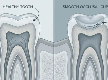
Dental professionals frequently encounter patients who present with subtle, bowl-shaped depressions on the occlusal chewing surfaces of their molars and premolars during routine examinations. This phenomenon, clinically referred to as occlusal cupping or dental cupping, represents a hallmark manifestation of intrinsic and extrinsic acid exposure rather than traditional bacterial decay. To fully grasp the clinical implications of tooth cupping vs cavity, we must first deeply examine the biomechanical and chemical processes that drive progressive enamel dissolution and structural compromise, as outlined in Mayo Clinic's guidelines on tooth erosion.
Free calculatorHeadache Location ChartIdentify headache types by locationOpen the calculatorThe Pathophysiology of Acid-Induced Wear
Tooth enamel is the most highly mineralized and resilient tissue in the human body, composed primarily of tightly packed hydroxyapatite crystals arranged in complex rod and interrod structures. However, these crystalline matrices are highly susceptible to demineralization when repeatedly exposed to acidic environments below a critical pH threshold of approximately 5.5. When dietary acids, chronic gastric reflux, or prolonged exposure to low-pH beverages consistently contact the tooth surface, the protective enamel matrix begins to chemically soften and gradually dissolve into the surrounding oral environment. Over time, this relentless chemical erosion preferentially weakens the anatomical cuspal slopes, leading directly to the characteristic concave depressions that clinically define cupping. Unlike bacterial decay, which requires a complex microbial biofilm matrix to initiate, colonize, and progressively destroy tooth structure, cupping occurs through direct, unforgiving chemical interaction. The entire process is frequently accelerated by parafunctional habits such as nocturnal bruxism or awake clenching, which mechanically abrade already softened and compromised enamel, creating a devastating synergistic effect that rapidly hastens structural loss and increases the risk of cusp fracture.
Primary Triggers and Risk Factors
Identifying and eliminating the root cause of enamel erosion is the foundational first step toward halting its relentless progression and preventing irreversible damage. Patients experiencing cupping frequently present with one or more of the following clinically documented risk profiles:
- Dietary Acid Exposure: Frequent, habitual consumption of citrus fruits, commercial sports drinks, highly carbonated beverages, and vinegar-heavy foods significantly lowers intraoral pH levels throughout the entire waking day. Slowly sipping these beverages over extended periods is particularly damaging to the dentition because it creates a continuous acid bath that completely prevents natural saliva from adequately buffering and neutralizing the environment.
- Gastroesophageal Reflux Disease (GERD) and LPR: Silent reflux and laryngopharyngeal reflux repeatedly bring highly concentrated hydrochloric gastric contents into the oral cavity, primarily targeting the lingual and occlusal surfaces of posterior teeth. This systemic condition is strongly correlated with severe occlusal cupping, enamel thinning, and eventual dentin hypersensitivity.
- Eating Disorders and Chronic Vomiting: Conditions such as bulimia nervosa, binge eating disorder, or cyclic vomiting syndrome repeatedly expose the entire dentition to highly corrosive gastric acid, dramatically accelerating enamel loss and contributing to rapid structural collapse if left untreated.
- Occupational and Environmental Factors: Individuals working in chemical manufacturing, battery production, or industries with high exposure to acidic fumes or vaporized acids face significantly elevated erosion risks. Additionally, those who frequently consume acidic medications, chewable vitamin C supplements, or iron-based formulations often develop localized erosion patterns.
Recognizing the Early Warning Signs
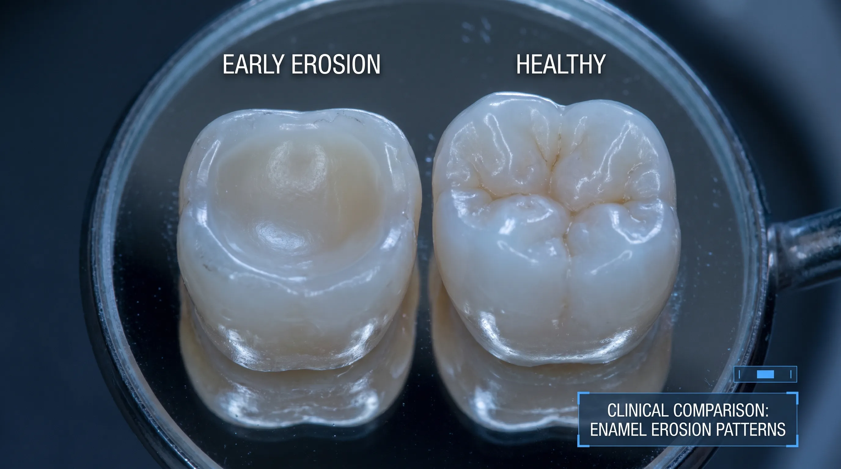
Cupping typically progresses insidiously and silently, often going unnoticed until significant structural loss has already occurred. Because the enamel loss occurs through a purely chemical mechanism without active bacterial infection or inflammatory response, noticeable pain is typically absent during the early stages of the condition. Patients may first report increased tooth sensitivity to thermal temperature changes, sweet foods, or a subtle visual change in the chewing surfaces that appears dull or flattened. The cupped areas usually maintain a glossy, smooth, or translucent appearance, clearly reflecting the progressive loss of the opaque outer enamel layer and subsequent show-through of the underlying yellowish dentin tissue. As the condition advances unchecked, the depressions deepen considerably, potentially leading to catastrophic fracture of the remaining thin enamel bridges during normal mastication. Early clinical recognition is absolutely critical because once the structural integrity of the functional cusp is compromised, conservative management becomes impossible and definitive restorative intervention becomes medically necessary to prevent tooth failure.
Decoding Dental Cavities: The Reality of Tooth Decay
While enamel erosion represents a purely chemical-mechanical degradation process, dental caries, universally known as cavities, is a complex, biofilm-driven infectious disease process that affects billions globally, according to recent WHO oral health fact sheets. Understanding the intricate microbiology and ecological dynamics of caries formation is absolutely fundamental when clinically evaluating tooth cupping vs cavity in both professional assessment and patient self-monitoring contexts.
The Caries Process Explained
Tooth decay absolutely does not occur spontaneously or without cause; it requires a sustained, highly specific interplay between cariogenic bacteria, readily available fermentable carbohydrates, susceptible crystalline tooth structure, and sufficient time for demineralization to dominate, as detailed by the CDC's oral health guidelines. The primary microbial culprits include highly acidogenic species such as Streptococcus mutans and various Lactobacillus strains, which aggressively metabolize dietary sugars and rapidly produce concentrated lactic acid as a primary metabolic byproduct. This potent acid continuously diffuses into the porous enamel matrix, aggressively dissolving hydroxyapatite crystals and creating microscopic subsurface pores. If the intraoral environment remains chronically acidic for prolonged daily periods, the relentless demineralization process significantly outpaces the mouth's natural remineralization capacity, inevitably leading to frank cavitation and structural breakdown. The caries process typically originates and spreads beneath the seemingly intact enamel surface directly at the dentinoenamel junction, where the lesion can rapidly expand laterally before finally breaking through to become clinically visible as a chalky white spot or open cavitated defect.
Common Signs and Clinical Progression
Dental cavities progress through highly predictable, distinct clinical stages, each presenting unique physical features that guide therapeutic decision-making:
- Initial Lesion (White Spot Stage): The absolute earliest detectable sign appears as a matte, opaque, chalky white area on the smooth or fissured enamel surface, clearly indicating active subsurface demineralization. Crucially, at this specific stage, the lesion remains fully reversible with targeted fluoride therapy, dietary modification, and improved plaque control.
- Enamel Cavitation: Continued, unmitigated acid production finally breaches the weakened enamel surface, creating a permanent, irreversible physical defect in the hard tissue. Patients often begin to experience mild, localized sensitivity or frequent food impaction directly within the affected anatomical area.
- Dentin Involvement: Once the bacterial invasion penetrates through the enamel and directly into the underlying dentin, the progression rate accelerates exponentially due to the dentin's significantly lower mineral content and highly organized network of fluid-filled tubules. Symptoms typically become pronounced, including sharp sensitivity to sweet stimuli, cold temperatures, and biting pressure.
- Pulpal Involvement: Advanced, untreated decay finally reaches the vascular and neural-rich dental pulp, triggering severe, sustained inflammatory responses that manifest clinically as spontaneous, lingering throbbing pain, extreme thermal hypersensitivity, and potential periapical abscess formation if left untreated.
Unlike cupping, which consistently presents as smooth, highly polished depressions, active cavities typically exhibit roughened, irregular, discolored brown or black margins, visibly soft dentin upon gentle probing, and distinctly demarcated lesion boundaries that trap plaque accumulation.
Systemic and Lifestyle Contributors
Modern dietary and lifestyle patterns significantly influence individual caries risk profiles. High-sugar, high-processed-carbohydrate diets, constant grazing or snacking behaviors, inconsistent oral hygiene routines, medication-induced xerostomia, and inadequate daily fluoride exposure all collaboratively create an intraoral environment highly conducive to pathogenic bacterial proliferation and continuous acid production. Understanding these complex variables directly helps patients accurately contextualize their personal susceptibility when carefully comparing tooth cupping vs cavity, thereby allowing for highly targeted, individualized preventive strategies rather than relying on generic, one-size-fits-all oral care routines that fail to address specific risk factors.
Tooth Cupping vs Cavity: A Comprehensive Clinical Comparison
Accurately distinguishing between these two prevalent conditions absolutely requires a highly systematic, multifaceted diagnostic approach. While both ultimately involve structural compromise of the hard dental tissues and increased patient sensitivity, their underlying etiology, microscopic progression patterns, clinical presentation features, and long-term management philosophies differ substantially and require distinct clinical pathways. The following detailed comparison explicitly outlines the critical differentiating factors that licensed dental professionals rigorously evaluate during routine screening and advanced diagnostic examinations.
| Feature | Tooth Cupping (Erosion) | Dental Cavity (Caries) |
|---|---|---|
| Primary Etiology | Chemical dissolution from direct acid exposure (dietary, gastric, environmental) | Bacterial metabolism of fermentable carbohydrates into concentrated organic acids |
| Surface Texture | Smooth, glossy, highly polished, or distinctly translucent appearance | Rough, chalky, leathery, or sticky to tactile probing; often catches instruments |
| Coloration | Maintains natural enamel translucency or appears darker/yellowish due to dentin exposure | Typically brown, black, or opaque yellow-brown with distinct lesion borders and demineralized margins |
| Pain Presentation | Often completely asymptomatic initially; later presents as generalized thermal or tactile sensitivity | Highly localized sensitivity that frequently progresses to sharp, lingering pain triggered by sweets, cold, or masticatory pressure |
| Biofilm Involvement | Absent or purely secondary; not causally driven by bacterial plaque colonization | Directly initiated, fueled, and maintained by pathogenic dental plaque biofilm activity |
| Progression Pattern | Broad, shallow, symmetrical depressions affecting occlusal cusps, facial, or lingual surfaces | Focal, highly localized defects frequently originating in deep fissures, tight interproximal areas, or cervical margins |
| Reversibility | Completely non-reversible once mineralized tissue is physically lost, but active progression can be successfully halted | Early subsurface white-spot lesions are clinically reversible; established cavitated lesions definitively require restorative intervention |
Distinguishing Features in Clinical Practice
Licensed dental professionals utilize several highly reliable diagnostic markers to confidently differentiate these conditions during a standard chairside examination. Tactile exploration with a sterile, blunt-tipped explorer probe remains a fundamental, time-tested clinical technique; cupped, eroded surfaces consistently glide smoothly and offer firm resistance under the instrument, whereas active carious lesions distinctly catch, stick, or yield to gentle probing pressure due to the presence of severely softened, demineralized dentin. Radiographic evaluation also plays a highly pivotal, irreplaceable role. Surface cupping rarely presents as distinct radiolucencies on standard two-dimensional bitewing radiographs unless extremely severe, progressive structural loss has already occurred, whereas active carious lesions typically appear as well-defined, triangular radiolucent shadows that clearly penetrate through the enamel layer and expand into the underlying dentin matrix.
Furthermore, comprehensive patient history provides absolutely invaluable diagnostic context. A patient with thoroughly documented medical histories of GERD, chronic high-acid beverage consumption, or recurrent vomiting disorders is highly, predictably likely to present with advanced erosion-based cupping patterns. Conversely, a patient presenting with consistently poor oral hygiene habits, frequent daily sugary intake, and documented xerostomia faces a substantially elevated risk for active carious lesions. Recognizing these distinct, highly predictable clinical patterns directly enables practitioners to develop precise, highly effective treatment plans that definitively address the true root cause rather than merely patching over superficially visible defects. When carefully evaluating tooth cupping vs cavity, the seamless integration of clinical examination, digital diagnostic imaging, and comprehensive patient history guarantees accurate diagnosis, appropriate intervention selection, and optimal, long-lasting clinical outcomes.
Diagnostic Protocols: How Professionals Differentiate the Two Conditions
Absolute diagnostic accuracy stands as the undisputed cornerstone of all effective, long-term dental care. Contemporary clinical dentistry rigorously employs a sophisticated combination of time-tested traditional clinical techniques and highly advanced technological diagnostic tools to comprehensively evaluate hard tissue mineral loss and definitively distinguish between erosive wear and active carious progression.
Visual-Tactile Examination and Caries Detection Devices
The universally accepted standard of care invariably begins with a meticulous visual inspection conducted under exceptionally bright, high-contrast dental operatory lighting. Clinicians carefully air-dry the teeth to completely remove the saliva film, which frequently masks or obscures early mineral changes from the unaided eye. Cupping characteristically reveals highly uniform, concave depressions with distinctly intact but noticeably thinned enamel margins, while active caries display irregular, undermined borders, localized discoloration, and obviously soft dentin that yields under gentle pressure. The essential tactile component involves carefully rolling a blunt explorer over the specific surface in question; consistent, firm resistance to penetration strongly indicates intact, heavily mineralized structure, while immediate yielding or softness unequivocally indicates active demineralization.
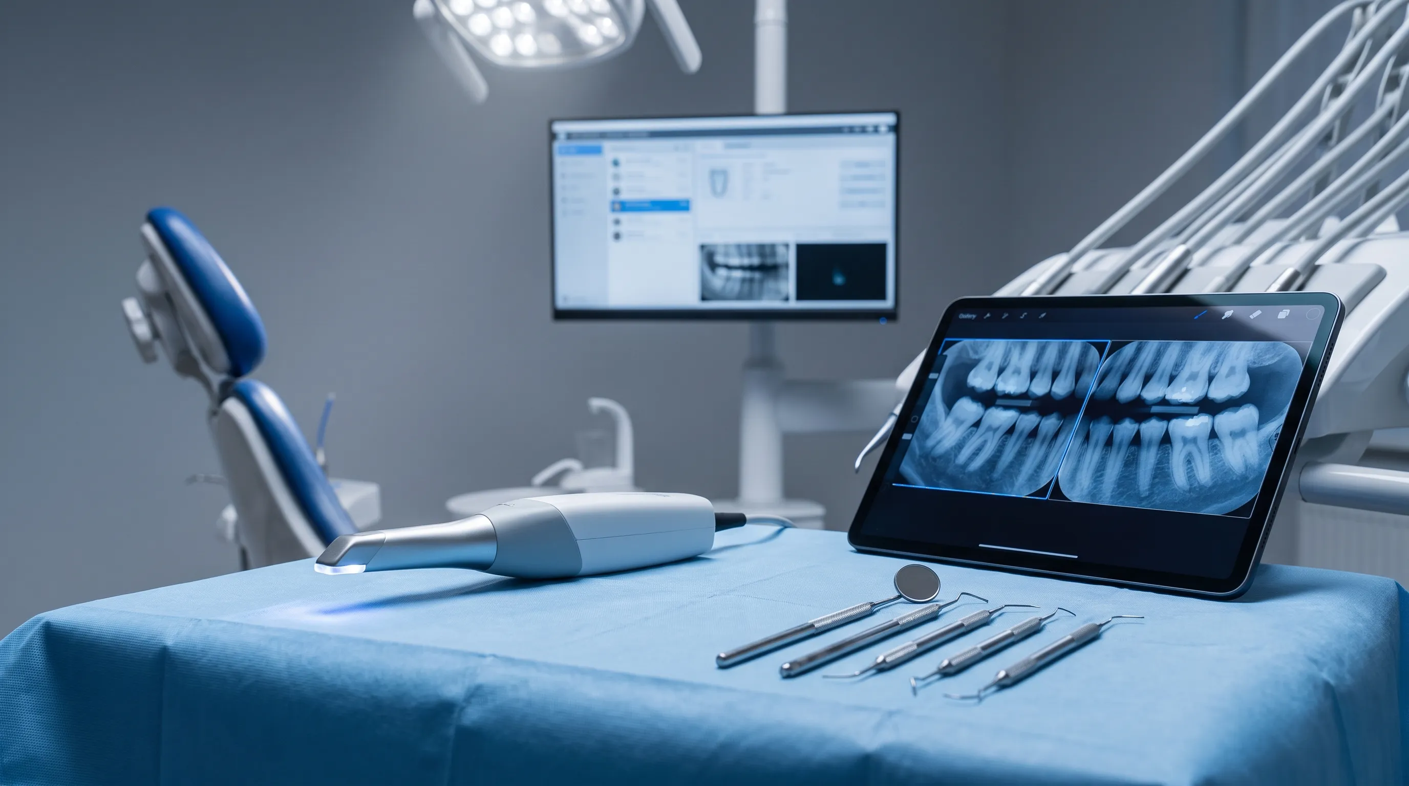
Advanced digital technologies have significantly and measurably improved diagnostic accuracy across all patient demographics. Laser fluorescence diagnostic devices (such as commercially available DIAGNOdent systems) accurately measure the highly specific fluorescent response of tooth structure when exposed to specific light wavelengths, providing clinicians with precise, quantitative data regarding the exact degree of mineral loss. Quantitative Light-Induced Fluorescence (QLF) imaging technology allows practitioners to visually track and quantify early subsurface demineralization in real-time while objectively monitoring remineralization progress across multiple recall appointments. These highly sensitive diagnostic tools prove particularly invaluable when systematically evaluating tooth cupping vs cavity, as they accurately quantify baseline mineral density changes and reliably help differentiate superficial chemical wear from active, spreading carious progression that demands immediate intervention.
Radiographic and Advanced Imaging Modalities
Standardized bitewing radiographs continue to serve as the absolute clinical gold standard for accurately detecting hidden interproximal caries, primarily because they clearly reveal active demineralization and bacterial spread that remains completely invisible during standard clinical visual examination. However, traditional two-dimensional X-ray technology possesses inherent, well-documented limitations in definitively detecting occlusal erosion or early cupping patterns, which typically progress horizontally parallel to the X-ray beam and therefore blend with healthy enamel density. Advanced modalities such as cone-beam computed tomography (CBCT) and digital volume tomography are occasionally utilized for highly complex, diagnostically challenging cases, providing exceptional three-dimensional visualization of exact enamel thickness measurements, direct dentin exposure locations, and comprehensive structural integrity assessments. State-of-the-art intraoral scanners further enable exceptionally high-resolution digital mapping of precise tooth morphology changes, enabling clinicians to accurately track and measure progressive surface changes across multiple six-month recall appointments with unprecedented accuracy.
The Critical Role of Patient History and Salivary Testing
A truly comprehensive diagnosis absolutely must extend far beyond the visible structures inside the mouth. Clinicians routinely and systematically assess objective salivary flow rate, natural buffering capacity, and baseline resting pH levels using validated chairside testing kits that deliver immediate, actionable results. Consistently low salivary flow severely compromises the mouth's entirely natural, highly effective biological ability to rapidly neutralize dietary acids and actively remineralize compromised enamel, dramatically accelerating the progression of both chemical erosion and bacterial caries. Thoroughly evaluating complete medication history, diagnosed systemic conditions, daily dietary patterns, and hydration habits directly helps contextualize the clinical findings within the patient's broader physiological framework. For individuals actively navigating the diagnostic uncertainty of tooth cupping vs cavity, this holistic, systems-based diagnostic approach definitively ensures that prescribed treatment addresses not just the immediately visible defect, but comprehensively targets and modifies the underlying physiological, behavioral, and environmental drivers.
Evidence-Based Treatment Pathways
Once a definitive, highly accurate diagnosis is thoroughly established and confirmed, clinicians promptly implement targeted, condition-specific therapeutic strategies designed to halt disease progression, restore function, and optimize long-term outcomes. The clinical management philosophy for progressive enamel erosion differs fundamentally from traditional caries treatment, heavily emphasizing aggressive preservation, continuous stabilization, and targeted risk modification rather than solely relying on immediate restorative intervention to mask the problem.
Managing Tooth Cupping Effectively
The absolute primary clinical goal in successfully treating active cupping is to completely halt further acid exposure, aggressively protect all remaining healthy enamel, and systematically restore functional anatomy only when structurally necessary. Conservative, non-invasive management comprehensively includes:
- Dietary and Behavioral Modification: Patients receive thorough, evidence-based education on significantly reducing direct acid contact, consistently utilizing drinking straws positioned past the lips for all acidic beverages, thoroughly rinsing the oral cavity with plain water or a mild baking soda neutralization solution immediately following acid exposure, and strictly waiting a minimum of thirty to sixty minutes before attempting to brush teeth, which strategically allows naturally produced saliva to successfully buffer acids and initiate early remineralization on temporarily softened enamel surfaces.
- Topical Remineralization Agents: In-office applied high-concentration professional fluoride varnishes, advanced calcium phosphate-based desensitizing pastes (specifically formulations utilizing CPP-ACP technology or highly reactive bioactive glass), and prescription-strength stannous fluoride therapeutic toothpastes work synergistically to actively stabilize the vulnerable enamel surface, reduce fluid movement in exposed dentin tubules, and significantly reduce daily sensitivity, in alignment with CDC recommendations on topical fluoride.
- Advanced Restorative Intervention: In severe clinical cases featuring significant, functionally compromising structural compromise, modern adhesive dentistry techniques are promptly deployed. Highly durable composite resin direct build-ups, precisely milled porcelain onlays, or full-coverage ceramic crowns may be strictly indicated to successfully restore proper occlusal function, redistribute masticatory forces, and actively prevent imminent cusp fracture. Biomimetic and minimally invasive bonding techniques are universally preferred whenever anatomically possible to aggressively preserve every micron of remaining healthy, mineralized tooth structure.
Treating Dental Cavities
Caries management unequivocally focuses on thoroughly removing all actively infected and necrotic tissue, completely eliminating the pathogenic biofilm reservoir, and hermetically sealing the prepared and restored tooth to aggressively prevent future bacterial recurrence.
- Non-Restorative Disease Management: Early, strictly non-cavitated white spot lesions are conservatively treated with intensive professional fluoride varnish applications, targeted silver diamine fluoride treatments specifically reserved for high-risk, pediatric, or medically compromised patients, and customized prescription remineralizing toothpaste regimens specifically designed to aggressively encourage natural hydroxyapatite reformation.
- Minimally Invasive Restorations: Once frank cavitation definitively occurs, all visibly decayed and structurally compromised tissue is carefully, completely excavated using specialized hand instruments, rotary burs, or water-driven air abrasion systems. The meticulously prepared, clean site is definitively sealed with highly aesthetic composite resin materials, traditional glass ionomers, or advanced resin-modified glass ionomer cements. These advanced restorative materials continuously release therapeutic fluoride ions directly into the surrounding tooth structure over months and years while chemically bonding directly to both enamel and dentin matrices, drastically reducing microleakage and secondary caries risk.
- Advanced Restorative and Endodontic Procedures: Extensive, deeply penetrating caries directly involving multiple tooth surfaces or advancing dangerously close to the vital dental pulp invariably require precision indirect restorations (laboratory-fabricated inlays, onlays, or monolithic crowns) or definitive root canal therapy if irreversible pulpal inflammation or bacterial necrosis has clinically developed.
Deeply understanding the entirely distinct treatment philosophies governing each condition powerfully reinforces precisely why accurate, timely diagnosis matters so profoundly in modern dentistry. Mislabeling progressive cupping as a standard bacterial cavity could tragically lead to unnecessary, destructive drilling of structurally sound tissue, while incorrectly treating an active, spreading cavity as simple chemical erosion would catastrophically allow unchecked bacterial progression to continue destroying the tooth. When thoroughly discussing tooth cupping vs cavity, patients should fully expect their licensed clinician to explicitly explain and justify the precisely chosen treatment pathway directly based on objective diagnostic findings, not merely superficial visual appearance alone.
Proactive Prevention and Lifestyle Optimization
Prevention remains unequivocally the most clinically effective, biologically sound, and economically efficient strategy for successfully maintaining lifelong, optimal dental health. Both chronic enamel erosion and active dental caries are highly preventable conditions that respond exceptionally well to targeted lifestyle modifications, meticulously consistent oral hygiene practices, and highly regular professional maintenance care, as emphasized by the World Health Organization's oral health initiatives.
Daily Oral Care Protocols
Brushing twice daily using a fluoride-containing toothpaste and meticulously flossing at least once daily form the absolute, non-negotiable foundation of modern preventive dentistry. Patients must consistently use extra-soft or soft-bristled brushes and aggressively avoid harsh, horizontal scrubbing techniques, which actively accelerate and exacerbate existing enamel wear patterns. Modern electric toothbrushes equipped with built-in pressure sensors are particularly, highly beneficial for individuals clinically diagnosed with early cupping or generalized erosion sensitivity, as they automatically alert the user when excessive force is being applied. Meticulous daily flossing actively removes tightly packed interproximal plaque that even the best brushing techniques cannot possibly reach, directly targeting and disrupting the primary microbial habitat of cariogenic bacteria. Additionally, consistently using clinically tested, alcohol-free antimicrobial mouth rinses can effectively help positively modulate the complex oral microbiome balance without inadvertently contributing to medication-induced dry mouth or tissue irritation.
Nutritional and Dietary Strategies
Diet profoundly and directly influences intraoral microbial ecology and baseline enamel resilience. Strategically reducing the absolute frequency of daily carbohydrate and refined sugar intake directly limits the available metabolic substrate for highly acid-producing bacterial colonies. For those actively managing documented acid erosion, aggressively limiting consumption of highly acidic citrus, sugary sports drinks, carbonated sodas, and vinegar-heavy prepared foods is absolutely crucial for structural preservation. Consistently consuming highly bioavailable calcium-rich foods (quality dairy products, dark leafy greens, calcium-fortified plant alternatives) alongside daily xylitol-containing mints or gums can directly enhance natural enamel remineralization rates and actively inhibit initial bacterial adhesion to clean tooth surfaces. Drinking adequately fluoridated municipal water and maintaining exceptional daily systemic hydration directly supports optimal salivary gland flow, which serves as the mouth's most powerful, naturally occurring biological defense mechanism.
Professional Maintenance and Risk Stratification
Strictly adhering to regular dental examinations and professional prophylactic cleanings every six months, or more frequently for medically established high-risk patients, enables exceptionally early clinical detection and timely, minimally invasive intervention. Modern dental professionals universally utilize validated caries and erosion risk assessment frameworks (such as the widely recognized CAMBRA protocol) to systematically categorize individual patients based on comprehensive biological, behavioral, environmental, and clinical factors. High-risk individuals consistently benefit from quarterly professional fluoride varnish applications, customized high-strength prescription toothpastes, and highly targeted, individualized dietary counseling sessions. Patients experiencing chronic GERD, diagnosed eating disorders, or severe xerostomia directly benefit from actively coordinated, interdisciplinary care alongside gastroenterologists, primary care physicians, and licensed mental health professionals, ensuring that complex systemic factors are comprehensively managed alongside targeted dental interventions, as recommended by the Cleveland Clinic's preventive dental guidelines.

By proactively integrating these thoroughly evidence-based practices into consistent daily routines, individuals can effectively and successfully mitigate the relentless progression of both conditions. Whether you are carefully monitoring very early signs of mineral loss or actively managing thoroughly diagnosed structural changes, deeply understanding the precise nuances of tooth cupping vs cavity powerfully empowers you to partner exceptionally effectively with your clinical dental team and aggressively prioritize long-term structural preservation, functional comfort, and aesthetic confidence.
Frequently Asked Questions
What exactly is tooth cupping and how does it differ from a regular cavity?
Tooth cupping, clinically known as occlusal erosion, refers to the gradual wearing away of enamel on the chewing surfaces of teeth due to chemical acid exposure. It presents as smooth, concave depressions and is driven by dietary acids, gastric reflux, or environmental factors. A cavity, or dental caries, is an infectious disease caused by bacteria metabolizing sugars into acid, leading to localized structural breakdown, discoloration, and roughened margins. Understanding the difference ensures appropriate clinical management and prevents unnecessary restorative procedures.
Can tooth cupping reverse itself or heal without dental intervention?
No, once enamel is physically lost through cupping, it cannot regenerate or reverse naturally. Unlike early-stage white spot lesions from cavities that can remineralize with fluoride and dietary changes, eroded enamel is permanently missing. However, the progression of cupping can be effectively halted through acid reduction strategies, topical remineralizing agents, and behavioral modifications. If structural integrity is compromised, restorative options like composite bonding or onlays can rebuild the lost anatomy and protect the underlying dentin.
How do dentists accurately diagnose cupping versus decay during a routine exam?
Dental professionals combine visual inspection, tactile exploration, diagnostic imaging, and patient history to differentiate these conditions. Cupping appears as smooth, translucent depressions that glide under an explorer probe, while cavities feel rough, catch the instrument, and often present with brown or black discoloration. Bitewing X-rays effectively detect interproximal decay, whereas occlusal erosion is primarily diagnosed through clinical examination and digital mapping tools. A thorough medical and dietary history further clarifies the underlying etiology.
Does using mouthwash help prevent or worsen tooth cupping?
It depends entirely on the formulation. Mouthwashes with a high alcohol content or highly acidic pH can exacerbate enamel softening and contribute to erosion. Alcohol-free, neutral-pH antimicrobial rinses or fluoride-containing mouthwashes are far safer and can help strengthen remaining enamel, reduce bacterial load, and alleviate sensitivity. Always check the product label for pH levels and alcohol content, and consult your dentist for personalized recommendations that align with your specific oral health profile and risk factors.
Are there any over-the-counter treatments that effectively manage early cupping or cavities?
For early non-cavitated white spot lesions, prescription-strength fluoride toothpaste and casein phosphopeptide-amorphous calcium phosphate pastes can promote significant remineralization. For mild sensitivity associated with cupping, stannous fluoride or bioactive glass toothpastes help occlude dentin tubules and stabilize the enamel surface. Over-the-counter nano-hydroxyapatite toothpastes also show promising clinical results for enamel repair and sensitivity reduction. However, once a cavity has cavitated or cupping has progressed to deep dentin exposure, professional dental intervention is necessary to restore structural integrity and prevent pulpal involvement.
Conclusion
Navigating the complexities of long-term dental health absolutely requires a clear, scientifically grounded understanding of how different conditions manifest, progress, and uniquely respond to targeted treatment. While the very initial visual signs of tooth cupping vs cavity may appear superficially similar to an untrained observer, their underlying biological mechanisms, diagnostic markers, clinical examination findings, and comprehensive management pathways are fundamentally, irreversibly distinct. Enamel erosion primarily driven by continuous chemical exposure demands a highly preventive, protective clinical approach heavily focused on consistent acid neutralization, strict dietary modification, and structural preservation strategies. Dental caries, deeply rooted in complex microbial activity and biofilm ecology, definitively requires targeted biofilm management, rigorous carbohydrate reduction, and timely, precise restorative intervention to eliminate infection and seal compromised tissue. By consistently recognizing early clinical warning signs, meticulously implementing thoroughly evidence-based oral hygiene routines, maintaining optimal systemic hydration, and adhering to highly consistent professional preventive care schedules, you can exceptionally effectively protect your natural dentition from irreversible, progressive damage. Your natural smile represents a lifelong physiological and aesthetic investment, and making highly informed, proactive decisions today will directly yield lasting structural integrity, optimal functional comfort, and unwavering aesthetic confidence for decades to come. Always consult a licensed, experienced dental professional for accurate, personalized diagnosis and comprehensive treatment planning precisely tailored to your unique clinical presentation, medical history, and long-term oral health goals.
How we work
Researched and drafted with AI assistance, then reviewed by a human editor against the cited sources.
This article is general health information, not medical advice. Consult a qualified healthcare professional about your own situation.