Safe and Clinically Proven Ways to Get a Tooth Out: A Complete Guide
The prospect of needing to remove a tooth can provoke significant anxiety, whether you are managing a stubbornly loose primary molar in a young child or dealing with a severely compromised adult tooth that has finally succumbed to decay or trauma. Understanding the clinically appropriate and safe ways to get a tooth out is essential for preserving oral health, preventing unnecessary complications, and ensuring a smooth recovery. While the internet is flooded with anecdotal DIY methods and old-fashioned folklore, dental professionals universally emphasize evidence-based protocols that prioritize patient safety, proper wound healing, and long-term occlusal stability, as detailed in comprehensive clinical guidelines from the Mayo Clinic. Navigating the spectrum from natural exfoliation in children to surgical interventions in adults requires a nuanced understanding of dental anatomy, pain management, and post-procedural care. This comprehensive guide breaks down the medically endorsed approaches, highlights critical warning signs, and provides actionable steps to help you make informed decisions when facing dental extraction.
Understanding the Need for Dental Extraction
Tooth removal is rarely a first-line intervention. Modern restorative dentistry prioritizes preservation whenever feasible, utilizing crowns, root canal therapy, periodontal grafts, and regenerative procedures to save compromised structures. However, clinical indications for extraction arise when pathological progression outweighs the benefits of conservation. Severe dental caries that has penetrated the pulp chamber and caused irreversible pulpal necrosis often necessitates removal when endodontic retreatment fails. Similarly, advanced periodontal disease destroys the alveolar bone and periodontal ligament fibers that anchor teeth in place, resulting in pathological mobility that compromises masticatory function and adjacent tooth stability.
Free calculatorHeadache Location ChartIdentify headache types by locationOpen the calculatorImpacted third molars represent another frequent clinical scenario. When developing wisdom teeth lack adequate eruption space, they may become trapped within the mandibular or maxibone, leading to pericoronitis, cyst formation, or resorption of neighboring second molar roots. Clinical management of these cases aligns with surgical guidelines from the Cleveland Clinic. Orthodontic treatment plans also occasionally require the strategic removal of specific teeth to alleviate severe crowding and achieve optimal alignment. Trauma, such as complicated crown-root fractures extending subgingivally or severe luxation injuries, frequently mandates extraction to prevent chronic infection and maintain jawbone integrity.
Clinical Indications and Diagnostic Evaluation
Before any removal occurs, a comprehensive clinical and radiographic assessment is mandatory. Digital periapical and panoramic radiographs allow clinicians to evaluate root morphology, proximity to vital anatomical structures like the inferior alveolar nerve or maxillary sinus, and the extent of periapical pathology. Cone-beam computed tomography may be utilized for complex surgical cases requiring three-dimensional spatial orientation. Blood coagulation panels, cardiovascular assessments, and medication reviews are equally critical, as patients on anticoagulants, bisphosphonates, or immunosuppressive therapies require specialized perioperative management protocols to mitigate hemorrhage or osteonecrosis risk, following CDC recommendations for dental patient screening.
When Self-Removal Crosses a Dangerous Line
While exploring ways to get a tooth out at home may seem financially convenient or time-saving, unsupervised attempts carry substantial medical risks. Forcing out a tooth that remains firmly integrated into the periodontal ligament can cause alveolar bone fractures, root tip retention, and severe gingival lacerations. Introducing unsterile objects into the oral cavity dramatically increases the risk of localized abscess formation, bacteremia, and systemic infection. Moreover, improperly managed extraction sites frequently develop dry socket, a highly painful condition that prolongs recovery and requires professional clinical intervention per the National Institute of Dental and Craniofacial Research. Recognizing when a tooth truly requires professional management is a cornerstone of responsible oral healthcare.
Clinically Approved Ways to Get a Tooth Out
When extraction becomes medically necessary, modern dentistry offers highly refined, minimally traumatic techniques designed to preserve surrounding tissue architecture and accelerate healing. Understanding these procedural pathways empowers patients to participate actively in their care planning and reduces procedural anxiety through informed consent and clear expectations.
Simple Extraction Procedures
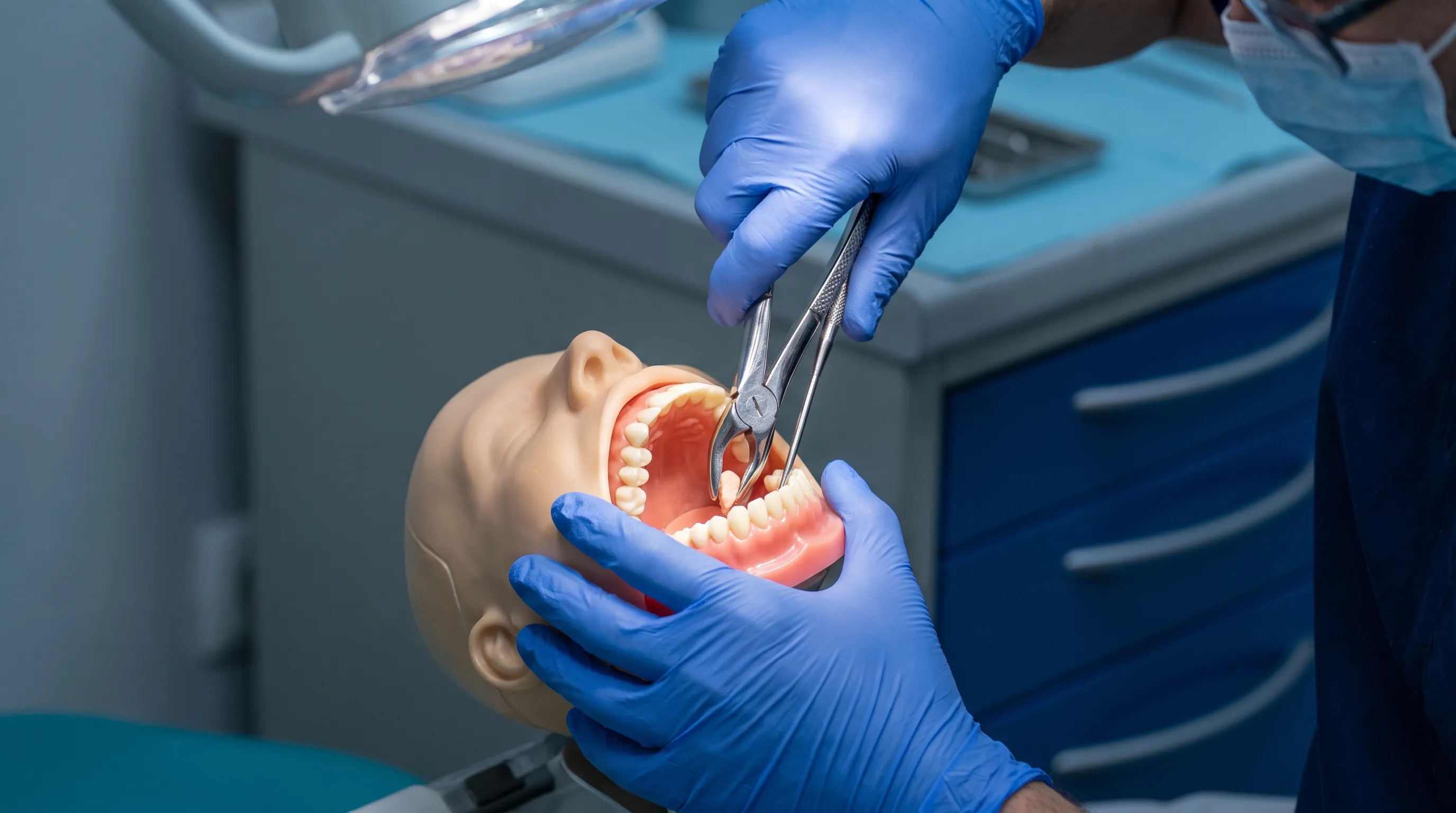
Simple extractions are indicated for fully erupted teeth with intact clinical crowns and predictable root anatomy. Following local anesthetic administration to achieve profound pulpal and periodontal anesthesia, the clinician utilizes specialized elevators to gently disrupt and sever the periodontal ligament fibers connecting the tooth to the alveolar socket. Once initial mobility is achieved, forceps are strategically positioned at the cementoenamel junction to grasp the crown while distributing traction forces evenly along the long axis of the tooth. Controlled rotational, buccal-lingual, or axial forces gradually expand the socket walls, allowing the tooth to be delivered with minimal bone disruption.
This technique prioritizes tissue preservation. The clinician avoids excessive lateral forces that could fracture thin alveolar plates or damage adjacent neurovascular bundles. Following tooth removal, the socket is carefully debrided of any pathological tissue, irrigated with sterile saline, and often contoured to ensure smooth bony margins. Sutures may be placed if primary closure is achievable or if soft tissue flaps were elevated for improved access. The entire procedure typically requires fifteen to thirty minutes, with immediate postoperative instructions provided before discharge.
Surgical Interventions and Impacted Teeth
When teeth are partially erupted, severely fractured below the gumline, or impacted within bone, surgical extraction becomes necessary. This procedure involves the reflection of a mucoperiosteal flap, precise bone removal using sterile rotary instruments with continuous water cooling, and potential crown sectioning to separate multi-rooted teeth into retrievable segments. The surgical approach allows direct visualization of root apices, proximity to the mandibular canal, and the extent of cystic or granulomatous lesions. Advanced techniques like piezoelectric surgery utilize ultrasonic vibrations for selective bone cutting while sparing soft tissues, nerves, and vascular structures, significantly reducing postoperative morbidity.
Anesthesia and Sedation Options
Pain control and anxiety management are integral components of any extraction protocol. Local anesthetics such as lidocaine, articaine, or mepivacaine effectively block nociceptive transmission at the surgical site. For patients with extreme dental phobia, complex surgical cases, or medically compromised conditions requiring stress reduction, moderate sedation or monitored anesthesia care may be administered. Nitrous oxide, oral benzodiazepines, or intravenous propofol infusions allow patients to remain deeply relaxed while maintaining protective airway reflexes. These protocols are administered by licensed professionals trained in airway management, hemodynamic monitoring, and emergency response.
Natural Progression: When Teeth Come Out on Their Own
Understanding physiological tooth exfoliation and pathological loosening provides crucial context for determining whether intervention is necessary or if observation is appropriate. Natural processes follow predictable biological timelines, while premature adult mobility signals underlying disease requiring immediate attention.
Pediatric Dental Development
Primary teeth naturally loosen as permanent successors develop beneath them. The eruption pathway of permanent premolars and canines exerts pressure on primary root structures, triggering clastic activity that resorbs dentin and cementum. As root structure diminishes, the periodontal attachment weakens, resulting in physiological mobility. Children should be encouraged to gently wiggle loose primary teeth with clean hands or tongues, avoiding forceful pulling with threads or unsterile instruments. Forcing out a primary tooth before the successor has adequately resorbed the root can cause traumatic hemorrhage, premature space loss, and potential damage to the developing permanent follicle, a process consistent with WHO pediatric oral health standards.
Adult Looseness and Underlying Pathology
Permanent teeth should not exhibit noticeable mobility under normal physiological conditions. When adults experience spontaneous loosening, it typically indicates advanced periodontal disease, traumatic occlusion, bruxism-induced alveolar bone loss, or severe periapical infection compromising the lamina dura. Unlike pediatric exfoliation, adult tooth mobility rarely resolves without intervention. Splinting techniques, periodontal therapy, or strategic extraction followed by implant placement may be required. Ignoring progressive mobility allows bacterial infiltration to deepen, accelerates bone resorption, and increases the risk of systemic inflammatory responses linked to cardiovascular and metabolic disorders.
Safe At-Home Practices for Mildly Loose Teeth
While professional intervention remains the gold standard, certain scenarios involving naturally mobile primary teeth or completely detached adult teeth may permit cautious, hygienic at-home management. These approaches emphasize gentleness, sterility, and vigilant observation rather than aggressive manipulation.
Preparing for a Gentle Removal
Before attempting any removal, thoroughly wash hands with antimicrobial soap and dry them completely. Ensure adequate lighting and a clean, stable environment. If the tooth is a child's primary molar, offer reassurance and explain that gentle movement is preferable to sudden pulling. Provide soft, cold foods like yogurt or ice chips to naturally soothe gingival tissue and encourage minor movement during mastication. Avoid sticky, hard, or crunchy foods that could traumatize the periodontal ligament or cause uncontrolled tearing.
Stepwise Mechanical Assistance
When a tooth is already detached at the root and resting in the gingival crevice, a clean tissue or sterile gauze pad may be used to grasp the crown and apply gentle, steady traction. If resistance is encountered, immediately cease manipulation and allow natural progression. Wiggling the tooth side-to-side with clean fingers can further loosen the remaining connective fibers. Once the tooth releases, inspect the root structure to ensure it is intact. Fragment retention in the socket can impede healing and harbor bacterial colonization.
Absolute Contraindications for DIY Attempts
Certain scenarios absolutely prohibit home extraction. If the tooth exhibits significant resistance, sharp pain, or localized swelling, professional intervention is mandatory. Do not attempt removal if you have a bleeding disorder, are taking blood thinners, have a compromised immune system, or notice pus, foul taste, or throbbing pain radiating to the ear or jaw. Using string, door hinges, or unsterile pliers introduces tetanus risk, causes uncontrolled soft tissue tearing, and frequently results in fractured root tips that require complex surgical retrieval, posing significant infection risks highlighted by the CDC. Recognizing these red flags is essential when exploring ways to get a tooth out without endangering oral or systemic health.
Managing Pain and Recovery After Tooth Removal
Post-extraction care significantly influences healing outcomes, comfort levels, and long-term ridge preservation. Evidence-based protocols focus on hemorrhage control, inflammation management, infection prevention, and tissue regeneration.
Hemostasis and the First 24 Hours
Immediately following extraction, bite down firmly on a folded gauze pad positioned directly over the socket for thirty to sixty minutes. Replace with fresh gauze if bleeding persists, maintaining consistent pressure rather than repeatedly checking the site. The initial blood clot is the foundation for secondary bone and soft tissue healing. Avoid rinsing, spitting forcefully, or consuming hot liquids for at least twenty-four hours, as hydraulic pressure and thermal expansion can dislodge the clot and trigger dry socket, which the Mayo Clinic notes can be largely prevented with strict adherence to aftercare protocols.
Diet, Hygiene, and Healing Timeline
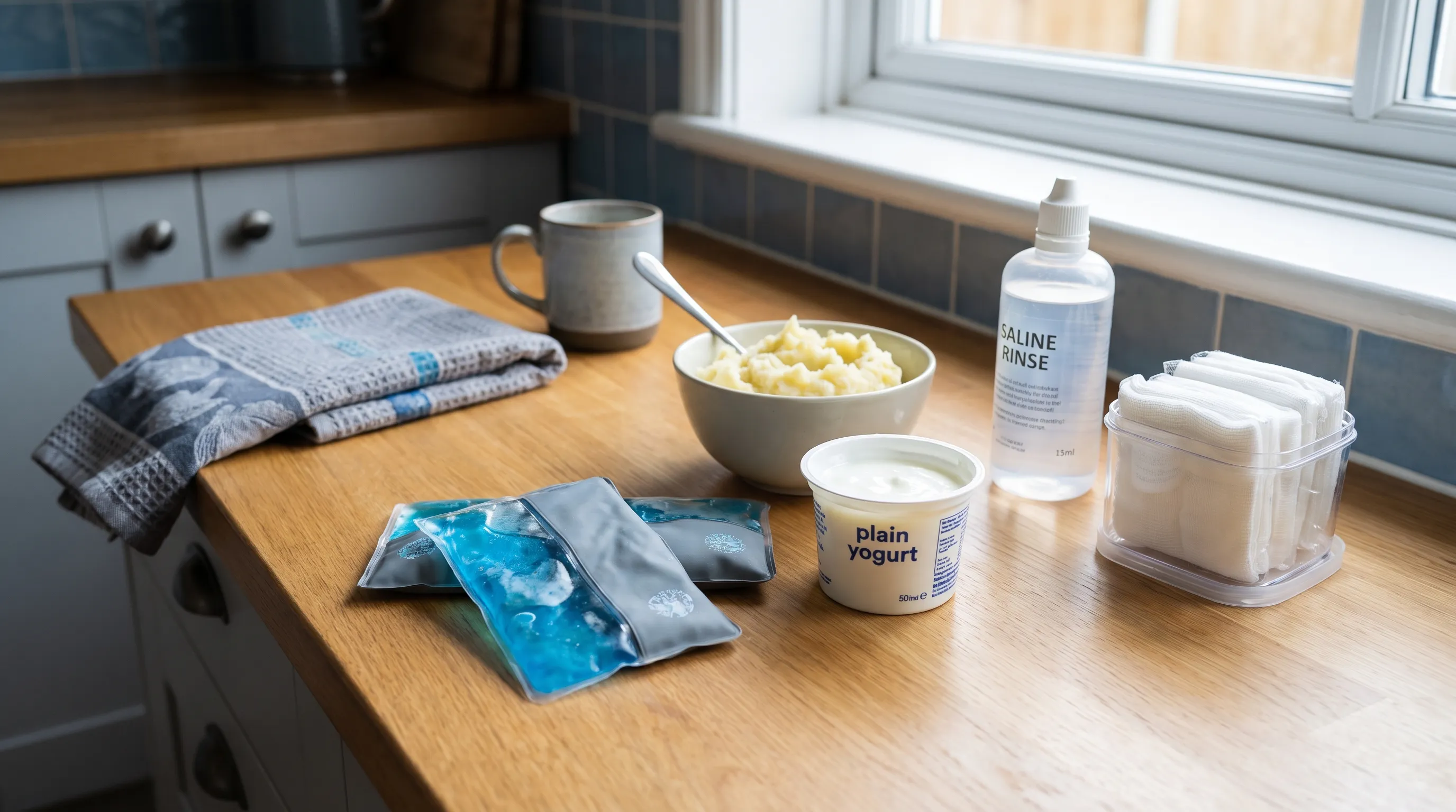
Transition to a soft, nutrient-dense diet for the first three to five days. Prioritize cool or room-temperature foods like mashed potatoes, smoothies, scrambled eggs, and oatmeal. Avoid using a straw, as suction forces create negative intraoral pressure that destabilizes the clot. After twenty-four hours, begin gentle saline rinses using half a teaspoon of dissolved sea salt in eight ounces of warm water. Perform these rinses after meals and before bedtime to clear debris and maintain a balanced oral microbiome. Resume normal brushing, carefully avoiding the surgical site for the first few days. Gradual bone remodeling occurs over three to six months, while soft tissue maturation typically completes within four weeks.
Recognizing Complications and When to Seek Help
Normal postoperative symptoms include mild oozing, moderate swelling peaking at forty-eight to seventy-two hours, and manageable discomfort responsive to prescribed or over-the-counter analgesics. However, seek immediate professional evaluation if bleeding soaks through multiple gauze pads hourly, pain intensifies sharply after day three, swelling spreads to the neck or floor of the mouth, fever develops, or you experience trismus preventing mouth opening beyond two finger widths. These signs indicate potential infection, hematoma formation, or alveolar osteitis requiring clinical debridement, antibiotic therapy, or specialized wound packing.
Financial Considerations and Finding the Right Provider
Accessing professional dental care requires navigating insurance structures, understanding cost variables, and selecting qualified clinicians who prioritize evidence-based practice and patient education.
Typical Cost Ranges and Payment Options
Simple extractions typically range from one hundred fifty to three hundred fifty dollars per tooth, while surgical removals of impacted or fractured teeth may cost three hundred to one thousand two hundred dollars, depending on anatomical complexity, anesthesia requirements, and geographic location. Dental insurance frequently covers fifty to eighty percent of medically necessary extractions after meeting annual deductibles. For uninsured patients, community health centers, dental schools, and charitable clinics offer significantly reduced fees through resident-supervised treatment programs. Flexible spending accounts, healthcare credit cards with promotional financing, and in-house membership plans can also alleviate upfront financial burdens.
How to Choose a Qualified Dental Provider
Selecting a clinician requires verifying board certification, reviewing patient testimonials, and assessing facility accreditation. General dentists routinely perform simple extractions and straightforward surgical removals. Oral and maxillofacial surgeons, who complete four to six additional years of hospital-based residency training after dental school, manage complex impactions, medically compromised patients, and full-mouth rehabilitations. Verify that the practice utilizes digital radiography, adheres to strict sterilization protocols compliant with CDC infection control guidelines, and provides comprehensive postoperative care instructions. When researching ways to get a tooth out safely, prioritizing clinician expertise over convenience ultimately protects your long-term oral health and financial investment.
Comparing Extraction Approaches: Clinical Overview
| Approach | Ideal Indication | Average Procedure Time | Pain Level | Healing Timeline | Cost Range |
|---|---|---|---|---|---|
| Simple Clinical Extraction | Fully erupted, intact crown | 15-30 minutes | Mild-Moderate | 14-21 days soft tissue | $150-$350 |
| Surgical Extraction | Impacted, fractured, multi-rooted | 30-90 minutes | Moderate (managed) | 3-6 months bone remodel | $300-$1,200+ |
| Natural Exfoliation (Pediatric) | Physiologically mobile primary teeth | N/A (Gradual) | Minimal | Immediate after shedding | $0 |
| At-Home Care (Mild Mobility) | Fully detached, non-resisting teeth | Variable | Low-Moderate | 7-14 days | Minimal |
This comparative framework illustrates why professional evaluation remains indispensable when exploring ways to get a tooth out. While natural and mild cases may tolerate observation or minimal assistance, complex pathological scenarios demand surgical precision, radiographic guidance, and pharmacological support that cannot be replicated outside a clinical environment.
Frequently Asked Questions
Can I safely remove a loose tooth at home without a dentist?
If a tooth is naturally mobile, completely detached at the root, and shows no signs of infection or swelling, gentle wiggling or twisting may allow it to fall out naturally. However, if it is firmly anchored, painful, or partially attached, attempting removal at home risks nerve damage, severe bleeding, and infection. Professional evaluation is always recommended to preserve alveolar bone and prevent complications.
How long does it take to heal after a professional tooth extraction?
Initial soft tissue closure typically occurs within fourteen to twenty-one days, though complete alveolar bone remodeling takes three to six months. The first twenty-four hours are critical for blood clot stabilization. Pain and swelling generally peak on days two through three before gradually subsiding with proper care and prescribed anti-inflammatory medications. Adherence to postoperative instructions significantly accelerates this timeline.
What is dry socket and how can I prevent it after extraction?
Dry socket, or alveolar osteitis, occurs when the protective blood clot dislodges or dissolves prematurely, exposing underlying bone and nerve tissue. It causes intense, radiating pain, foul odor, and delayed healing. Prevention includes avoiding straws, smoking, vigorous rinsing, and hot beverages for at least seventy-two hours post-procedure, while strictly following your clinician's oral hygiene and medication protocols. Smoking cessation is particularly crucial, as nicotine impairs microvascular circulation and delays tissue repair.
When should I seek emergency dental care after removing a tooth?
Seek immediate professional care if bleeding persists beyond twenty-four hours despite firm gauze pressure, if you develop a fever above one hundred point four degrees Fahrenheit, experience severe pain unresponsive to prescribed medication, notice purulent discharge, encounter difficulty breathing or swallowing, or if the surrounding gum tissue exhibits rapid, worsening swelling, erythema, or numbness. These symptoms indicate potential infection, hematoma expansion, or neurovascular compromise requiring prompt clinical intervention.
Does dental insurance typically cover tooth extractions?
Most comprehensive dental insurance plans cover medically necessary extractions, typically reimbursing fifty to eighty percent of the procedure cost after the annual deductible is met. Coverage limits often distinguish between simple and surgical extractions, with surgical codes requiring additional pre-authorization. Medicaid, Medicare Advantage plans, and private dental networks vary significantly by region and policy tier. Contacting your insurer directly with specific CDT codes before scheduling ensures transparent financial expectations and prevents unexpected out-of-pocket expenses.
Conclusion
Navigating the various ways to get a tooth out requires a balanced approach that respects both biological healing processes and clinical necessity. While children's primary teeth typically exfoliate naturally and mildly mobile adult teeth may occasionally require gentle assistance, severe decay, advanced periodontal disease, and impacted structures demand professional intervention. The distinction between physiological shedding and pathological mobility determines whether observation, minimally invasive extraction, or advanced surgical management is appropriate. Prioritizing sterile techniques, evidence-based pain management, and strict adherence to postoperative protocols minimizes complications, accelerates tissue regeneration, and preserves the structural integrity of the dental arch.
Ultimately, safe tooth removal is not merely about extracting a compromised structure; it is about creating the optimal biological environment for future rehabilitation, whether that involves implant placement, fixed prosthetics, or orthodontic alignment. Consulting a licensed dental professional ensures accurate diagnosis, appropriate anesthesia, and comprehensive recovery support that home methods simply cannot provide. By integrating clinical expertise with informed self-care, patients can confidently navigate dental extraction procedures while safeguarding their long-term oral health and overall well-being.
How we work
Researched and drafted with AI assistance, then reviewed by a human editor against the cited sources.
This article is general health information, not medical advice. Consult a qualified healthcare professional about your own situation.