Understanding Bone Growth in the Mouth: Causes, Types, and Clinical Management

The human jaw is a remarkably dynamic structure, continuously adapting to mechanical demands, nutritional inputs, and systemic physiological shifts. When patients notice unusual hard lumps, raised ridges, or asymmetrical bumps along the gums, palate, or inner cheeks, the natural concern often centers around the possibility of disease. In reality, most of these presentations represent benign bone growth in mouth structures that develop over years as adaptive responses to bite forces, genetic programming, or localized trauma. Understanding why these formations occur, when they require clinical intervention, and how to maintain optimal skeletal health in the oral cavity is essential for long-term dental wellness. This comprehensive guide explores the cellular mechanisms behind oral bone development, categorizes common clinical presentations, outlines evidence-based diagnostic protocols, and provides actionable strategies for monitoring and management. Whether you have noticed a smooth, painless bump on the roof of your mouth, are preparing for implant surgery, or simply want to preserve jawbone density through midlife, the information presented here synthesizes current maxillofacial research with practical clinical guidance to help you make informed decisions about your oral health journey.
Understanding Oral Bone Anatomy and Physiology
The oral cavity is anchored by two primary skeletal components: the maxilla (upper jaw) and the mandible (lower jaw). Unlike the static bone found in the long appendicular skeleton, the jawbone is highly responsive to functional loads, making it uniquely susceptible to both adaptive thickening and pathological resorption. The alveolar process, the thickened ridge of bone that contains the tooth sockets, undergoes continuous remodeling throughout adulthood. This dynamic equilibrium is governed by two specialized cell types: osteoclasts, which resorb mineralized tissue, and osteoblasts, which lay down new osteoid matrix that eventually mineralizes into mature bone.
Free calculatorHeadache Location ChartIdentify headache types by locationOpen the calculatorThe Alveolar Ridge and Jaw Architecture
The mandible consists of a dense cortical outer shell surrounding a cancellous (spongy) inner network rich in hematopoietic marrow spaces and vascular channels. The maxilla shares a similar composition but features thinner cortical plates and higher trabecular density, which explains why the upper jaw typically responds more rapidly to mechanical stress. When teeth exert occlusal forces during mastication or parafunctional habits like bruxism, mechanotransduction pathways trigger localized microdamage. In response, signaling molecules such as sclerostin, RANKL, and BMPs (bone morphogenetic proteins) coordinate the recruitment of osteoprogenitor cells to the affected site. This physiological cascade is the foundation of bone growth in mouth structures, particularly in regions subjected to repetitive stress. The periosteum, a highly innervated and vascularized connective tissue membrane enveloping the bone, plays a critical role in sensing strain and initiating adaptive thickening. When this system functions optimally, the jaw maintains structural integrity, supports periodontal attachment, and distributes masticatory loads efficiently.
Cellular Dynamics: Osteoblasts and Osteoclasts
The balance between bone formation and resorption is not static; it fluctuates in response to dietary mineral availability, hormonal fluctuations, systemic inflammation, and localized mechanical stimuli. Parathyroid hormone (PTH), calcitonin, and vitamin D metabolites regulate calcium homeostasis, directly influencing osteoblast differentiation from mesenchymal stem cells. When mechanical strain exceeds baseline thresholds, osteocytes embedded within the mineralized matrix detect fluid shear stress in the canaliculi and downregulate sclerostin, a natural inhibitor of the Wnt signaling pathway. Sclerostin reduction unleashes the Wnt cascade, promoting osteoblast proliferation and matrix deposition. This molecular mechanism explains why individuals with heavy bite forces, malocclusion, or habitual teeth grinding frequently develop compensatory bone growth in mouth tissues. Research consistently demonstrates that jawbone adaptation follows Wolff's Law: bone remodels precisely along lines of mechanical stress. Consequently, areas experiencing chronic pressure often exhibit localized thickening, while unloaded regions (such as post-extraction sites) undergo rapid resorption. Maintaining this delicate equilibrium requires adequate protein synthesis, micronutrient availability, and proper occlusal distribution, all of which will be detailed in subsequent management sections.
Common Types of Bone Growth in the Mouth
Clinical presentations of oral skeletal proliferation vary significantly in etiology, location, and histological composition. Distinguishing between physiological adaptation, developmental anomalies, and reactive proliferations is essential for appropriate triage. The following subsections outline the most frequently encountered benign formations, their clinical characteristics, and their typical natural history.
Mandibular and Palatal Tori
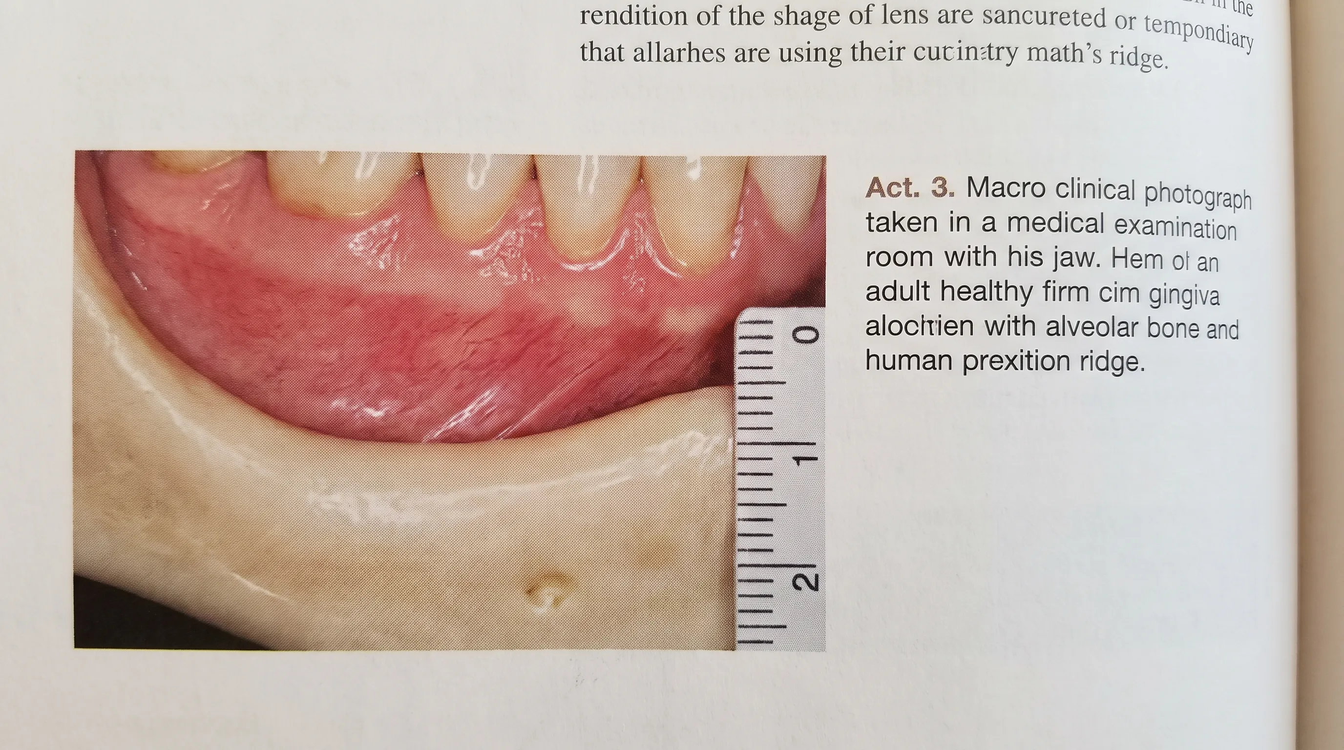
Torus mandibularis and torus palatinus represent the most prevalent forms of non-pathological bone growth in mouth. Mandibular tori appear as bilateral, symmetrical, nodular protrusions along the lingual surface of the lower jaw, typically premolar region. Palatal tori present as midline, dome-shaped bony masses on the hard palate, varying from subtle ridges to large, lobulated structures that extend toward the posterior pharynx. Both conditions are classified as developmental exostoses with strong hereditary components. Studies published by the National Institutes of Health (NIH) indicate a prevalence rate of 8% to 15% in the general population, with significant variation across ethnic groups. Tori are characteristically asymptomatic, slow-growing, and covered by thin, tightly adherent mucosa. They rarely require intervention unless they interfere with denture fabrication, cause recurrent mucosal ulceration from sharp foods, or obstruct airway space in severe obstructive sleep apnea cases. Clinicians monitor these formations using periodic measurements and clinical photography to document stability.
Dental Exostoses and Benign Osteomas
Exostoses differ from tori primarily in their location and unilateral presentation. These smooth, pedunculated or broad-based bony outgrowths frequently arise on the buccal (cheek-facing) aspect of the maxillary or mandibular arch, often near the molars or along the alveolar ridge. Unlike tori, which are genetically programmed and symmetrically distributed, exostoses frequently develop in response to chronic friction, ill-fitting prosthetics, or localized periodontal inflammation. Histologically, both consist of dense lamellar bone with minimal medullary space, classifying them as osteogenic lesions rather than neoplasms. True osteomas, though rare in the oral cavity, present as solitary, well-circumscribed, slow-growing masses that may attach to the cortical plate via a stalk (pedunculated) or blend seamlessly with the underlying bone (sessile). While osteomas are almost invariably benign, multiple facial osteomas can indicate Gardner syndrome, a rare autosomal dominant condition associated with colonic polyposis and increased malignancy risk. Routine screening ensures early identification of syndromic patterns.
Reactive Hyperplasia vs True Bone Formation
Not all hard tissue enlargements represent genuine osseous proliferation. Some clinically palpable masses result from reactive soft tissue hyperplasia, fibrous dysplasia, or calcified lesions such as sialoliths or phleboliths. Fibrous dysplasia replaces normal marrow with fibrous stromal tissue, creating a characteristic ground-glass appearance on radiographs. True bone growth in mouth exhibits distinct radiopacity matching or exceeding adjacent cortical bone, with well-defined borders and intact trabecular architecture. Differentiating these conditions requires correlating clinical examination with advanced imaging. When uncertainty persists, incisional biopsy remains the gold standard for definitive histopathological diagnosis. Clinicians emphasize patient education regarding self-monitoring: any rapid expansion, pain upon palpation, mucosal discoloration, or paresthesia warrants prompt evaluation to exclude aggressive pathology.
Causes and Risk Factors for Abnormal Oral Bone Growth
The development of localized skeletal enlargements in the oral cavity rarely stems from a single causative factor. Instead, it typically emerges from a complex interplay of genetic predisposition, biomechanical loading, and systemic metabolic influences. Understanding these contributors allows patients and providers to implement targeted preventative measures and modify environmental triggers that exacerbate compensatory proliferation.
Genetic Blueprint and Hereditary Patterns
Population studies consistently demonstrate a strong familial aggregation for tori and exostoses. Twin studies reveal heritability estimates exceeding 60%, suggesting polygenic inheritance patterns involving multiple loci regulating osteogenesis. Specific single nucleotide polymorphisms in genes encoding bone morphogenetic proteins (BMP2, BMP4), transforming growth factor-beta (TGF-β), and collagen type I alpha 1 (COL1A1) have been implicated in heightened osteoblast responsiveness to mechanical stimuli. Individuals of Asian, Inuit, and certain African descent exhibit significantly higher prevalence rates, underscoring evolutionary adaptations to dietary textures and masticatory demands in ancestral environments. Genetic counseling is rarely indicated unless multiple osteomas suggest syndromic involvement, but family history remains a valuable clinical marker for anticipating developmental trajectories. Patients with first-degree relatives exhibiting prominent tori should undergo baseline documentation during adolescence to establish individualized monitoring protocols.
Biomechanical Stress and Occlusal Forces
Chronic mechanical loading represents the most modifiable driver of compensatory bone growth in mouth tissues. Bruxism, clenching, malocclusion, and premature tooth contacts generate microtrauma that stimulates periosteal apposition. The mandible, subjected to approximately 70 kilograms of bite force during normal chewing and significantly higher loads during parafunctional habits, responds through localized cortical thickening. Malpositioned teeth create uneven force distribution, causing specific regions to bear disproportionate stress. Over time, osteocytes signal for additional mineral deposition to reinforce overloaded segments. Orthodontic interventions that correct occlusal discrepancies can halt progression by redistributing forces evenly across the arch. Custom-fabricated night guards further mitigate nocturnal clenching, reducing cyclical microfracture and subsequent reparative bone formation. Biomechanical assessment using occlusal analysis systems and digital bite registration provides objective data to guide therapeutic intervention.
Systemic Influences: Hormones and Medications
Endocrine fluctuations profoundly impact oral skeletal metabolism. Estrogen deficiency during menopause accelerates alveolar bone resorption, paradoxically increasing fracture risk while potentially triggering compensatory reactive growth in unloaded areas. Conversely, hyperparathyroidism elevates systemic PTH, causing generalized demineralization that may mask localized proliferations. Certain pharmacological agents also modulate bone turnover. Selective serotonin reuptake inhibitors (SSRIs), proton pump inhibitors (PPIs), and long-term corticosteroid use negatively impact mineral absorption and osteoblast viability. Interestingly, antiresorptive medications like bisphosphonates and denosumab, prescribed for osteoporosis, profoundly suppress osteoclast activity. While they reduce overall jawbone remodeling, they concurrently increase the risk of medication-related osteonecrosis of the jaw (MRONJ), a distinct clinical entity from benign bone growth in mouth that requires strict dental surveillance before invasive procedures. Patients on these therapies must maintain meticulous oral hygiene and undergo pre-surgical risk stratification to prevent necrotic complications.
When to Seek Professional Evaluation
Distinguishing between benign developmental anomalies and pathologically significant skeletal changes requires clinical expertise, advanced imaging, and systematic monitoring. While most oral bone formations remain asymptomatic, certain clinical presentations mandate prompt evaluation to rule out aggressive lesions, malignancies, or systemic disorders manifesting in the maxillofacial region.
Recognizing Clinical Warning Signs
Patients should schedule an immediate consultation if they experience rapid enlargement (visible growth over weeks to months), spontaneous pain without mechanical provocation, mucosal ulceration that fails to heal within two weeks, bleeding upon light contact, or radiating numbness suggestive of inferior alveolar nerve compression. Benign tori and exostoses grow imperceptibly over years, remaining painless and mucosa-intact. In contrast, aggressive neoplasms like osteosarcoma or chondrosarcoma present with accelerated expansion, paresthesia, tooth mobility, and radiographic ill-defined borders. For comprehensive guidance on identifying abnormal oral lesions and warning signs, refer to the Mayo Clinic. Soft tissue infections, abscesses, or periodontal cysts can also create firm, tender masses that mimic osseous growth. Any asymmetrical swelling crossing the midline, accompanied by systemic symptoms like unexplained weight loss, fatigue, or cervical lymphadenopathy, requires urgent maxillofacial referral. Early diagnosis dramatically improves treatment outcomes and preserves functional anatomy.
Advanced Diagnostic Workflows
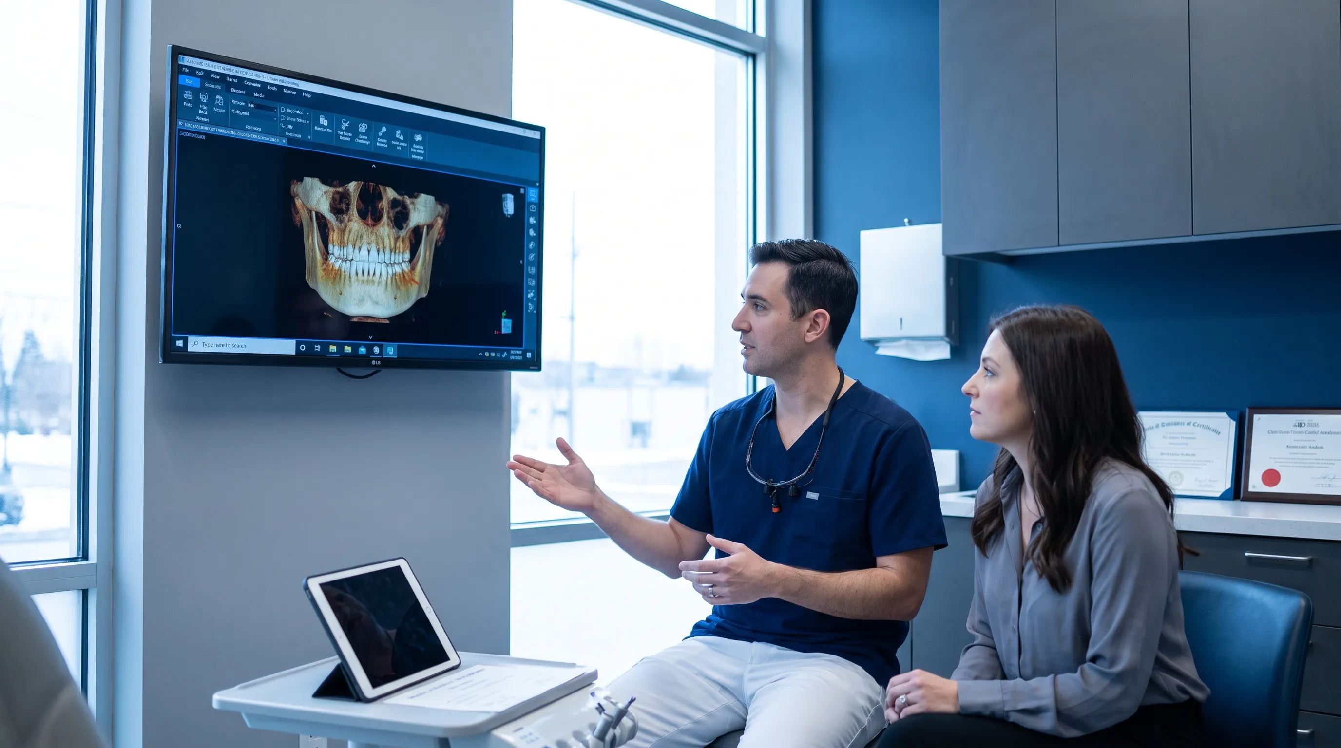
The diagnostic algorithm begins with comprehensive clinical examination, including visual inspection, palpation for mobility and tenderness, and documentation of dimensions using calibrated periodontal probes. Panoramic radiography provides an initial overview of mandibular and maxillary architecture, identifying gross anatomical variations. For precise volumetric assessment, cone-beam computed tomography (CBCT) delivers high-resolution 3D reconstructions, enabling accurate measurement of cortical thickness, medullary cavity involvement, and proximity to vital structures like the mental foramen and maxillary sinus. Histopathological analysis remains definitive when imaging reveals atypical features. Incisional biopsy under local anesthesia extracts representative tissue for microscopic evaluation, confirming benign lamellar bone versus fibro-osseous dysplasia, giant cell granuloma, or malignant transformation. Dental and oral surgery guidelines emphasize radiographic correlation with clinical findings before any surgical intervention for suspected bone growth in mouth lesions.
Treatment Options and Management Strategies
Management strategies for oral skeletal proliferations depend entirely on clinical presentation, symptom severity, and functional impact. Asymptomatic lesions typically require conservative monitoring, while mechanically obstructive or diagnostically ambiguous formations warrant procedural intervention. Modern oral surgery techniques prioritize minimally invasive approaches, accelerated healing protocols, and long-term structural preservation.
Observation vs Surgical Contouring
For patients with small, stable tori or exostoses causing no functional limitation, watchful waiting represents the standard of care. Clinicians document baseline measurements during routine hygiene visits and capture intraoral photographs annually to detect subtle changes. Surgical intervention becomes indicated when the bony mass interferes with denture fabrication, causes recurrent mucosal trauma from sharp foods, obstructs proper speech articulation, or creates significant aesthetic concerns. The surgical procedure, termed toriectomy or exostosectomy, involves local anesthesia administration, mucoperiosteal flap reflection, precise osteotomy using piezoelectric handpieces or carbide burs, and tension-free primary closure with resorbable sutures. Piezoelectric surgery selectively cuts mineralized tissue while preserving neurovascular bundles, significantly reducing postoperative edema and paresthesia risk. Postoperative protocols emphasize soft dietary progression for 10 to 14 days, chlorhexidine rinses to prevent surgical site infection, and avoidance of positive pressure activities like blowing or sucking that could disrupt the clot. Healing typically completes within three to six weeks, with bone remodeling gradually restoring cortical continuity.
Bone Grafting and Guided Tissue Regeneration
In scenarios where bone growth in mouth coexists with significant alveolar resorption, simultaneous grafting procedures restore architectural integrity. Autogenous bone harvested from the chin, retromolar pad, or iliac crest provides optimal osteogenic potential, containing live osteoblasts, growth factors, and structural scaffolding. Allografts (donor bone), xenografts (bovine-derived), and synthetic calcium phosphates serve as osteoconductive matrices, allowing host cells to infiltrate and deposit new bone. Guided bone regeneration (GBR) techniques utilize resorbable collagen membranes to exclude soft tissue ingrowth while stabilizing the graft material during the critical consolidation phase. Vascular endothelial growth factor (VEGF) and platelet-rich fibrin (PRF) applications accelerate angiogenesis and cellular proliferation, shortening integration timelines. Successful grafting requires strict compliance with postoperative restrictions, smoking cessation (nicotine profoundly impairs neovascularization), and systemic optimization of mineral nutrition. Implant placement typically occurs after a four to six month healing interval, confirmed via follow-up CBCT demonstrating radiographic integration and trabecular bridging.
Orthodontic and Prosthodontic Considerations
Orthodontic therapy must account for underlying skeletal morphology to prevent iatrogenic damage. Large tori alter arch form and complicate bracket placement, often requiring surgical reduction before fixed appliance bonding. Palatal expanders may inadvertently fracture thin mucosa overlying prominent tori, necessitating prior surgical modification or customized appliance design. In edentulous patients, implant-supported overdentures frequently require ridge augmentation to compensate for extensive bone loss, yet concurrent tori complicate prosthetic path of insertion. Digital workflow integration, including intraoral scanning and virtual surgical planning, enables clinicians to simulate implant trajectories, design patient-specific cutting guides, and anticipate soft tissue limitations before entering the operating theater. Prosthodontic management of patients with bone growth in mouth emphasizes customized relief channels in denture bases, pressure-indicating paste verification during try-in, and periodic relining to accommodate natural bone remodeling over decades. Collaborative care between oral surgeons, prosthodontists, and orthodontists ensures predictable, functionally optimized outcomes.
| Clinical Feature | Mandibular/Palatal Tori | Dental Exostoses | Fibrous Dysplasia |
|---|---|---|---|
| Typical Location | Lingual mandible, midline palate | Buccal maxilla/mandible, molar region | Maxilla > mandible, diffuse expansion |
| Symmetry | Usually bilateral | Unilateral or multifocal | Often unilateral, asymmetric |
| Radiographic Appearance | Homogenous radiopacity, well-defined borders | Focal radiopacity, pedunculated or sessile | Ground-glass opacification, ill-defined margins |
| Growth Rate | Extremely slow, decades | Slow, linked to mechanical stress | Variable, may accelerate during puberty |
| Malignant Potential | None | None | Very low (<1%), typically benign |
| Indication for Surgery | Prosthetic obstruction, ulceration, airway compromise | Functional interference, diagnostic uncertainty | Pain, asymmetry, nerve compression, malignancy exclusion |
Lifestyle Modifications and Preventative Care
While genetics and developmental factors establish baseline skeletal architecture, daily habits profoundly influence long-term bone quality, remodeling balance, and susceptibility to pathological proliferation. Implementing evidence-based lifestyle strategies supports optimal oral skeletal health, reduces compensatory bone growth in mouth driven by mechanical stress, and preserves alveolar bone density critical for tooth retention and implant success.
Targeted Nutrition for Oral Skeletal Integrity
Bone metabolism relies on precise micronutrient availability. Calcium remains foundational, but modern nutrition science emphasizes the synergistic roles of vitamin D3, vitamin K2, and magnesium. Vitamin D3 facilitates intestinal calcium absorption and upregulates osteocalcin production, while vitamin K2 activates matrix Gla protein, directing calcium into bone matrix rather than soft tissues or vascular walls. Magnesium serves as a cofactor for over 300 enzymatic reactions, including ATP production required for osteoblast activity and vitamin D activation. Dietary sources include leafy greens (kale, collards), fermented foods (natto, aged cheese for K2), fatty fish (salmon, mackerel for D3), nuts and seeds (pumpkin, almonds for magnesium), and fortified dairy or plant milks. Supplementation should be guided by serum testing, as excessive calcium without D3/K2 can precipitate ectopic calcification. For detailed intake recommendations and deficiency guidelines, consult the NIH Office of Dietary Supplements. Anti-inflammatory dietary patterns, such as the Mediterranean or Okinawan models, reduce systemic cytokines like TNF-α and IL-6 that accelerate osteoclastogenesis. Limiting refined sugars, processed carbohydrates, and excessive alcohol intake mitigates chronic low-grade inflammation that disrupts bone turnover equilibrium. Adequate protein intake (1.0 to 1.2 g/kg body weight) provides essential amino acids for collagen matrix synthesis, the organic framework upon which hydroxyapatite mineralization occurs.
Daily Habits to Preserve Jawbone Density
Oral hygiene extends beyond caries prevention; it directly safeguards periodontal attachment and underlying alveolar bone. Plaque biofilm harbors Porphyromonas gingivalis and Tannerella forsythia, pathogenic species that trigger chronic periodontitis, releasing matrix metalloproteinases that degrade periodontal ligament and stimulate localized bone resorption. For evidence-based brushing and flossing protocols, refer to CDC oral health guidelines. Twice-daily mechanical plaque disruption with soft-bristled brushing, interdental brushes, and water flossing removes inflammatory triggers before they penetrate gingival sulci. Antimicrobial mouth rinses containing essential oils or cetylpyridinium chloride supplement mechanical cleaning, but prolonged chlorhexidine use requires periodic discontinuation to prevent staining and taste alteration. Nighttime occlusal guard fabrication for bruxism patients significantly reduces parafunctional loading, halting progressive cortical thickening and microfracture cycles. Stress management techniques, including cognitive behavioral therapy, meditation, and progressive muscle relaxation, reduce nocturnal clenching frequency by lowering sympathetic nervous system hyperarousal. Regular dental prophylaxis every three to six months, combined with annual radiographic evaluation, enables early detection of bone growth in mouth lesions before they progress to functional compromise. Patients committed to these evidence-based practices consistently demonstrate superior periodontal stability, longer tooth retention, and more predictable prosthetic rehabilitation outcomes.
Frequently Asked Questions
Is bone growth in the mouth a normal part of aging?
Mild bone remodeling throughout adulthood is normal, but distinct bone growth in mouth like tori or exostoses is not strictly age-related. These benign formations typically develop gradually in early adulthood and may become more noticeable over decades due to mechanical stress, genetic factors, and cumulative occlusal forces.
Can mandibular or palatal tori become cancerous?
No. Tori and dental exostoses are benign bone proliferations with virtually zero malignant transformation potential. However, any rapid growth, ulceration, bleeding, or asymmetrical expansion warrants immediate clinical evaluation to rule out pathological conditions unrelated to normal bone growth in mouth.
What is the standard treatment for large tori that interfere with dentures?
When bone growth in mouth physically obstructs prosthetic seating, surgical removal (toriectomy) is indicated. The procedure involves a localized flap, careful bone contouring with surgical burs or piezoelectric devices, and suturing. Recovery typically requires 2 to 4 weeks of soft diet and meticulous oral hygiene.
Do dietary changes actually stop abnormal jawbone growth?
Nutrition cannot reverse established bony protrusions, but optimizing calcium, vitamin D3, K2, magnesium, and protein intake supports healthy bone remodeling and may reduce excessive compensatory growth triggered by mechanical stress. Anti-inflammatory diets also mitigate periodontal bone loss.
How do dentists diagnose whether bone growth in mouth is benign or pathological?
Diagnosis begins with clinical palpation and intraoral examination, followed by panoramic radiography or cone-beam computed tomography (CBCT) to assess trabecular architecture, cortical thickness, and relationship to vital structures like the inferior alveolar nerve. Sudden onset, pain, or neurological changes prompt biopsy referral.
Conclusion
Recognizing the distinction between physiological adaptation, developmental anomalies, and pathological skeletal changes empowers patients to navigate oral health decisions with confidence. Most instances of bone growth in mouth represent harmless, genetically influenced, or biomechanically driven adaptations that require nothing more than periodic monitoring and conservative care. When functional impairment, diagnostic uncertainty, or aesthetic concerns arise, modern oral surgery and regenerative techniques offer safe, predictable interventions that restore form and function without compromising structural integrity. By prioritizing comprehensive clinical evaluation, adhering to personalized management protocols, and implementing targeted nutritional and lifestyle strategies, individuals can maintain robust jawbone architecture well into later life. Always consult a licensed dental or medical professional before initiating treatment, particularly when considering surgical modification of oral skeletal structures or managing systemic medications that influence bone metabolism. Proactive monitoring, evidence-based care, and informed lifestyle choices remain the cornerstone of lifelong maxillofacial health.

How we work
Researched and drafted with AI assistance, then reviewed by a human editor against the cited sources.
This article is general health information, not medical advice. Consult a qualified healthcare professional about your own situation.