Blood Clot After Tooth Extraction: Complete Healing Guide & Care Tips
A tooth extraction is one of the most common dental procedures performed worldwide, yet the days following the removal of a tooth require careful attention to ensure optimal recovery. At the center of this healing process is a small but vital biological structure: the blood clot that forms in the empty socket. Understanding how to protect this delicate formation is the key to preventing complications, minimizing discomfort, and accelerating tissue regeneration. When the blood clot after tooth extraction remains undisturbed, it acts as a natural protective barrier against oral bacteria, foreign debris, and painful nerve exposure. However, everyday habits, dietary choices, or certain medications can inadvertently disrupt this clot, leading to a condition known as alveolar osteitis, commonly referred to as dry socket. This comprehensive guide explores the science behind post-extraction healing, outlines evidence-based care protocols, and provides actionable strategies to safeguard your recovery. By combining clinical insights with practical, everyday recommendations, you will gain the knowledge necessary to navigate the healing timeline with confidence. Whether you are preparing for an upcoming extraction or currently in the midst of recovery, understanding the intricate role of the clot will empower you to make informed decisions, communicate effectively with your dental care team, and achieve a smooth, complication-free recovery.
Understanding the Healing Process: What Is a Dental Blood Clot?
The Biological Mechanism of Socket Closure
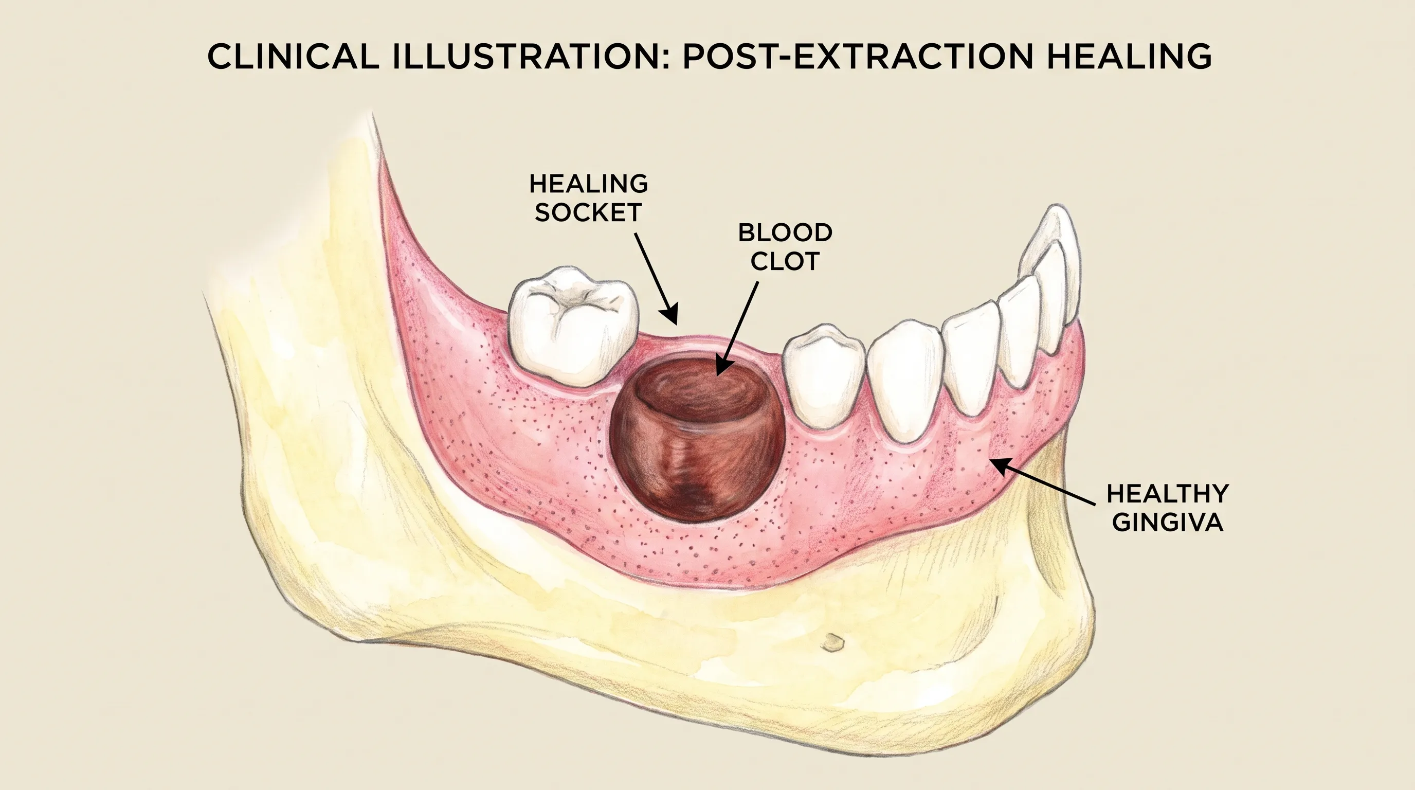
When a tooth is removed, the surrounding bone, periodontal ligament, and gingival tissue are left exposed to the highly dynamic oral environment. Within minutes of the procedure, circulating platelets begin to aggregate at the wound margins, releasing a cascade of clotting factors that trigger rapid coagulation. This biochemical sequence transforms liquid blood into a semi-solid fibrin matrix, effectively sealing the extraction socket. The blood clot after tooth extraction is not merely a temporary mechanical plug; it functions as a highly active biological scaffold. Within this provisional matrix, fibroblasts, osteoblasts, and endothelial cells migrate to initiate angiogenesis, collagen synthesis, and bone remodeling. According to clinical guidelines from the American Dental Association, this initial clot is indispensable because it prevents bacterial colonization and creates a protected microenvironment where granulation tissue can develop safely. Without this protective barrier, the underlying alveolar bone and nerve endings would remain vulnerable to microbial invasion, severe inflammation, and intense neuropathic pain. The structural stability of this clot depends heavily on localized perfusion, adequate systemic hydration, and the absolute absence of mechanical trauma or chemical interference during the critical first seventy-two hours.
Free calculatorWater Intake CalculatorCalculate daily water needsOpen the calculatorTimeline of Normal Healing After Tooth Removal
The post-extraction healing trajectory follows a highly predictable physiological sequence. During the initial twenty-four hours, the clot undergoes contraction and stabilization, while minor serosanguineous oozing is entirely normal as coagulation factors finalize their enzymatic work. By day three, fibroblast activity accelerates, and the clot begins organizing into early vascularized granulation tissue. This phase typically coincides with a noticeable reduction in localized swelling and throbbing discomfort. Between days five and seven, epithelial cells begin migrating across the granulation tissue, progressively covering the socket opening from the periphery inward. By the second week, the gingival architecture appears clinically healed, though underlying osseous remodeling continues for several additional months. The blood clot after tooth extraction remains functionally active throughout this transitional period, gradually integrating with newly formed connective tissue. Maintaining optimal oral pH levels, adhering to a nutrient-dense soft-food regimen, and avoiding cardiovascularly strenuous activities are clinically proven methods to support this biological timeline. Peer-reviewed research published in the Journal of Oral and Maxillofacial Surgery emphasizes that strict adherence to post-operative instructions reduces complication rates by over sixty percent, particularly in complex surgical extractions involving impacted mandibular third molars or concurrent bone grafting procedures.
How to Identify a Healthy Blood Clot vs. Dry Socket
Visual and Sensory Differences
Distinguishing between a properly healing socket and a developing pathological condition requires careful attention to subtle clinical signs and sensory feedback. A healthy blood clot after tooth extraction typically presents as a dark red, deep maroon, or occasionally yellowish-white fibrinous coating. The surface may appear slightly irregular, moist, and semi-translucent, which reflects normal protein deposition and early cellular migration. Conversely, a dry socket occurs when the clot either dislodges prematurely, undergoes accelerated fibrinolysis, or fails to form entirely. When this protective layer is compromised, the underlying cortical and cancellous bone becomes directly exposed to the oral atmosphere, food debris, and microbial flora. Visually, an empty socket will appear stark, dry, and often pale gray or yellowish-white, lacking the moist, cohesive appearance of healthy granulation tissue. Patients frequently report an abrupt transition from dull, manageable post-surgical soreness to sharp, radiating pain that extends toward the temporomandibular joint, ear, or lateral neck region. The presence of an intact clot ensures that nociceptive nerve fibers remain adequately insulated, while its premature loss exposes them to environmental stimuli, resulting in severe, unremitting discomfort that standard analgesics often fail to control.
Pain Patterns and Odor Indicators
The temporal progression of pain serves as one of the most reliable clinical indicators of clot stability. Normal post-operative discomfort typically peaks within the first twenty-four to forty-eight hours and steadily declines with appropriate nonsteroidal anti-inflammatory drug (NSAID) therapy, localized cold application, and head elevation. However, if the blood clot after tooth extraction becomes compromised, pain characteristically intensifies between days three and five, directly contradicting the expected recovery curve. This delayed pain escalation represents the pathognomonic hallmark of alveolar osteitis. Additionally, clot failure frequently introduces a distinctive foul odor or persistent metallic taste within the oral cavity. This olfactory symptom results from anaerobic bacterial colonization, trapped food debris, and subsequent proteolytic breakdown of exposed tissue. A healthy clot maintains a relatively neutral oral environment and does not generate noticeable halitosis beyond standard post-surgical breath. Patients should also monitor for localized erythema extending beyond the surgical margin, excessive swelling, or purulent exudate, as these manifestations may indicate a secondary bacterial infection requiring prompt antimicrobial intervention and professional debridement.
Comparative Table: Healthy Clot vs. Alveolar Osteitis
| Clinical Feature | Healthy Blood Clot Presentation | Alveolar Osteitis (Dry Socket) |
|---|---|---|
| Visual Appearance | Dark red, maroon, or moist yellowish-white fibrin layer | Dry, hollow socket with exposed grayish/white alveolar bone |
| Pain Trajectory | Peaks at 24-48 hours, steadily decreases with medication | Sudden escalation on days 3-5, often radiating to ear/jaw |
| Odor & Taste | Neutral to mild post-surgical breath | Persistent foul odor and bad taste despite routine rinsing |
| Pharmacological Response | Effective pain control with NSAIDs or acetaminophen | Poor response to standard OTC or prescription analgesics |
| Socket Moisture | Consistently moist, covered by protective tissue | Appears desiccated, with possible purulent drainage if infected |
Essential Care Protocols to Preserve Your Blood Clot
Immediate Post-Operative Guidelines (First 24 Hours)
The initial twenty-four hours constitute the most critical physiological window for clot stabilization and vascular hemostasis. Immediately following the extraction, your oral surgeon will position a sterile gauze pad directly over the surgical site. Applying gentle but consistent biting pressure for thirty to sixty minutes promotes rapid platelet aggregation and minimizes post-operative hemorrhage. After this period, carefully remove the gauze and avoid repositioning replacement materials unless active, uncontrolled bleeding resumes. Spitting forcefully, inhaling through a straw, or rinsing vigorously generates significant negative intraoral pressure that can easily shear the fragile blood clot after tooth extraction from the socket walls. Instead, allow saliva and minor oozing to pool naturally or swallow gently. Applying an external ice pack in twenty-minute cycles for the first eighteen hours constricts superficial vasculature, substantially reducing post-operative edema and thermal nerve sensitivity. Maintain a reclined resting position with your head elevated on multiple pillows to decrease cephalic hydrostatic pressure, which otherwise promotes localized swelling. Avoid consuming hot beverages, engaging in heavy physical exertion, or bending forward abruptly, as elevated core temperature and increased cardiac output promote vasodilation that can destabilize the forming clot matrix.
Dietary Modifications and Hydration Strategies
Optimal nutrition directly influences tissue regeneration velocity, immune function, and coagulation efficiency. During the first three to five days, prioritize soft, lukewarm, or room-temperature foods that require minimal masticatory effort. Clinically recommended options include mashed potatoes, scrambled eggs, blended oatmeal, full-fat Greek yogurt, unsweetened applesauce, smoothies consumed with a spoon, and thoroughly cooked lentils. Avoid hard, crunchy, spicy, highly acidic, or extremely hot culinary items, as these can mechanically traumatize the surgical site or trigger localized inflammatory cascades. Systemic hydration plays an equally vital role in maintaining plasma volume, supporting salivary gland function, and facilitating nutrient transport to the healing socket. Consuming two to three liters of filtered water daily ensures adequate tissue perfusion. Conversely, carbonated beverages, alcoholic cocktails, and high-caffeine energy drinks should be strictly avoided, as they induce mild dehydration, alter oral pH, and interfere with prostaglandin-mediated healing pathways. If you are concurrently taking prescription analgesics or prophylactic antibiotics, adhere strictly to dietary contraindications to prevent gastrointestinal mucosal irritation or reduced drug bioavailability. Consistent intake of foods rich in ascorbic acid, bioavailable zinc, and complete amino acids significantly accelerates fibroblast mitosis and strengthens the collagenous network that ultimately replaces the temporary clot.
Oral Hygiene Practices That Promote Healing
Maintaining a clean oral ecosystem without disrupting the surgical site requires a carefully modified hygiene protocol. On the day of your extraction, refrain from brushing, flossing, or applying any mechanical cleaning devices near the treated quadrant. Beginning twenty-four hours post-procedure, resume gentle tooth brushing on adjacent dentition while meticulously steering the bristles away from the extraction socket. To control microbial proliferation, warm saline irrigation should commence on day two. Dissolve exactly one-half teaspoon of pharmaceutical-grade non-iodized salt into eight fluid ounces of sterilized warm water. Allow the solution to passively bathe the oral cavity before tilting your head to facilitate gentle drainage; avoid vigorous swishing or suction. This isotonic rinse reduces localized inflammation, draws out minor serous exudate, and establishes an alkaline microenvironment that actively discourages pathogenic bacterial colonization. Commercial antiseptic mouthwashes containing ethanol or harsh surfactants must be avoided during the first seven days, as they cause gingival desiccation and delay epithelial migration. If chlorhexidine gluconate was prescribed by your provider, utilize it precisely twice daily to maintain aseptic conditions while the blood clot after tooth extraction matures into resilient vascularized tissue.
Risk Factors That Complicate Blood Clot Formation
Tobacco Use and Vasoconstriction
Cigarette smoking and electronic vaporization represent two of the most heavily documented risk factors for clot failure and delayed wound closure. Nicotine induces rapid systemic and localized vasoconstriction, drastically reducing capillary blood flow to the extraction site and impairing the delivery of oxygen, leukocytes, and essential micronutrients required for tissue repair. The physical mechanics of inhalation generate a pronounced suction force within the oral vestibule, creating negative pressure that frequently dislodges newly stabilized clots. Furthermore, combustion byproducts and aerosolized chemical compounds act as direct tissue irritants, upregulating pro-inflammatory cytokines and significantly slowing fibroblast migration. Clinical epidemiological studies consistently demonstrate that patients who smoke or vape within seventy-two hours of extraction experience dry socket incidence rates up to tenfold higher than nonsmokers. If immediate tobacco cessation proves challenging, utilizing transdermal nicotine replacement patches instead of inhalation or oral delivery methods substantially mitigates local tissue damage and preserves clot architecture. Always disclose your smoking history transparently to your surgical provider, enabling them to implement prophylactic interventions such as medicated socket packing or targeted anti-inflammatory regimens.
Hormonal Contraceptives and Clot Stability
Estrogen-dominant combined oral contraceptives have been extensively linked to increased endogenous fibrinolytic activity, meaning the physiological environment breaks down fibrin networks more rapidly than typical. This pharmacological effect elevates the probability of premature clot dissolution during the crucial initial post-operative week. Women actively taking combination birth control formulations should proactively inform their oral surgeon prior to scheduling any surgical tooth removal, as clinicians may strategically time the procedure or implement additional clot preservation protocols. Research published in The Journal of Dental Research highlights that estrogenic fluctuations influence capillary permeability, platelet aggregability, and localized vascular tone, making meticulous post-operative care particularly vital for this patient demographic. Temporary discontinuation of oral contraceptives is rarely medically necessary, but heightened clinical vigilance, strict adherence to activity restrictions, and proactive communication with prescribing physicians ensure optimal surgical outcomes and uninterrupted healing progression.
Systemic Conditions and Medication Interactions
Pre-existing medical pathologies profoundly influence coagulation capacity, immune responsiveness, and overall tissue repair efficiency. Patients with poorly managed diabetes mellitus frequently experience impaired angiogenesis, dysfunctional neutrophil chemotaxis, and elevated susceptibility to opportunistic infections, all of which critically undermine clot integrity and stability. Autoimmune rheumatological disorders, hereditary bleeding diatheses such as hemophilia or von Willebrand disease, and chronic corticosteroid therapy further complicate the physiological healing cascade. Additionally, widely prescribed medications including aspirin, nonsteroidal anti-inflammatory agents, direct oral anticoagulants, and selective serotonin reuptake inhibitors inherently alter platelet function or prolong bleeding times. It is absolutely imperative to disclose your complete medical history, surgical background, and current medication inventory to your dental professional before any invasive intervention. Never discontinue prescribed anticoagulant or antiplatelet therapy without explicit guidance from your cardiologist or primary care physician, as the systemic risks of thromboembolic events or cardiovascular complications far outweigh localized dental concerns. Instead, your dental team will coordinate a multidisciplinary perioperative management plan that safely balances systemic hemostatic requirements with localized socket preservation strategies.
What to Do If You Suspect a Lost or Dislodged Clot
Emergency First Steps at Home
If you observe a sudden, unexplained escalation in pain intensity, visible bone exposure within the socket, or a persistent foul metallic taste around the third post-operative day, the blood clot after tooth extraction may have become dislodged or prematurely dissolved. While prompt professional evaluation remains essential, several immediate at-home interventions can significantly mitigate acute discomfort and reduce secondary infection risk until you reach your dental clinic. Begin by gently irrigating the mouth with lukewarm saline solution to carefully clear trapped food particles without generating forceful hydrostatic pressure. Administer an appropriate therapeutic dose of ibuprofen or acetaminophen according to your provider's previous instructions, deliberately avoiding aspirin derivatives unless explicitly directed. Apply a chilled external compress over the affected facial region for fifteen-minute intervals to modulate localized inflammatory response and reduce nociceptive signaling. Position a fresh, sterile, lightly moistened gauze pad over the socket and apply gentle, steady occlusal pressure for twenty to thirty minutes to temporarily stabilize the exposed area. Under no circumstances should you attempt to pack the socket with cotton swabs, household cloths, clove oil, or unverified home remedies, as these materials frequently introduce environmental contaminants or become irreversibly embedded within the wound bed, severely complicating professional debridement and prolonging recovery timelines.

Professional Treatments and Dressings
Upon clinical examination, your dentist will carefully debride the compromised socket using sterile instruments and sterile irrigation to thoroughly remove necrotic tissue, accumulated debris, and mature bacterial biofilm that actively sustain pain and delay epithelialization. Following meticulous antiseptic irrigation, the practitioner will typically introduce a specialized medicated dressing containing eugenol, benzocaine, or iodoform-impregnated gauze directly into the alveolar cavity. This targeted packing delivers immediate, localized analgesic effects by physically insulating exposed neurovascular bundles and providing sustained therapeutic release of antiseptic and anti-inflammatory compounds. Guidelines referenced by the National Institutes of Health indicate that these clinical dressings generally require replacement every forty-eight to seventy-two hours until healthy granulation tissue adequately bridges and covers the exposed osseous structures. In advanced or complex cases, clinicians may apply autologous platelet-rich fibrin (PRF) membranes or bioactive collagen scaffolds to dramatically accelerate cellular proliferation and stabilize the microenvironment. Systemic antibiotics are prescribed selectively, reserved strictly for presentations demonstrating clear clinical signs of spreading infection, such as purulent exudate, systemic fever, or regional lymphadenopathy, as routine prophylactic antimicrobial use does not prevent alveolar osteitis development.
Expected Recovery Timeline After Intervention
Once targeted professional treatment commences, patients typically experience profound pain relief within twenty-four hours of initial dressing application. The characteristic severe, radiating throbbing subsides substantially, though mild localized soreness may persist for an additional three to five days as active epithelialization progresses. Complete clinical resolution typically occurs within ten to fourteen days when supported by consistent follow-up appointments and appropriate dressing modifications. Throughout this accelerated healing phase, maintaining stringent oral hygiene compliance, adhering to restrictive dietary protocols, and completely avoiding tobacco exposure remain non-negotiable prerequisites for successful tissue restoration. The protective matrix will gradually reestablish and seamlessly integrate with newly synthesized connective tissue, following a slightly modified but highly predictable biological recovery trajectory. Routine clinical evaluations enable your dental provider to monitor tissue regeneration, safely remove residual medicated packing, and proactively address any developing complications before they escalate. Patience and disciplined self-care during this critical window guarantee optimal functional restoration and long-term periodontal stability.
When to Call Your Dentist: Red Flags You Shouldn't Ignore
Recognizing the boundary between normal post-operative recovery and emerging clinical complications requires vigilance and symptom awareness. While mild swelling, localized stiffness, and controlled discomfort are expected, certain signs demand immediate professional assessment. Persistent bleeding that fails to resolve after applying continuous pressure for forty-five minutes warrants urgent evaluation. Fever exceeding one hundred degrees Fahrenheit, chills, or systemic malaise often indicate a developing infection that may compromise the blood clot after tooth extraction and require targeted antimicrobial therapy. Difficulty swallowing, compromised airway, or rapidly expanding neck swelling represent severe emergencies that necessitate immediate medical attention. Additionally, if your prescribed pain medication provides zero relief after twenty-four hours, or if pain intensifies significantly after initially improving, contact your dental office immediately. Early intervention for complications like infection, nerve involvement, or retained root fragments drastically reduces long-term morbidity and preserves alveolar bone volume for future prosthetic rehabilitation.
Frequently Asked Questions
How long does it take for the blood clot to fully stabilize?
The initial coagulation phase occurs within minutes, but full structural stabilization typically requires three to four days. During this critical window, the fibrin matrix strengthens and begins transitioning into vascularized granulation tissue. By the conclusion of the first week, the socket surface is usually covered by early migrating epithelial cells, establishing the foundation for complete closure.
Can I rinse with mouthwash during the first week of healing?
You should strictly avoid alcohol-based or highly concentrated commercial mouthwashes during the initial healing phase, as they can desiccate delicate tissues and actively disrupt clot formation. Instead, utilize gentle warm saline rinses beginning twenty-four hours post-extraction, allowing the fluid to passively wash over the area. If chlorhexidine gluconate was specifically prescribed, adhere precisely to the dosage and frequency outlined by your clinician.
Is it normal for the blood clot to look white or yellowish?
Yes, this visual transformation is entirely physiological and expected. As healing progresses, the fibrin network within the blood clot after tooth extraction naturally takes on a yellowish or whitish hue due to salivary protein deposition, early keratinocyte migration, and normal tissue remodeling. This appearance should not be misinterpreted as purulent discharge, which invariably presents alongside escalating pain, significant swelling, and distinct foul odor.
What activities create negative pressure that can dislodge the clot?
Any oral or respiratory action that generates suction or vacuum force poses a severe mechanical threat to clot retention. This encompasses drinking liquids through straws, forceful spitting, vigorous mouth rinsing, smoking or vaping inhalation techniques, and playing wind instruments. Maintaining completely neutral intraoral pressure during the first seventy-two hours is clinically essential for preserving the clot's structural integrity.
Should I continue taking prescribed antibiotics after surgery?
You must complete the entire prescribed antibiotic course exactly as directed by your dental provider, regardless of whether visible symptoms improve or minor bleeding ceases. Premature medication discontinuation permits residual pathogenic bacteria to proliferate unchecked, significantly increasing the likelihood of secondary infections that actively compromise the healing microenvironment and destabilize the regenerating tissue matrix.
Conclusion
Navigating the recovery period following a dental extraction demands informed vigilance, disciplined self-care, and proactive communication with your healthcare providers. The blood clot after tooth extraction functions as far more than a temporary mechanical seal; it represents a highly dynamic, biologically active matrix that orchestrates cellular migration, protects sensitive neurovascular structures, and establishes the foundational architecture for long-term periodontal health. By thoroughly understanding the physiological mechanisms at play, recognizing early clinical warning signs of alveolar osteitis or infection, and rigorously adhering to evidence-based care protocols, you dramatically minimize complication risks and accelerate tissue regeneration. Prioritize gentle, modified oral hygiene routines, maintain consistent hydration with nutrient-dense soft foods, completely avoid suction-inducing behaviors and tobacco exposure, and never hesitate to seek professional evaluation when symptoms deviate from normal expectations. When complications do arise, timely clinical intervention ensures rapid symptom resolution and uninterrupted biological healing. With dedicated self-management and expert dental guidance, your extraction site will heal efficiently, restoring optimal masticatory function, comfort, and a healthy, resilient smile.
How we work
Researched and drafted with AI assistance, then reviewed by a human editor against the cited sources.
This article is general health information, not medical advice. Consult a qualified healthcare professional about your own situation.