Understanding a Black Tooth: Causes, Diagnosis, and Evidence-Based Treatments
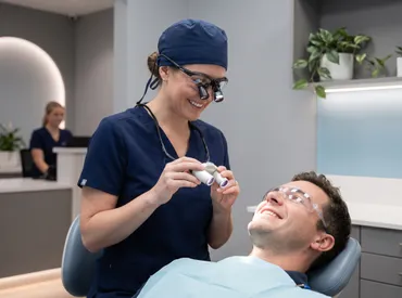
Noticing a sudden or gradual shift in the color of one of your teeth can be deeply unsettling, especially when the change progresses to a dark, opaque appearance. When a tooth turns gray or develops a prominent black spot, it is rarely a mere cosmetic concern that will fade with improved brushing. A black tooth often signals underlying structural changes, pulp compromise, or advanced bacterial activity that requires professional evaluation. The oral cavity functions as a complex biological ecosystem, and visible tooth discoloration is typically the final clinical manifestation of a cascade of biochemical events occurring beneath the surface. Whether the darkening stems from years of dietary habits, an old facial injury you barely remember, or progressive decay advancing through the dentin, understanding the precise root cause is the foundational step toward effective intervention. This comprehensive guide explores the pathophysiology behind tooth discoloration, outlines evidence-based diagnostic pathways, and details modern treatment protocols that prioritize both function and aesthetics. By separating clinical facts from popular misinformation and focusing on actionable dental health strategies, you can protect your smile from irreversible damage and restore your oral confidence with medically sound, professionally supervised interventions.
Understanding What Causes a Black Tooth
The development of a black tooth is rarely an instantaneous event. Instead, it typically emerges as the visible endpoint of prolonged chemical, physical, or pathological processes that have been quietly altering the tooth's internal or external architecture. Dental professionals classify tooth discoloration into two primary clinical categories: extrinsic and intrinsic. Extrinsic staining occurs on the outermost protective layer of the tooth, known as the enamel, and is heavily influenced by environmental exposure, lifestyle habits, and dietary choices. In contrast, intrinsic discoloration penetrates deeper into the dentin layer and often reflects internal structural damage, vascular compromise, metabolic changes, or developmental anomalies that occurred during tooth formation. When a black tooth appears in your mouth, it is crucial to determine which anatomical layer is primarily affected, as the clinical approach differs drastically between the two classifications. Surface stains might respond well to professional prophylaxis and chemical bleaching, while internal darkening usually requires restorative dentistry, endodontic therapy, or prosthetic rehabilitation.
Free calculatorHeadache Location ChartIdentify headache types by locationOpen the calculatorExtrinsic Staining vs. Intrinsic Discoloration
Extrinsic staining accumulates gradually as chromogenic compounds adhere to the dental pellicle, a thin proteinaceous film that constantly coats your teeth throughout the day. Foods and beverages with intense color profiles, such as red wine, black tea, dark sodas, and certain deeply pigmented berries, contain polyphenols and tannins that bind stubbornly to the microscopic irregularities of enamel. Tobacco products, including cigarettes, cigars, and smokeless varieties, introduce nicotine and tar that oxidize into dark brown or blackish pigments over time. While these extrinsic stains can appear severe and highly visible, they generally remain superficial and do not compromise the structural integrity or vitality of the underlying tooth. Intrinsic discoloration, however, tells a fundamentally different clinical story. This type of discoloration occurs when pigmentation molecules become embedded within the hydroxyapatite crystalline matrix of the dentin. Common causes include excessive systemic fluoride exposure during childhood dental development (dental fluorosis), certain broad-spectrum antibiotics like tetracycline taken during active tooth calcification, or metabolic conditions that alter calcium and phosphate homeostasis. When a black tooth results from intrinsic factors, conventional polishing and over-the-counter whitening agents prove completely ineffective because the pigment molecules reside beneath the translucent enamel layer, shielded from topical chemical oxidation.
Dental Trauma and Pulp Necrosis
Blunt force impact to the face, jaw, or teeth—even if it occurred years or even decades earlier—can trigger a silent, progressive chain reaction within the tooth's vascular and nervous tissue. Following a significant mechanical impact, the microvasculature supplying the dental pulp may rupture, kink, or undergo thrombosis, effectively cutting off the nutrient and oxygen supply to the internal chamber. Without adequate circulation, the pulpal tissue undergoes coagulative necrosis, a biological process where living cells die and break down into necrotic debris. The metabolic byproducts of this cellular degradation, primarily iron sulfide and hemoglobin breakdown compounds, slowly diffuse through the microscopic dentinal tubules and accumulate inside the coronal structure. As these dark pigments migrate outward toward the clinical crown, they cause the visible tooth to take on a grayish, bluish, or distinctly black hue. This phenomenon is particularly common in maxillary central incisors, which are the most vulnerable to direct facial impact. A black tooth stemming from unmonitored trauma requires immediate professional assessment because the necrotic tissue can rapidly become a breeding ground for anaerobic oral bacteria, potentially leading to chronic apical periodontitis, radicular cysts, or a painful dental abscess.
Untreated Decay and Cavities
Dental caries remain the most prevalent chronic infectious disease globally, and they represent a leading cause of localized, progressive tooth darkening. When cariogenic oral bacteria metabolize fermentable dietary carbohydrates, they produce organic acids that rapidly demineralize the highly structured enamel surface. As the protective outer shell breaks down and loses mineral density, the decay advances into the softer, more organic-rich dentin layer. The affected dentin undergoes a profound biochemical transformation, turning from a healthy yellowish-brown to a leathery dark brown, and eventually to black or charcoal gray as bacterial colonies, necrotic tissue, and metallic sulfides accumulate. In advanced stages, a carious lesion can create a large, visible black spot, shadow, or actual hole in the tooth structure. If left untreated, the decay severely compromises the tooth's load-bearing capacity, increases susceptibility to catastrophic fracture under normal masticatory forces, and exposes the pulp to oral flora. The presence of a black tooth in this context is a clear clinical red flag indicating active, progressive disease that will not arrest or resolve without operative dental intervention and professional restoration.
Medications, Supplements, and Systemic Factors
Certain pharmacological agents, long-term supplements, and underlying systemic conditions directly influence tooth pigmentation through complex biochemical pathways. Chlorhexidine gluconate mouthwashes, while highly effective against pathogenic bacteria and gingivitis, can cause pronounced extrinsic staining when used chronically beyond the recommended two-week period, often appearing as dark brown or blackish lines near the gingival margin. Iron supplements, bismuth subsalicylate, and certain antihypertensive medications are notorious for depositing dark metallic pigments on the enamel surface. On a systemic level, hematological conditions affecting hemoglobin metabolism, such as sickle cell anemia or thalassemia, can cause intrinsic tooth discoloration due to altered iron deposition and bilirubin accumulation in developing dental tissues. Additionally, genetic disorders like dentinogenesis imperfecta or amelogenesis imperfecta affect collagen synthesis and enamel matrix formation, resulting in teeth that appear translucent, blue-gray, or dark amber from early childhood development. Recognizing the intricate connection between systemic health, pharmacological exposure, and oral manifestation is essential for accurate diagnosis, comprehensive patient management, and appropriate interdisciplinary referrals.
Recognizing the Signs and Symptoms
Identifying the early warning signs of tooth discoloration, structural compromise, and pulpal compromise allows patients to seek professional care before irreversible tissue loss occurs. A black tooth rarely presents as an isolated aesthetic anomaly; it is frequently accompanied by other clinical indicators that directly reflect the underlying pathological process. Patients should actively monitor not only the visual appearance of their teeth but also any accompanying sensory changes, soft tissue reactions, or functional limitations. Early intervention significantly improves treatment success rates, reduces procedural complexity, and preserves maximum natural tooth structure. Proactive symptom recognition transforms dental care from reactive emergency management to proactive health maintenance.
Visual Changes and Color Progression
The progression of tooth darkening typically follows a predictable, clinically observable pattern that provides valuable diagnostic clues. Initially, subtle yellowing or light brown patches may appear, usually localized to the deep grooves, fissures, or cervical margins near the gumline. Over weeks or months, these areas frequently darken into distinct gray, brown, or black zones that become increasingly opaque. The specific distribution of the color change is highly informative for clinicians: uniform darkening across the entire clinical crown often suggests widespread pulpal necrosis, developmental hypocalcification, or intrinsic metabolic changes, while localized black pits, shadowing, or distinct lines along the occlusal surfaces strongly indicate active carious lesions. In cases of dental trauma, the discoloration may emerge weeks, months, or even years after the initial impact, gradually intensifying as necrotic breakdown products continue to accumulate within the pulp chamber. Patients should also closely monitor changes in tooth translucency; healthy enamel allows a slight, natural translucent quality that reveals the underlying dentin color, whereas necrotic or severely decayed teeth typically appear opaque, flat, and lifeless under direct lighting.
Pain, Sensitivity, and Associated Discomfort
While some cases of tooth discoloration are entirely asymptomatic and discovered only during routine examinations, many are accompanied by significant sensory feedback that indicates active tissue involvement. Early enamel decay may cause sharp, transient pain specifically triggered by consuming sweet, hot, or cold substances, as the demineralized enamel allows rapid fluid shifts within the dentinal tubules that stimulate pulpal nerve endings. As the lesion approaches or breaches the pulp chamber, this fleeting sensitivity typically evolves into lingering, dull discomfort that persists long after the stimulus is removed. When a black tooth results from complete pulp necrosis, patients often experience a deep, persistent ache, a heavy sensation of pressure, or pain that intensifies noticeably with biting forces or when lying flat at night. Visible swelling of the adjacent gingival tissue, the appearance of a parulis (commonly known as a gum boil), or unilateral facial asymmetry strongly indicates that the infection has extended beyond the tooth apex into the surrounding alveolar bone and connective tissues. These symptoms demand prompt professional intervention, as untreated odontogenic infections can rapidly progress to deep fascial space involvement, systemic bacteremia, or life-threatening sepsis if consistently neglected.
When to Seek Immediate Dental Attention
Certain clinical presentations constitute genuine dental emergencies that require same-day or next-day professional evaluation and management. The sudden onset of severe, throbbing pain that does not respond appropriately to standard over-the-counter analgesics, visible swelling that spreads to the cheek, neck, or under-eye region, difficulty swallowing or breathing, or a persistent fever accompanying noticeable tooth discoloration are all unequivocal warning signs of advanced, uncontrolled infection. Additionally, if a tooth has recently experienced significant trauma and rapidly begins to change color, immediate vitality testing and radiographic assessment are necessary to determine the exact pulpal status and initiate timely intervention. Delaying professional care during acute inflammatory phases dramatically increases the likelihood of complete tooth loss, necessitates more extensive surgical procedures, and complicates future restorative options. Establishing a reliable relationship with a trusted dental professional, understanding local emergency protocols, and maintaining dental insurance that covers urgent care can prevent minor discoloration from escalating into complex, costly, and biologically devastating clinical scenarios.
Professional Diagnosis and Examination
Accurate, systematic diagnosis forms the absolute cornerstone of effective treatment planning in modern restorative dentistry and endodontics. Dental professionals employ a comprehensive, multi-modal approach to thoroughly evaluate a black tooth, seamlessly combining meticulous clinical observation with advanced diagnostic technologies and specialized testing protocols. This structured assessment ensures that the underlying etiology is correctly identified, effectively preventing misdirected treatments, unnecessary tissue removal, and optimizing long-term clinical outcomes. A standardized diagnostic workflow eliminates guesswork and replaces it with objective, measurable data.
Clinical Assessment and Medical History
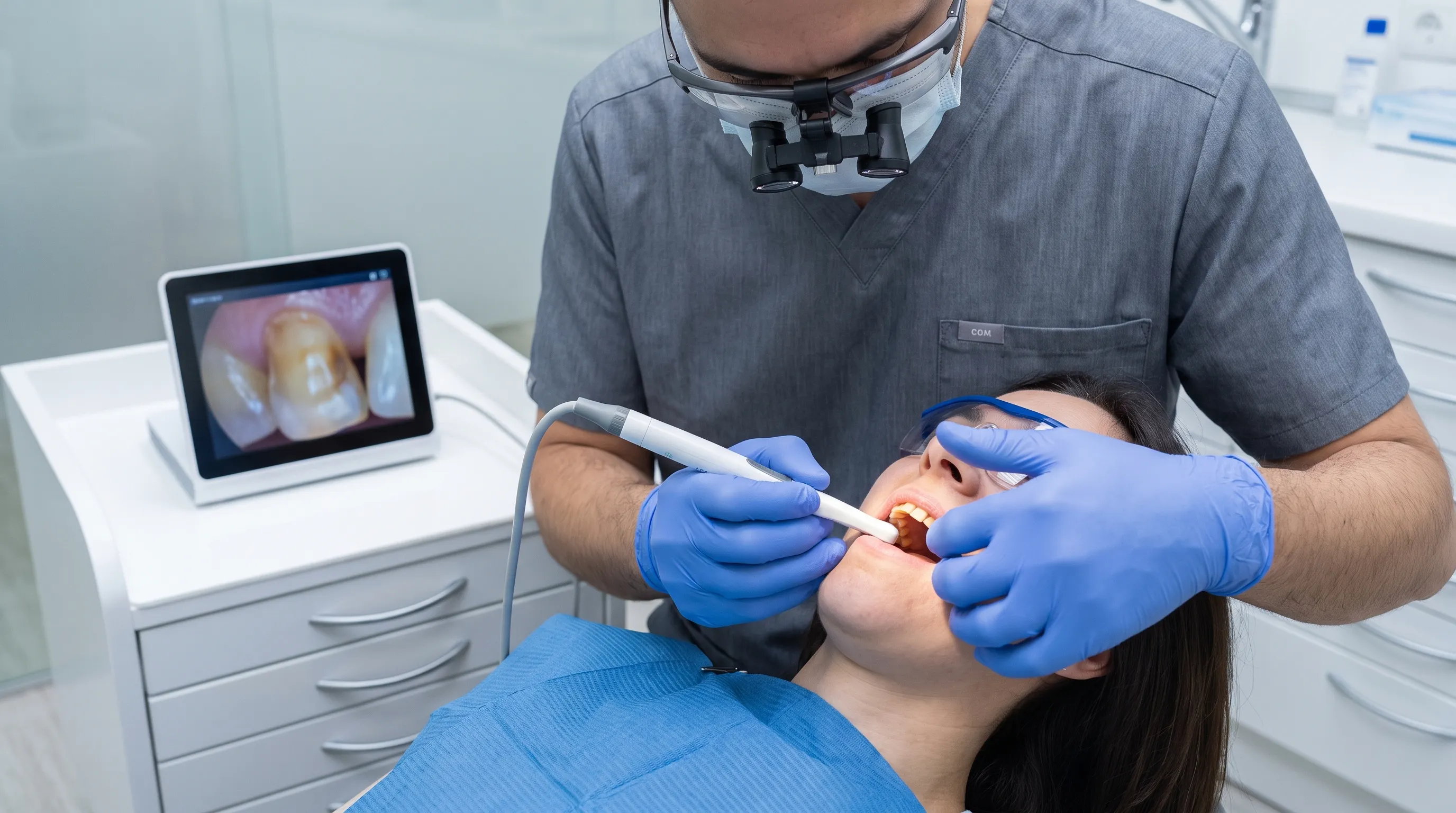
A thorough dental examination always begins with an in-depth, patient-centered medical and dental history review. Clinicians will carefully inquire about past facial trauma, chronic medication use, dietary patterns, oral hygiene routines, and underlying systemic conditions that might directly influence tooth vitality or enamel development. Visually, the dentist utilizes high-intensity clinical lighting, intraoral cameras, and dental operating loupes to evaluate enamel integrity, assess gingival health, and detect subtle subsurface defects or microfractures. Periodontal probes help determine if the dark discoloration corresponds to a soft, easily penetrable surface indicative of active decay, or if it feels hard, glassy, and polished, suggesting an inactive, arrested lesion or dense extrinsic calculus. Comprehensive periodontal pocket measurements assess the health of the supporting soft tissues and definitively rule out subgingival tartar deposits as a primary contributing factor. This meticulous, hands-on clinical evaluation provides the essential initial diagnostic framework before proceeding to instrumental and imaging-based testing.
Radiographic Imaging and Vitality Testing
Bitewing, periapical, and occlusal radiographs are absolutely indispensable for visualizing internal structures and hidden pathologies that are completely invisible to the naked eye during a surface examination. Radiographs clearly reveal the precise depth and proximal extent of carious lesions, assess the morphology and patency of the pulp chamber, and identify periapical radiolucencies or widened periodontal ligament spaces that indicate bone resorption from chronic infection. In complex or anatomically atypical cases, cone-beam computed tomography (CBCT) may be utilized to provide precise, three-dimensional visualization of root canal anatomy, accessory canals, and surrounding bone pathology. Pulp vitality testing perfectly complements radiographic findings by assessing physiological function rather than just structural appearance. Thermal testing using standardized cold sprays or heated gutta-percha, combined with electric pulp testing, directly measures the neurological and vascular responsiveness of the pulp tissue. A definitively negative or severely delayed vitality test in a noticeably discolored tooth strongly suggests complete pulpal necrosis, immediately guiding the clinician toward endodontic therapy rather than passive observation or conservative monitoring. Combining these diagnostic modalities ensures a highly accurate, evidence-based treatment pathway tailored to the patient's unique clinical presentation.
Evidence-Based Treatment Options
Contemporary dentistry offers an extensive array of scientifically validated treatment modalities specifically tailored to the precise etiology, anatomical location, and severity of tooth discoloration. From minimally invasive, non-operative preventive strategies to comprehensive restorative protocols and advanced endodontic micro-procedures, clinicians prioritize interventions that aggressively preserve natural tooth structure while fully restoring optimal function, occlusion, and aesthetics. Selecting the most appropriate clinical intervention requires carefully balancing biological necessity, realistic patient expectations, financial considerations, and long-term prognostic data derived from peer-reviewed clinical trials.
Conservative Approaches for Staining and Decay
For teeth exhibiting purely extrinsic staining, professional dental prophylaxis followed by targeted, in-office bleaching protocols often yields rapid and highly significant aesthetic improvement. Dental clinics utilize clinically formulated, high-concentration hydrogen peroxide or carbamide peroxide gels, frequently enhanced by safe LED light activation or controlled thermal protocols, to rapidly oxidize and neutralize deeply embedded chromogenic pigments within the enamel matrix. Professionally fabricated, custom-fitted take-home trays containing calibrated, lower-concentration whitening agents provide gradual, highly controlled results for mild to moderate staining while minimizing gingival irritation. Early-stage decay, particularly initial enamel demineralization or clinically arrested carious lesions, can often be managed effectively through strictly non-invasive techniques. These include professional application of silver diamine fluoride (SDF) to halt bacterial metabolism, prescription-strength remineralizing varnishes rich in bioavailable calcium and phosphate, and strict, supervised dietary modification protocols. These conservative measures actively halt disease progression without requiring mechanical tooth preparation or local anesthesia, making them exceptionally safe and ideal for pediatric patients, geriatric populations, and individuals classified as high caries risk.
Restorative Procedures and Root Canal Therapy
When a black tooth exhibits definitive structural compromise, large cavitated lesions, or diagnosed pulp necrosis, direct restorative and endodontic interventions become clinically mandatory. Direct composite resin restorations effectively restore small to moderate cavities by precisely replacing lost tooth structure with highly aesthetic, nano-hybrid composite materials that bond micromechanically to the remaining sound enamel and dentin through advanced adhesive dentistry systems. For more extensive decay or when cuspal coverage is required to prevent fracture, CAD/CAM milled porcelain or high-strength zirconia inlays and onlays provide superior durability, wear resistance, and exceptional longevity. In cases of irreversible pulpitis, symptomatic pulp necrosis, or confirmed periapical pathology, nonsurgical root canal therapy is the absolute clinical standard of care. The procedure involves the careful, aseptic removal of infected or necrotic pulpal tissue, meticulous three-dimensional disinfection of the root canal system using ultrasonic activation and antimicrobial irrigants, and permanent obturation of the space with biocompatible gutta-percha and sealer to definitively prevent reinfection. Modern endodontic practice routinely utilizes dental operating microscopes, rubber dam isolation, and nickel-titanium rotary instrumentation to achieve unprecedented precision and predictability, significantly improving long-term success rates for teeth previously considered hopeless.
Cosmetic Solutions for Permanent Discoloration
Teeth that remain stubbornly dark or grayish after conventional surface whitening or successful root canal treatment require advanced, highly specialized cosmetic dental rehabilitation to achieve optimal aesthetics. Internal bleaching, clinically known as the walking bleach technique, is exceptionally effective and minimally invasive for endodontically treated anterior teeth. The clinician carefully accesses the treated pulp chamber, places a controlled, sodium perborate-based bleaching paste directly inside the empty space, seals the access with a temporary restorative material, and allows the oxidizing agent to gradually lighten the darkened dentin from the inside out over several carefully monitored appointment intervals. For severe cases where internal bleaching yields insufficient aesthetic correction, or when significant tooth structure is missing, custom porcelain veneers or full-coverage ceramic crowns provide highly predictable, permanent aesthetic masking. Ultra-thin veneers require minimal, enamel-sparing preparation and are ideal for structurally intact teeth, while full-coverage crowns completely encompass the clinical crown, offering maximum fracture resistance and structural reinforcement for severely compromised or heavily restored teeth. Advanced digital smile design software, spectrophotometer shade matching, and layered ceramic characterization techniques ensure seamless visual integration with the patient’s natural surrounding dentition, consistently achieving harmonious, highly lifelike, and radiographically indistinguishable results.
Preventive Care and Long-Term Maintenance
Preventing recurrent tooth discoloration, new carious lesions, and progressive structural compromise requires a proactive, highly disciplined, and multi-faceted approach that extends far beyond the clinical dental chair. Sustainable, lifelong oral health fundamentally hinges on the consistent application of evidence-based daily habits, informed nutritional awareness, and strategic, long-term professional oversight. Implementing these scientifically validated preventive strategies significantly reduces the biological likelihood of future dental pathology and preserves natural dentition well into advanced age.
Daily Oral Hygiene Best Practices
The absolute foundation of oral disease prevention lies in proper, thorough, and consistent mechanical plaque disruption and removal. Patients should brush a minimum of twice daily using a soft-bristled manual toothbrush or a smart electric toothbrush, paired with fluoride toothpaste containing at least 1450 ppm fluoride, and employ a modified Bass technique that carefully angles the bristles at a precise 45 degrees toward the gingival sulcus to effectively sweep debris from beneath the gumline. Clinical trials consistently demonstrate that oscillating-rotating electric toothbrushes equipped with pressure sensors remove plaque more effectively and reduce gingivitis more reliably than traditional manual brushing methods. Flossing or using interdental cleaning devices remains completely non-negotiable in a comprehensive hygiene routine; it actively disrupts the mature, pathogenic biofilm that rapidly accumulates in tight interdental spaces where brush filaments simply cannot penetrate. For individuals experiencing dexterity limitations due to arthritis, neurological conditions, or advanced age, pressurized water flossers or flexible interdental brushes offer highly effective, gentle, and accessible alternatives. Consistent, daily mechanical disruption absolutely prevents the calcification of soft plaque into hardened calculus, which actively traps dark pigments, accelerates gingival inflammation, and creates a protected niche for cariogenic bacteria to thrive.
Dietary Adjustments to Protect Enamel
Nutritional intake profoundly influences enamel mineral density, salivary composition, and overall susceptibility to both decay and staining. Frequent, repetitive consumption of highly fermentable carbohydrates and refined sugars creates a chronically acidic oral environment that heavily favors the proliferation and metabolism of aciduric, cariogenic bacteria. Patients should consciously limit sugary snacks, sticky confections, and highly acidic beverages, deliberately opting instead for filtered water, calcium-rich dairy products, and fibrous, crunchy vegetables that actively stimulate robust salivary flow and promote continuous natural remineralization. Saliva functions as a powerful, multifaceted physiological buffer that rapidly neutralizes harmful plaque acids, physically washes away food debris, and continuously supplies free calcium and phosphate ions that actively repair microscopic enamel lesions before they cavitate. When consuming known stain-producing beverages, utilizing a reusable drinking straw strategically minimizes direct contact time with the facial surfaces of anterior teeth, and thoroughly rinsing the mouth with plain water immediately afterward significantly reduces pigment adherence and acid exposure. Additionally, regularly chewing sugar-free gum containing clinically proven xylitol actively inhibits bacterial adhesion, reduces cariogenic microbial load, and further supports a healthy, resilient oral microbiome balance.
The Role of Routine Dental Checkups
Professional dental evaluations scheduled consistently every six months serve as an absolutely essential, irreplaceable safety net for comprehensive oral health management. During these systematic visits, clinicians perform thorough extraoral and intraoral soft tissue cancer screenings, accurately measure periodontal attachment levels, and utilize diagnostic bitewing and periapical radiographs to definitively detect interproximal pathology and bone loss in its earliest, completely reversible stages. Professional dental cleanings utilizing ultrasonic scaling and air-polishing meticulously remove stubborn, calcified deposits, heavy extrinsic stains, and tenacious bacterial biofilms that stubbornly accumulate despite the most diligent home care efforts. Early clinical detection of micro-fractures, recurrent marginal decay around existing composite or crown restorations, or subtle, early vitality changes allows for immediate, minimally invasive interventions that aggressively preserve maximum remaining tooth structure. Establishing a continuous, long-term care model with a trusted, highly trained dental team ensures the consistent application of personalized preventive strategies and enables rapid, coordinated responses to emerging clinical concerns, effectively safeguarding against the silent progression of a black tooth or other devastating dental complications.
Debunking Common Myths About Tooth Discoloration
The modern internet landscape is unfortunately saturated with persistent misinformation regarding oral health, and tooth discoloration is consistently surrounded by highly popular but completely unscientific claims, anecdotal testimonials, and dangerously aggressive DIY practices. Relying heavily on unverified social media trends and viral home remedies routinely delays appropriate clinical care, actively exacerbates tissue damage, and ultimately multiplies long-term treatment costs significantly. Separating rigorously tested, peer-reviewed dental science from widespread commercial myths is absolutely essential for safe, effective, and biologically sound dental management.
At-Home Remedies and DIY Risks
Many well-intentioned patients attempt to aggressively treat a black tooth or severe staining using abrasive household products such as activated charcoal powders, highly alkaline baking soda scrubs, or dangerously undiluted hydrogen peroxide swabs. While these substances may occasionally produce temporary, superficial surface lightening, they completely lack the chemical specificity, controlled pH buffering, and clinical safety profiles of professionally formulated dental products. Activated charcoal, heavily marketed as a natural whitener, primarily operates through coarse mechanical abrasion that irreversibly scratches and erodes the delicate enamel surface, creating microscopic surface roughness that actually attracts and traps more food pigments over time, worsening the original problem. Excessive, unmonitored use of baking soda rapidly disrupts the delicate oral pH balance, potentially softening and weakening the enamel hydroxyapatite structure and drastically increasing dentin hypersensitivity. Unsupervised, repeated application of high-concentration, industrial-strength bleaching agents can easily cause severe chemical burns to the gingival tissues, irreversible pulp irritation, and profound inflammatory damage to the periodontal ligament and cementum. Licensed dental professionals universally and strongly advise against DIY chemical or abrasive interventions for persistent internal discoloration, as they merely mask superficial symptoms, completely ignore the underlying active pathology, and frequently result in highly costly, complex restorative complications that could have been entirely avoided.
Natural Healing and Enamel Regeneration
A highly pervasive and dangerous myth circulating in alternative wellness communities suggests that the human body possesses the inherent biological capacity to naturally regenerate completely lost enamel tissue or spontaneously reverse established, cavitated tooth decay solely through specialized dietary supplements, herbal mouth rinses, or alkaline water protocols. Unlike highly vascularized living tissues such as bone, muscle, or skin, mature dental enamel is entirely acellular, highly mineralized, and completely lacks any direct blood supply or nerve network, meaning it possesses absolutely zero intrinsic regenerative or self-repair capacity once fully developed and erupted. While controlled remineralization can scientifically strengthen early, demineralized subsurface lesions at the microscopic level by actively delivering supplemental calcium, phosphate, and fluoride ions from saliva or prescription pastes, it absolutely cannot physically reconstruct grossly lost tooth structure, close cavitated lesions, or eliminate an infected, necrotic pulp chamber. Bold claims that specific proprietary essential oil blends, aggressive daily oil pulling with coconut oil, or extreme dietary alkalization can permanently cure a black tooth or reverse advanced decay are entirely scientifically unfounded and contradict fundamental principles of human biology. When the enamel barrier is definitively breached, or when pulpal tissue has completely died due to trauma or infection, natural biological processes cannot spontaneously restore the original complex anatomy or eliminate anaerobic bacterial colonization. Professional restorative dentistry, adhesive techniques, and endodontic interventions remain the only clinically validated, legally recognized, and scientifically proven methods for definitively resolving advanced discoloration, halting active infection, and safely restoring compromised tooth structure.
Frequently Asked Questions
Can a black tooth indicate an infection that requires antibiotics?
Yes, in many clinical scenarios. When complete pulp necrosis or deeply advancing decay allows aggressive oral bacteria to infiltrate the periapical tissues and surrounding alveolar bone, an acute, symptomatic infection frequently develops. Licensed dentists and oral surgeons typically prescribe targeted antibiotics like amoxicillin, penicillin V, or clindamycin for patients presenting with systemic involvement, including high fever, diffuse facial swelling, regional lymphadenopathy, or spreading cellulitis. However, antibiotics alone are completely incapable of permanently eliminating a localized dental infection because the blood supply to the necrotic pulp chamber is severed, preventing the medication from reaching the primary bacterial source. Definitive, source-control treatment such as nonsurgical root canal therapy or surgical extraction is absolutely required to physically remove the contaminated tissue, drain the abscess, and prevent dangerous recurrence.
Is tooth whitening safe for teeth that have undergone root canal therapy?
Yes, internal bleaching is specifically designed, extensively researched, and highly effective for endodontically treated teeth that darken post-operatively. The procedure is considered exceptionally safe when performed under strict professional supervision using rubber dam isolation and internal sealing techniques. Clinicians strategically avoid exposing the delicate periodontal ligament to potentially irritating high-concentration bleaching agents by placing a robust internal barrier, such as resin-modified glass ionomer cement, precisely at the cervical margin and root canal orifice before applying the oxidizing paste. Standard over-the-counter external whitening strips or gels will absolutely not affect a necrotic or successfully root-canal-treated tooth because the dark pigmentation molecules are trapped internally within the dentinal tubules. Patients should always consult a certified endodontist or accredited cosmetic dentist to determine the safest, most predictable whitening protocol tailored to their exact anatomical and clinical situation.
How long does it take for a traumatized tooth to turn black?
The biological timeline varies significantly depending on the initial severity of the vascular disruption, the patient's age, and individual healing responses. Some severely compromised teeth begin displaying subtle grayish or yellowish discoloration within just two to four weeks of the original impact, while others may take several months, or even many years, to gradually darken to a noticeable degree. The progressive color change directly correlates with the continuous rate of hemoglobin degradation, iron sulfide formation, and the steady diffusion of breakdown products through the dentinal tubules toward the outer enamel. Consistent, routine vitality monitoring by a dental professional for at least one to two years following significant trauma is absolutely essential to accurately detect delayed pulpal necrosis early and intervene appropriately, regardless of whether immediate, visible color changes initially appear to the patient or clinician.
Can poor oral hygiene cause a tooth to appear black even without active cavities?
Yes, absolutely. Chronic, unmanaged plaque accumulation and hardened calculus formation can create extremely dense, dark deposits that closely mimic the clinical appearance of an internal black tooth. Mature supragingival calculus frequently absorbs dietary chromogens, bacterial metabolic byproducts, and environmental pigments over time, transforming from a pale yellow into a distinctly blackish-brown, rock-hard substance. Subgingival calculus located below the gumline can also cast a noticeable dark shadow through the thin gingival tissue or translucent enamel margins. While this extrinsic, hygiene-related staining does not indicate internal bacterial infection, pulp death, or active dentin decay, it still requires prompt professional ultrasonic scaling, meticulous root planing, and significantly improved daily home hygiene protocols to effectively reverse the discoloration, prevent progressive gingivitis, halt periodontal bone loss, and eliminate the protected environment that accelerates future caries development.
What is the cost difference between saving a black tooth versus extracting it?
The long-term financial investment and biological consequences differ drastically between tooth preservation and extraction. Saving a compromised natural tooth typically involves upfront costs for diagnostic imaging, root canal therapy, core build-up, and a custom-fabricated full-coverage crown, but this approach permanently maintains natural jawbone density, precise occlusal proprioception, and harmonious dental arch alignment. Initial extraction procedures are comparatively inexpensive, but the subsequent mandatory replacement of the missing tooth using a multi-unit fixed bridge or a single-tooth dental implant routinely exceeds the total cumulative cost of conservative endodontic treatment. Far more critically, permanent tooth extractions inevitably trigger adjacent teeth to tilt into the empty space, cause opposing teeth to supra-erupt, initiate progressive alveolar bone resorption, and frequently lead to complex bite collapse and temporomandibular joint dysfunction. The overwhelming consensus among evidence-based dental professionals strongly recommends actively preserving the natural dentition through appropriate intervention whenever the clinical prognosis and remaining structure remain feasible.
Conclusion
A black tooth is never a minor cosmetic inconvenience that should be passively ignored, self-treated with dangerous DIY chemicals, or dismissed as harmless aging. It consistently serves as a highly visible, unmistakable clinical marker of underlying pathological changes, whether those changes involve stubborn extrinsic pigment accumulation, aggressively advancing bacterial decay, complete pulpal necrosis following old trauma, or profound systemic influences. Contemporary dentistry provides highly predictable, rigorously tested, evidence-based diagnostic protocols and treatment modalities that can accurately pinpoint the exact biological etiology and safely restore both optimal chewing function and natural aesthetics. Seeking early, comprehensive professional evaluation definitively prevents silent disease progression, drastically minimizes the future need for complex surgical interventions, and actively preserves maximum irreplaceable tooth structure for decades of continued use. By prioritizing rigorous daily mechanical hygiene, making highly informed dietary choices that protect enamel mineralization, and maintaining consistent, biannual dental checkups, patients can effectively prevent dangerous discoloration, halt active bacterial colonization, and rigorously protect their overall systemic and oral health. If you observe any persistent darkening, unexpected structural shifts, or new sensory discomfort in your teeth, immediately schedule a thorough clinical examination with a licensed dental professional. Proactive, disciplined care remains the single most reliable, scientifically proven strategy for maintaining a vibrant, structurally resilient, and profoundly healthy smile for life.
How we work
Researched and drafted with AI assistance, then reviewed by a human editor against the cited sources.
This article is general health information, not medical advice. Consult a qualified healthcare professional about your own situation.