How to Get Rid of Pimple Inside Lip: Complete Guide to Safe Relief & Healing
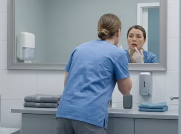
Discovering a tender, raised bump on the delicate inner surface of your mouth can be surprisingly uncomfortable and disruptive to daily life. While many people instinctively assume they have a simple facial breakout, learning how to get rid of pimple inside lip requires a fundamentally different approach because oral mucosa operates under entirely different biological conditions than the keratinized skin on your cheeks or forehead. The constant moisture, unique bacterial flora, and mechanical friction from chewing, speaking, and swallowing create a distinct microenvironment where standard acne treatments can actually cause severe chemical burns and prolonged inflammation. In this comprehensive, evidence-based guide, we will explore the most medically sound methods for soothing, treating, and eliminating these internal bumps safely. We will differentiate true comedonal lesions from mucoceles, aphthous ulcers, and minor salivary gland cysts, ensuring you apply the correct remedy for the precise condition you are experiencing. By combining immediate first-aid strategies, scientifically backed oral hygiene adjustments, nutritional support, and clear clinical warning signs for professional intervention, you will gain complete control over this common yet frustrating issue. Read on to discover exactly how to get rid of a pimple inside lip efficiently while protecting your long-term oral health and tissue integrity.
Understanding What You’re Actually Dealing With
Before attempting any treatment protocol, it is crucial to accurately identify the lesion in question. The term "pimple inside lip" is widely used in casual conversation, but medically speaking, true comedonal acne cannot form inside the mouth. Skin on the face contains sebaceous glands and hair follicles that produce the keratin and sebum necessary for blackheads and whiteheads. In contrast, the oral mucosa lacks pilosebaceous units entirely. Therefore, when patients report searching for how to get rid of pimple inside lip, they are typically experiencing mucoceles, aphthous ulcers, traumatic fibromas, blocked minor salivary glands, or viral lesions like cold sores. Each of these conditions follows a distinct pathophysiological pathway and demands a specific management strategy. Misidentifying the lesion often leads to prolonged irritation, secondary bacterial infections, or unnecessary tissue damage from aggressive over-the-counter acne products.
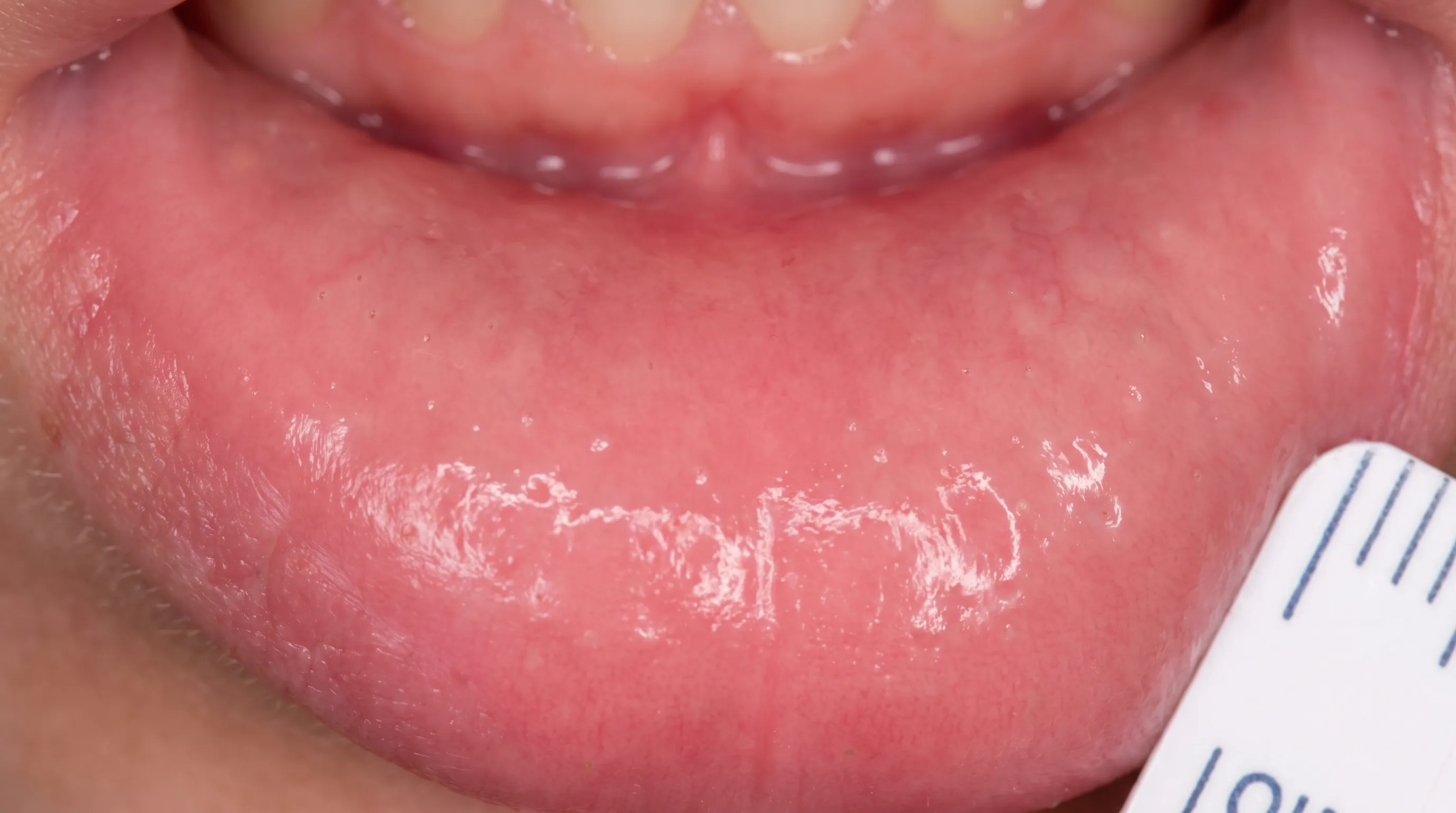
Free calculatorHeadache Location ChartIdentify headache types by locationOpen the calculatorThe inner lip contains hundreds of minor salivary glands responsible for maintaining baseline oral lubrication. Trauma from accidental biting, sharp dental appliances, or abrasive foods can disrupt the delicate ducts that transport saliva into the oral cavity. When a duct becomes obstructed or severed, saliva accumulates within the soft tissue, creating a fluid-filled cyst known as a mucocele. These lesions frequently appear as translucent or bluish bumps that fluctuate in size and are often mistaken for pimples. Conversely, canker sores develop due to minor immune responses triggered by stress, nutritional deficiencies, or localized tissue injury. They present as painful, shallow ulcers with a white or yellow center surrounded by an erythematous halo. Viral infections such as herpes simplex manifest as clustered vesicles that rupture into crusted sores, predominantly along the vermilion border but occasionally extending inward. Accurate differentiation is the foundational step in determining exactly how to get rid of a pimple inside lip without exacerbating underlying inflammation. For detailed clinical descriptions of common oral lesions, refer to resources from the Mayo Clinic.
Common Causes of Inside-Lip Bumps
Several primary factors contribute to the development of raised lesions on the inner lip. Mechanical trauma remains the most prevalent trigger. Biting down during sleep, chewing on the inside of the cheek or lip habitually, and poorly fitted orthodontic devices create repeated micro-injuries. These injuries initiate localized inflammatory cascades, prompting immune cells like macrophages and neutrophils to migrate to the site, which manifests as visible swelling. Food-related allergies or sensitivities can also provoke mucosal eruptions. Certain acidic, spicy, or highly processed foods contain chemical irritants that compromise the epithelial barrier, allowing opportunistic oral bacteria to colonize and trigger papule formation. For authoritative guidance on managing food sensitivities that affect oral tissues, visit the National Institute of Dental and Craniofacial Research (NIH).
Hormonal fluctuations, particularly during puberty, pregnancy, or menstrual cycles, influence mucosal hydration levels and immune responsiveness. While acne itself does not form intraorally, systemic hormonal shifts can alter salivary viscosity, making salivary ducts more prone to blockage. Additionally, nutritional imbalances involving iron, vitamin B12, folate, and zinc compromise epithelial regeneration. When the oral mucosa cannot repair micro-tears efficiently, persistent bumps develop. Finally, viral and bacterial agents play a significant role. Streptococcus species or candida overgrowth in immunocompromised states can present as focal nodules. Understanding these etiological factors empowers individuals to modify environmental triggers and implement targeted interventions that align with the biological reality of how to get rid of pimple inside lip effectively.
Pimples vs. Mucoceles vs. Canker Sores
Distinguishing between these three commonly confused conditions is essential for appropriate management. The table below outlines key clinical differences to guide accurate self-assessment.
| Feature | Facial Pimple | Mucocele | Canker Sore |
|---|---|---|---|
| Primary Cause | Clogged hair follicle/sebaceous gland | Blocked or severed minor salivary duct | Localized tissue injury or immune trigger |
| Appearance | Red bump with white/black head | Clear, bluish, or translucent fluid-filled dome | |
| Location | Only on keratinized facial skin | Exclusively on wet oral mucosa | Inner cheeks, lips, tongue base, or gums |
| Sensation | Tender, may itch or throb | Usually painless, occasional pressure sensation | Sharp, burning, highly painful |
| Healing Time | 3-7 days | Variable; may rupture and refill | 7-14 days spontaneous resolution |
| Response to Acne Cream | Positive | Negative/irritating | Highly irritating/damaging |
Mucoceles, however, are characterized by their fluid-filled nature. They feel soft, mobile, and typically dome-shaped, with a color ranging from clear to pale blue depending on depth and blood vessel proximity. Unlike inflammatory acne, mucoceles do not produce pus unless secondarily infected, and they frequently rupture temporarily before refilling as salivary flow continues. Canker sores present a completely different clinical picture. They begin as erythematous macules that rapidly ulcerate, forming a fibrin-covered necrotic base that is exquisitely tender, especially when exposed to salty or acidic stimuli. Pain typically peaks within 48 hours and gradually subsides over a 7 to 10-day period without intervention. Applying drying agents intended for facial acne directly onto a canker sore delays epithelial migration and intensifies discomfort. Recognizing these morphological differences guides appropriate therapeutic selection and prevents iatrogenic harm.
Step-by-Step Guide on How to Get Rid of a Pimple Inside Lip
Once you have assessed the nature of your lesion, implementing a structured care protocol becomes the priority. The mucosal environment requires gentle, antimicrobial, and moisture-balancing interventions rather than harsh drying agents. The following methodology outlines a sequential approach to reduce inflammation, prevent secondary infection, and promote natural tissue remodeling.
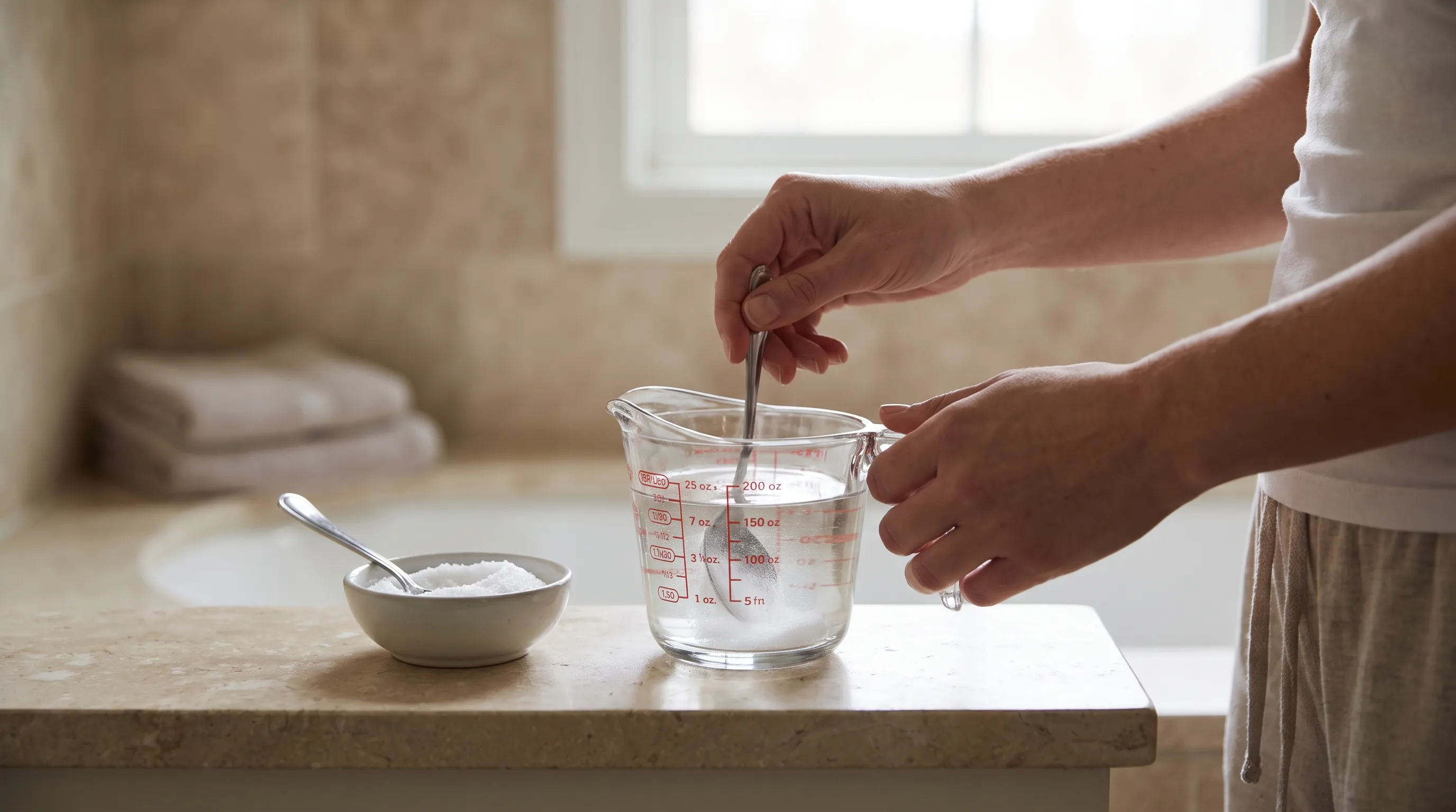
Begin with a conservative saline irrigation routine. Dissolve half a teaspoon of pharmaceutical-grade sodium chloride in eight ounces of warm distilled water. Gently swish the solution for thirty seconds, focusing the flow over the affected area without applying aggressive pressure. This osmotic technique draws out localized edema, neutralizes excess oral acids, and creates an environment less hospitable to anaerobic bacteria. Repeat this process three to four times daily, particularly after meals, to maintain optimal clearance of food debris and microbial biofilms. You can review detailed oral rinse protocols and safe hygiene practices at the Centers for Disease Control and Prevention (CDC).
Incorporate targeted antiseptic rinses that contain cetylpyridinium chloride or dilute hydrogen peroxide formulations specifically approved for intraoral use. These compounds reduce surface bacterial colonization without stripping the protective mucin layer that lines the oral cavity. Avoid mouthwashes containing high alcohol concentrations, as they desiccate the epithelium and trigger compensatory vasodilation, which paradoxically increases swelling. When exploring how to get rid of pimple inside lip safely, prioritize barrier-supporting agents over aggressive chemical exfoliants.
Apply a protective coating of oral-grade hyaluronic acid gel or pharmaceutical aloe vera specifically formulated for mucosal use. These substances contain polysaccharides that bind moisture to the tissue surface, accelerate fibroblast migration, and form a temporary physical shield against mechanical friction from teeth and food. Consistent application twice daily creates a microenvironment conducive to re-epithelialization. If the lesion demonstrates signs of localized bacterial overgrowth, consider introducing a probiotic lozenge containing Lactobacillus reuteri or Streptococcus salivarius strains. Research published in peer-reviewed dental journals indicates that these commensal organisms competitively exclude pathogenic microbes, modulate local immune responses, and shorten the duration of mucosal inflammation.
Proper Oral Hygiene Practices
Maintaining meticulous oral hygiene serves as both a therapeutic intervention and a preventive measure when dealing with internal lip lesions. Transitioning to an ultra-soft bristle toothbrush minimizes micro-trauma during brushing. Use a non-sodium lauryl sulfate (SLS) free toothpaste, as SLS is a known detergent irritant that disrupts mucosal desmosomes and delays ulcer healing. Brush using gentle circular motions, angling the bristles at forty-five degrees toward the gumline while avoiding direct contact with the raised bump. Interdental cleaning becomes equally important. Food particles trapped between the inner lip and teeth create a persistent nidus for inflammation. Use water flossing devices on the lowest pressure setting to flush out debris without mechanical disruption of the healing tissue. Replace your toothbrush every six to eight weeks, or immediately after an active infection, to prevent recolonization by residual bacterial strains.
Tongue scraping should be performed daily to remove biofilm that contributes to overall oral microbial load. A cleaner oral environment reduces the frequency of opportunistic infections that manifest as lip bumps. Additionally, avoid sharing utensils, drinking vessels, or lip products to prevent cross-contamination. Consistent adherence to these hygiene standards accelerates the resolution process and significantly lowers recurrence rates.
When to Use Over-the-Counter Treatments
Not every lesion requires immediate pharmaceutical intervention, but understanding when to deploy over-the-counter (OTC) options streamlines recovery. Topical benzocaine gels provide temporary analgesic relief by blocking voltage-gated sodium channels in peripheral nerve endings, reducing pain signals during chewing and speaking. However, these should be applied sparingly and only to intact surrounding tissue to avoid masking progressive symptoms. Hydrocortisone-based oral pastes may be indicated for severe inflammatory responses or autoimmune-mediated ulcerations. These formulations create a localized occlusive barrier while delivering mild corticosteroids to suppress leukocyte migration and cytokine release. Consult a pharmacist before initiating steroid use, as inappropriate application can impair microbial clearance.
For suspected salivary duct obstructions, applying a warm, moist compress to the external lip region for ten minutes encourages vasodilation and promotes gentle salivary flow past the blockage. Follow this with gentle intraoral massage, moving from the periphery of the gland toward the duct opening using a clean finger. Never apply excessive pressure, as this can force saliva into adjacent fascial planes. Integrating these OTC strategies at the appropriate stage ensures optimal therapeutic outcomes when determining how to get rid of a pimple inside lip.
What to Avoid During the Healing Process
Recovery depends as much on eliminating harmful behaviors as it does on implementing positive interventions. The oral mucosa possesses remarkable regenerative capacity, but continuous irritation disrupts cellular signaling and prolongs the inflammatory cascade. Recognizing and avoiding specific aggravating factors prevents secondary complications and ensures the lesion progresses through normal healing phases without regression.
The Danger of Popping or Picking
Attempting to express or puncture an intraoral bump carries significant risks. Unlike facial acne, which sits within a keratinized epithelial layer, inner lip lesions reside in non-keratinized stratified squamous epithelium rich in vasculature and nerve endings. Piercing this tissue introduces oral pathogens directly into deeper submucosal spaces, potentially triggering cellulitis or abscess formation. The act of squeezing also forces inflammatory mediators and potentially infectious material into adjacent capillaries, spreading localized irritation. Furthermore, mechanical disruption destroys the granulation tissue that bridges the wound bed. Each trauma episode resets the healing timeline, replacing organized collagen deposition with disordered scar tissue. In the case of mucoceles, rupturing the cyst wall without removing the damaged salivary duct guarantees recurrence, as the underlying anatomical defect remains unaddressed. Repeated picking can also induce traumatic fibroma development, where dense connective tissue proliferates in response to chronic low-grade injury. Maintaining strict discipline by refraining from manipulation protects both immediate comfort and long-term structural integrity.
Foods and Products That Worsen Irritation
Dietary choices exert a profound influence on mucosal inflammation. During active healing, eliminate citrus fruits, tomatoes, vinegar-based condiments, and carbonated beverages. The low pH of these substances denatures surface proteins and stimulates nociceptors, intensifying discomfort. Spicy foods containing capsaicin activate TRPV1 receptors on sensory neurons, creating a burning sensation that mimics and amplifies underlying tissue damage. Hard, abrasive foods such as pretzels, tortilla chips, and crusty bread create shear forces that scrape against the raised area. Switch temporarily to a soft, lukewarm diet incorporating cooked grains, steamed vegetables, and smooth protein sources. Avoid extremely hot beverages that cause thermal injury and delay re-epithelialization.
Certain personal care products also exacerbate mucosal irritation. Toothpastes containing sodium lauryl sulfate, artificial flavoring agents like cinnamaldehyde, and alcohol-based mouth rinses strip the protective mucin layer. Read ingredient labels carefully and select hypoallergenic, pH-balanced formulations until complete resolution occurs. Managing environmental exposures ensures the tissue receives uninterrupted support during its natural repair cycle.
When to See a Healthcare Professional
While most intraoral bumps resolve spontaneously within seven to fourteen days, certain clinical presentations necessitate expert evaluation. Persistent lesions that exceed three weeks in duration, progressively enlarge, or develop indurated bases require histopathological assessment to rule out premalignant or malignant transformations. Early identification of dysplastic changes significantly improves therapeutic outcomes and preserves function.
Warning Signs That Require Medical Attention
Several specific indicators should prompt immediate consultation with a dentist, oral surgeon, or primary care physician. Unexplained bleeding that does not cease after gentle pressure application suggests vascular involvement or coagulopathy. The presence of purulent exudate with a foul odor indicates secondary bacterial infection requiring targeted antimicrobial therapy. Systemic symptoms such as fever, lymphadenopathy, or malaise signal that localized inflammation has progressed beyond the oral cavity. Difficulty swallowing, speaking, or breathing, though rare with typical lesions, demands urgent care as it indicates potential airway compromise or deep fascial space involvement. Numbness or tingling radiating to adjacent facial structures suggests neural compression, which requires advanced imaging for accurate diagnosis. Recurrent lesions appearing in identical anatomical locations may point toward underlying salivary gland pathology or autoimmune conditions such as lupus erythematosus or Crohn’s disease. Documenting lesion progression through consistent photographic records assists clinicians in tracking morphological changes. Bringing this visual documentation alongside a list of current medications, supplements, and recent dietary modifications enables comprehensive differential diagnosis. Prompt professional intervention prevents complications and provides targeted treatments unavailable through self-care protocols.
Professional Treatment Options
Clinical management varies based on precise diagnosis. Mucoceles frequently require minor surgical excision or micro-marsupialization, a technique that creates a permanent drainage pathway while preserving surrounding glandular tissue. These procedures utilize precise electrocautery or laser ablation to minimize bleeding and accelerate wound closure. Recalcitrant lesions may necessitate cryotherapy, which applies controlled freezing temperatures to induce cellular apoptosis in abnormal tissue while stimulating regenerative pathways in adjacent healthy cells. For suspected infectious etiologies, clinicians prescribe targeted antifungal or antiviral medications alongside supportive debridement. Biopsy procedures provide definitive histological confirmation, examining cellular architecture for dysplasia, granulomatous inflammation, or specific pathogen presence. Advanced imaging such as high-resolution ultrasound or magnetic resonance imaging delineates the extent of deeper structural involvement when clinical palpation proves insufficient. Professional interventions address underlying anatomical or pathological mechanisms rather than merely alleviating surface symptoms, ensuring durable resolution and restoring optimal oral function. You can find detailed clinical guidelines on oral lesion management at the Cleveland Clinic.
Preventing Future Inside-Lip Breakouts
Long-term management focuses on eliminating triggers and fortifying mucosal resilience. Implementing consistent protective habits reduces the frequency of epithelial disruptions that initiate inflammatory responses. Understanding individual susceptibility patterns allows for proactive adjustments that maintain tissue homeostasis even under environmental or physiological stress.
Daily Habits That Protect Oral Mucosa
Establishing mindful chewing practices significantly reduces accidental trauma. Slow your eating pace and take smaller bites to maintain precise mandibular control. Individuals with dental anomalies or misaligned teeth should consult orthodontists about corrective appliances or protective night guards that redistribute occlusal forces. Addressing parafunctional habits such as lip biting, cheek chewing, or bruxism through cognitive behavioral strategies or occlusal splints prevents repetitive injury. Hydration maintenance remains foundational. Keep a reusable water bottle accessible throughout the day to ensure consistent fluid intake. Use alcohol-free, xylitol-based chewing gum to stimulate salivary flow without introducing irritants. Replace harsh dental floss with soft silicone picks if traditional fibers cause gingival or mucosal abrasion. Regular professional dental cleanings remove calculus deposits and bacterial reservoirs that contribute to chronic low-grade inflammation. Establish a nightly oral care routine that prioritizes gentle cleansing and tissue restoration. Remove all debris before applying any soothing barrier products. Maintain consistent sleep schedules to optimize circadian regulation of immune function and epithelial turnover. These integrated lifestyle modifications align with global preventive standards outlined by the World Health Organization (WHO) and create a resilient oral environment that resists breakdown and supports rapid recovery from incidental injuries.
Nutritional Support for Skin and Mouth Health
Dietary composition directly influences mucosal integrity and immune competence. Prioritize omega-3 fatty acids from fatty fish, flaxseeds, and walnuts to modulate inflammatory prostaglandin pathways and support cell membrane fluidity. Incorporate zinc-rich foods like pumpkin seeds, lentils, and lean meats, as zinc serves as a critical cofactor for epithelial proliferation and collagen synthesis. Vitamin C facilitates hydroxylation of proline and lysine residues during collagen formation, strengthening connective tissue scaffolding. Consume bell peppers, kiwifruit, and broccoli to maintain adequate antioxidant levels that neutralize reactive oxygen species generated during wound healing. B-complex vitamins, particularly riboflavin, niacin, and cobalamin, sustain epithelial turnover and prevent micronutrient deficiency-related ulcerations. Comprehensive nutritional guidelines for mucosal healing are available from the NIH Office of Dietary Supplements. Fermented foods containing active probiotic cultures support gastrointestinal-immune axis communication, reducing systemic inflammation that manifests in oral tissues. Limit refined carbohydrates and added sugars that feed opportunistic microbial populations and promote dysbiotic shifts. A nutrient-dense, whole-food diet provides the biochemical building blocks necessary for sustained tissue regeneration and optimal mucosal defense mechanisms.
Frequently Asked Questions
Can I safely use regular acne cream on a bump inside my mouth?
Applying topical acne formulations designed for facial skin directly to intraoral lesions is strongly discouraged. These products contain concentrated salicylic acid, benzoyl peroxide, or retinoids that rapidly desiccate keratinized facial skin but cause chemical mucositis when applied to delicate oral tissue. The resulting ulceration delays healing, intensifies pain, and increases infection risk. Always utilize products specifically formulated for oral mucosal application.
How long does a typical inside-lip bump take to heal?
Most benign intraoral lesions, including minor salivary cysts and traumatic ulcers, resolve spontaneously within seven to fourteen days when protected from further irritation. Larger mucoceles or lesions subjected to repeated trauma may persist for several weeks or require minor clinical intervention. If the bump shows no reduction after two weeks or continues to enlarge, professional evaluation is necessary to determine underlying pathology.
Does stress contribute to developing bumps on the inner lip?
Yes, chronic psychological stress significantly impacts mucosal health. Elevated cortisol levels suppress local immune surveillance, impair epithelial regeneration, and increase salivary viscosity, making ductal obstructions more likely. Stress also promotes parafunctional habits like unconscious lip biting, which introduces mechanical trauma. Implementing stress-reduction techniques such as mindfulness meditation, adequate sleep hygiene, and regular physical activity helps maintain hormonal balance and reduces recurrence frequency.
Can vitamin deficiencies cause recurrent inside-lip lesions?
Inadequate levels of iron, vitamin B12, folate, and zinc directly compromise oral mucosal integrity. These micronutrients are essential for rapid epithelial cell division, hemoglobin oxygen transport, and DNA synthesis. Deficiencies manifest as recurrent ulcerations, delayed wound closure, and increased susceptibility to secondary infections. Blood testing can identify specific nutritional gaps, allowing targeted supplementation that restores tissue resilience and prevents future breakouts.
Is it safe to apply ice directly to the affected area?
Applying ice can temporarily reduce swelling and numb discomfort, but direct contact with frozen objects causes thermal injury to non-keratinized mucosa. Instead, wrap a clean ice cube in a soft, damp gauze or use a chilled spoon to gently apply cold therapy. Limit application to ten-minute intervals with equal rest periods to prevent frostbite-like tissue damage and maintain healthy blood flow to the healing site.
Conclusion
Navigating the discomfort of an internal lip lesion requires a clear understanding of mucosal biology and a disciplined approach to gentle care. True intraoral bumps rarely represent standard facial acne, meaning conventional spot treatments often cause more harm than healing. By accurately identifying whether you are managing a mucocele, canker sore, or traumatic ulcer, you can apply targeted, evidence-based interventions that respect the delicate oral environment. Implementing consistent saline irrigation, switching to non-irritating dental products, avoiding mechanical manipulation, and supporting tissue regeneration through proper nutrition creates an optimal recovery pathway. Remember that persistent, enlarging, or repeatedly recurring lesions warrant professional assessment to exclude underlying anatomical or pathological conditions. Armed with these comprehensive strategies, you now possess a complete framework for understanding exactly how to get rid of a pimple inside lip safely, efficiently, and permanently. Prioritize gentle care, maintain vigilant hygiene, and consult qualified healthcare providers when necessary to ensure lasting oral health and comfort.
How we work
Researched and drafted with AI assistance, then reviewed by a human editor against the cited sources.
This article is general health information, not medical advice. Consult a qualified healthcare professional about your own situation.