Hole in Gum Above Tooth: Causes, Symptoms, Treatment & Prevention Guide
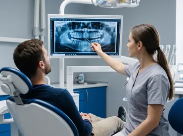
Discovering an unexpected hole in gum above tooth can be a startling and uncomfortable experience that immediately raises concerns about your oral health. Whether you notice it while brushing your teeth, feel a strange gap with your tongue, or experience sudden localized discomfort after eating, this symptom should never be dismissed as trivial. The oral cavity is a highly complex ecosystem where soft tissues, bone structures, and dental anatomy work in precise harmony. Any visible disruption, such as a hole in gum above tooth, often signals an underlying pathological process that requires prompt professional evaluation. From minor soft tissue trauma and localized inflammation to advanced periodontal disease, dental abscesses, or complications following orthodontic treatment, the potential causes vary widely in both severity and treatment requirements. Understanding the physiological mechanisms behind this condition is the first step toward finding effective relief, preventing systemic spread of infection, and preserving long-term dental function. This comprehensive guide will walk you through the medical causes, clinical warning signs, evidence-based diagnostic procedures, professional interventions, and practical self-care strategies that align with current dental standards. By following recommendations and maintaining consistent preventive habits, you can protect your gingival health and ensure that minor oral changes do not progress into complex dental emergencies.
What Causes a Hole in the Gum Above a Tooth?
The development of a hole in the gingival tissue is rarely random. The oral mucosa, including the attached and free gingiva, possesses remarkable regenerative capacity. However, when chronic stress, bacterial invasion, or mechanical trauma disrupts this balance, structural breakdown can occur. Understanding the primary etiologies helps patients recognize early warning signs and seek appropriate care before irreversible damage sets in.
Free calculatorHeadache Location ChartIdentify headache types by locationOpen the calculatorDental Infections and Abscess Formation
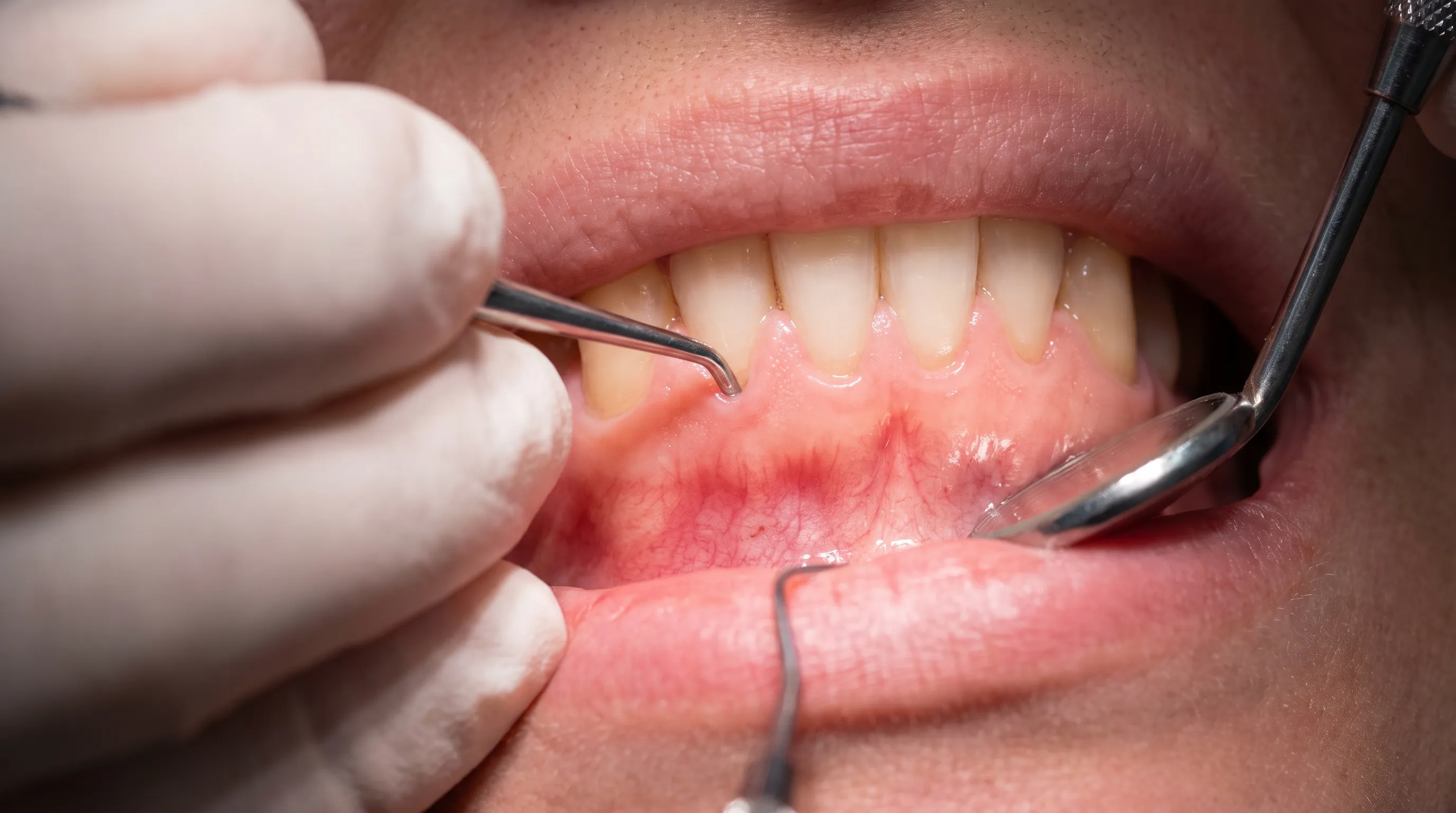
One of the most common reasons patients report a hole in gum above tooth is a periapical or periodontal abscess. Dental infections typically originate from untreated tooth decay that breaches the enamel and dentin, eventually reaching the dental pulp. As bacteria multiply within the root canal system, they produce inflammatory byproducts and toxins that migrate through the root apex into the surrounding alveolar bone and gingival tissue. The body's immune response creates a localized pocket of pus to isolate the infection. Over time, pressure builds within this pocket until it creates a natural drainage pathway through the soft tissue, forming a visible opening or sinus tract. This tract often appears as a small hole or pimple-like lesion on the gum tissue above the affected tooth. Without professional drainage and source elimination, the tract may temporarily close and reopen cyclically, causing recurring discomfort.
Advanced Periodontal (Gum) Disease
Chronic periodontitis represents a progressive inflammatory condition that destroys the connective tissue and bone supporting the teeth. When bacterial plaque accumulates below the gumline and hardens into calculus, it triggers a sustained immune response that gradually breaks down the periodontal ligament and alveolar bone. As bone loss progresses, deep periodontal pockets form between the tooth and the gum tissue. In severe cases, the loss of structural support causes the gingiva to detach, recede, or develop ulcerated openings. Patients with advanced gum disease frequently observe a hole in gum above tooth, particularly in areas where significant tissue recession and bone resorption have occurred. According to the Centers for Disease Control and Prevention, untreated periodontitis remains a leading cause of tooth loss in adults, making early intervention critical.
Trauma and Physical Injury
Mechanical trauma is a frequently overlooked cause of gingival defects. Aggressive tooth brushing with hard-bristled toothbrushes, improper flossing techniques, or accidental impacts from sports injuries can lacerate or puncture the delicate gum tissue. Additionally, habitual behaviors such as chewing on pens, fingernails, or ice can create micro-tears that enlarge over time. When the epithelial layer of the gingiva is compromised, it may heal with scar tissue or leave a permanent indentation or opening if the underlying connective tissue is severely damaged. The body's natural wound-healing process typically closes minor lacerations within one to two weeks, but deep or repeatedly traumatized areas may require professional assessment and possible surgical repair.
Orthodontic Appliances and Dental Prosthetics
Patients undergoing orthodontic treatment with fixed braces, clear aligners, or removable appliances occasionally experience localized gingival irritation. Brackets, wires, or poorly fitting dentures can rub continuously against the gum margin, causing chronic friction, inflammation, and eventual tissue breakdown. Over time, persistent pressure may lead to the formation of a decubitus ulcer or a small opening in the gingiva. Orthodontic specialists routinely monitor gum health during treatment and adjust appliances to minimize soft tissue contact. If you notice a hole developing while wearing orthodontic devices, consult your provider promptly to prevent secondary infection and ensure proper tissue preservation.
Post-Extraction Healing and Dry Socket Complications
Following a tooth extraction, the socket normally fills with a blood clot that protects underlying bone and nerves during the initial healing phase. If this clot dislodges prematurely due to suction, smoking, vigorous rinsing, or poor oral hygiene, the condition known as alveolar osteitis or dry socket can occur. This exposes bone and creates an open, painful cavity in the gum tissue. While technically a missing socket rather than a hole in healthy gum above tooth, the clinical presentation often mirrors similar symptoms of localized tissue disruption. Additionally, partially erupted wisdom teeth can develop a gingival flap (operculum) that traps food debris and bacteria, eventually leading to pericoronitis and tissue breakdown around the crown.
Recognizing the Symptoms and Warning Signs
Identifying the early clinical manifestations of gingival tissue breakdown is essential for timely intervention. Symptoms often progress gradually, but recognizing them early can prevent complications that require invasive procedures. Below are the most common indicators associated with this condition.
Localized Pain and Tenderness
Pain associated with a hole in gum above tooth typically presents as a dull ache that intensifies into sharp, throbbing discomfort when pressure is applied. Chewing, biting, or touching the area often exacerbates symptoms due to increased inflammation and fluid accumulation in the tissues. If the underlying tooth nerve is compromised, patients may experience heightened sensitivity to hot, cold, or sweet stimuli. Persistent pain that interferes with daily activities or sleep strongly suggests active infection or advanced tissue damage.
Visible Discharge and Pus Formation
When a bacterial infection establishes itself within the periodontal or periapical tissues, the immune system recruits white blood cells to combat the pathogens. This process generates purulent exudate that seeks an exit point through the path of least resistance, often forming a visible drainage tract on the gingiva. Patients may notice a yellowish or grayish fluid leaking from the opening, particularly upon waking or after eating. Pus drainage temporarily relieves pressure and reduces pain, creating a false sense of improvement while the infection continues to spread beneath the surface.
Gum Recession and Tissue Breakdown
Chronic inflammation causes the gingival margin to pull away from the tooth surface, exposing more of the root structure. This recession can create a visible gap or pocket above the tooth that resembles a hole. In advanced cases, the tissue becomes thin, translucent, and highly susceptible to bleeding. The Mayo Clinic notes that gum recession not only compromises aesthetics but also increases the risk of root caries, dentin hypersensitivity, and further periodontal attachment loss. Monitoring gum line position during routine brushing is a simple yet effective self-assessment method.
Halitosis and Unpleasant Taste
Persistent bad breath and a metallic or foul taste in the mouth are hallmark signs of active bacterial colonization within periodontal pockets or abscess cavities. Anaerobic bacteria thrive in oxygen-deprived environments beneath the gumline, releasing volatile sulfur compounds that cause characteristic oral malodor. When a drainage tract forms, the expelled exudate carries these compounds into the oral cavity, resulting in noticeable halitosis that resists conventional mouthwash or mints. Addressing the underlying infection typically resolves the odor, whereas masking agents provide only temporary relief.
Systemic Indicators of Oral Infection
In severe or untreated cases, local infection can progress to affect systemic health. Patients may develop low-grade fever, swollen submandibular or cervical lymph nodes, generalized malaise, or difficulty swallowing. These symptoms indicate that pathogens have breached local tissue barriers and are circulating through lymphatic or vascular pathways. Immediate medical attention is warranted if systemic signs accompany a hole in gum above tooth, as they suggest a potentially life-threatening spread of infection. As warned by the Cleveland Clinic, untreated dental infections can rapidly escalate into serious systemic complications if ignored.
When to See a Dentist or Seek Emergency Care
Not every gingival abnormality requires emergency intervention, but certain red flags indicate that waiting could result in permanent tissue damage or systemic complications. Knowing when to escalate care ensures that treatment is initiated at the optimal time.
Signs of Systemic Spread and Sepsis Risk
Dental infections are among the leading causes of odontogenic sepsis, a condition where bacteria enter the bloodstream and trigger a systemic inflammatory response. If you experience fever above 100.4°F (38°C), rapid heart rate, confusion, difficulty breathing, or severe facial swelling that extends toward the eye or neck, seek emergency medical care immediately. These symptoms suggest that the infection has progressed beyond local containment and requires intravenous antibiotics and possible surgical drainage in a hospital setting.
Timing Your Dental Appointment
For non-emergency cases where a hole in gum above tooth is accompanied by mild discomfort or occasional drainage, schedule a dental evaluation within 48 to 72 hours. Delaying care allows bacteria to multiply, increases bone loss, and reduces the likelihood of conservative treatment options. If symptoms worsen despite diligent oral hygiene or over-the-counter pain management, move your appointment forward or visit an urgent care dental clinic.
What to Expect During an Initial Consultation
A comprehensive dental evaluation begins with a thorough medical history review, including questions about recent dental procedures, medication usage, and symptom timeline. The dentist will perform a visual inspection of the oral cavity, use a periodontal probe to measure gum pocket depths, and assess tooth mobility. Dental radiographs, typically periapical or bitewing X-rays, are essential for evaluating bone density, identifying periapical lesions, and determining the extent of root involvement. In complex cases, a cone-beam CT scan may be ordered to provide three-dimensional visualization of the affected area.
Diagnosis and Professional Treatment Options
Once the underlying cause is confirmed, dentists recommend targeted interventions designed to eliminate infection, preserve natural tooth structure, and restore gingival integrity. Treatment selection depends on disease severity, patient health status, and long-term prognostic factors.
Clinical Examination and Diagnostic Imaging
Accurate diagnosis forms the foundation of effective treatment. Dentists correlate clinical findings with radiographic evidence to determine whether the hole in gum above tooth originates from a periapical abscess, periodontal pocket, traumatic laceration, or anatomical variation. Vitality testing, which assesses the tooth's response to thermal or electrical stimuli, helps determine pulp health. If the tooth tests non-vital, endodontic therapy is typically indicated. If vitality remains intact but deep pockets are present, periodontal intervention becomes the priority.
Incision and Drainage Procedures
When an acute abscess is confirmed, the first step involves establishing drainage to relieve pressure and remove purulent material. Under local anesthesia, the dentist makes a small incision in the swollen gingival tissue to allow complete evacuation of the infection. The area is thoroughly irrigated with sterile saline, and a small rubber drain may be temporarily placed to prevent premature closure and allow continued drainage. This procedure provides immediate pain relief and halts the progression of tissue destruction.
Endodontic Therapy (Root Canal Treatment)
For teeth with necrotic pulps and periapical infections, root canal therapy removes the infected nerve tissue, disinfects the root canal system, and seals it with biocompatible material. This procedure eliminates the source of bacterial toxins that cause the hole in gum above tooth. Following endodontic treatment, a crown is typically placed to restore structural integrity and prevent future fracture. Modern endodontic techniques boast success rates exceeding 90%, making root canal therapy a highly predictable and tooth-preserving intervention.
Periodontal Surgery and Tissue Grafting
When advanced gum disease has caused significant bone and tissue loss, non-surgical scaling and root planing may be insufficient. In such cases, periodontal flap surgery allows direct access to infected roots, enabling thorough debridement and bone contouring. For areas with severe recession or tissue deficiency, soft tissue grafting restores gingival volume, covers exposed roots, and creates a healthier anatomical barrier. Connective tissue grafts and free gingival grafts are the most commonly utilized techniques, both demonstrating excellent long-term integration when paired with meticulous maintenance protocols.
| Treatment Approach | Primary Indication | Procedure Overview | Expected Recovery Time |
|---|---|---|---|
| Incision and Drainage | Acute abscess with pus accumulation | Surgical opening, irrigation, and temporary drain placement | 3-7 days for initial tissue closure |
| Root Canal Therapy | Necrotic pulp with periapical lesion | Nerve removal, canal disinfection, and permanent sealing | 1-2 weeks for symptom resolution |
| Scaling and Root Planing | Moderate periodontal pockets (4-6mm) | Deep cleaning to remove calculus and smooth root surfaces | 2-4 weeks for gingival stabilization |
| Periodontal Flap Surgery | Advanced pocketing and bone loss | Tissue elevation, debridement, and bone reshaping | 4-8 weeks for complete tissue healing |
| Soft Tissue Grafting | Severe recession and tissue deficiency | Harvesting and suturing donor tissue to cover defects | 6-12 weeks for full integration |
At-Home Care and Management Strategies
While professional treatment addresses the underlying cause, diligent home care accelerates healing, minimizes discomfort, and prevents recurrence. These evidence-based self-management strategies should complement, not replace, clinical interventions.
Therapeutic Saltwater and Antiseptic Rinses
Rinsing with a warm saline solution (half teaspoon of non-iodized salt dissolved in 8 ounces of warm water) creates an osmotic environment that draws out excess fluid from inflamed tissues while reducing bacterial load. Swishing gently for 30 seconds, three to four times daily, promotes circulation and supports epithelial migration. For persistent bacterial colonization, dentists may prescribe chlorhexidine gluconate mouthwash, a potent antimicrobial agent proven to reduce plaque accumulation and gingival inflammation. However, prolonged use can cause temporary tooth staining and altered taste perception, so limit prescribed rinses to two weeks or as directed by your healthcare provider.
Safe Over-the-Counter Pain Management
Nonsteroidal anti-inflammatory drugs (NSAIDs) like ibuprofen effectively reduce both pain and localized swelling by inhibiting cyclooxygenase enzymes that produce inflammatory prostaglandins. Acetaminophen serves as an alternative for patients who cannot tolerate NSAIDs, though it does not reduce inflammation. Always follow label instructions and consult your physician if you have underlying kidney, liver, or cardiovascular conditions, as recommended by the National Institutes of Health. Topical oral gels containing benzocaine provide temporary numbing but should be used sparingly, as overapplication can cause methemoglobinemia or mask worsening symptoms.
Dietary Modifications and Soft Food Intake
During active healing, chewing on the affected side introduces mechanical stress and increases the risk of food impaction within the opening. Adopting a soft food diet rich in vitamins A, C, and D supports collagen synthesis and immune function. Foods like yogurt, mashed vegetables, scrambled eggs, and smoothies require minimal mastication while providing essential nutrients for tissue regeneration. Avoid extremely hot, spicy, acidic, or crunchy foods that can irritate exposed tissue or dislodge healing clots. Staying hydrated maintains salivary flow, which naturally buffers acids and contains antimicrobial peptides like lysozyme and lactoferrin.
Prevention and Long-Term Oral Health Maintenance
Preventing a hole in gum above tooth requires consistent adherence to proven oral hygiene practices and regular professional monitoring. The gingival tissues are highly resilient but vulnerable to cumulative neglect. Implementing these preventive strategies significantly reduces your risk of tissue breakdown, infection, and tooth loss.
Mastering Proper Brushing and Flossing Techniques
Effective plaque removal hinges on correct methodology rather than aggressive force. Use a soft-bristled toothbrush held at a 45-degree angle to the gumline, employing gentle circular or vibratory motions rather than horizontal scrubbing, which causes gingival recession and abrasion. Brush for two minutes, twice daily, ensuring all surfaces receive attention. Daily interdental cleaning removes plaque from areas a brush cannot reach. Traditional dental floss, when used with a C-shape technique along each tooth surface, effectively disrupts subgingival biofilm. For wider spaces, interdental brushes or water flossers provide comparable benefits while minimizing tissue trauma.
The Role of Nutrition in Gum Tissue Integrity
Systemic nutrition directly influences periodontal health. Vitamin C is essential for collagen synthesis and wound healing, while vitamin D modulates immune responses and supports calcium absorption. Omega-3 fatty acids exhibit anti-inflammatory properties that mitigate chronic gingival inflammation. Diets high in refined sugars and acidic beverages promote pathogenic bacterial proliferation and enamel demineralization, indirectly increasing the risk of secondary gum complications. Incorporating leafy greens, citrus fruits, fatty fish, nuts, and lean proteins provides the micronutrients necessary for robust gingival tissue maintenance.
Avoiding Tobacco and Alcohol Consumption
Smoking and smokeless tobacco use severely compromise periodontal health by constricting blood vessels, reducing oxygen delivery, impairing fibroblast function, and suppressing local immune responses. The World Health Organization notes that tobacco users experience significantly faster periodontal breakdown, delayed wound healing, and higher failure rates for dental surgeries. Alcohol consumption dehydrates oral tissues and alters the microbiome, creating favorable conditions for pathogenic overgrowth. Quitting tobacco and moderating alcohol intake represent two of the most impactful lifestyle modifications for preserving gingival integrity.

Frequently Asked Questions
Is a hole in the gum above a tooth always an infection?
Not always. While dental infections and abscesses are common culprits, a hole in gum above tooth can also result from physical trauma, aggressive brushing, orthodontic appliance irritation, gum recession, or normal healing after a dental extraction. A clinical examination is necessary to determine the exact cause.
Can a hole in the gum heal on its own?
Minor tissue injuries, such as small scratches from hard foods or temporary irritation from dental floss, can heal naturally within 7 to 14 days with proper oral hygiene. However, holes caused by deep infections, advanced periodontal disease, or bone involvement will not heal without professional dental intervention.
What does a gum abscess hole feel like?
A gum abscess hole typically presents as a small, tender bump or opening that may release pus or fluid when pressed. It often causes throbbing pain, localized swelling, sensitivity to temperature, and a foul taste in the mouth. The surrounding tissue may appear red, inflamed, or ulcerated.
How do dentists diagnose the cause of a hole in the gum?
Dentists use a combination of visual examination, periodontal probing, dental X-rays, and sometimes cone-beam computed tomography (CBCT) to assess bone loss, identify infection sources, evaluate gum pocket depths, and determine whether the tooth's nerve or supporting structures are compromised.
What should I do if I notice a hole in my gum before seeing a dentist?
Rinse gently with warm saltwater several times daily, maintain careful brushing and flossing around the area, avoid chewing on that side, use over-the-counter pain relievers if needed, and schedule a dental appointment promptly. Do not attempt to drain, squeeze, or puncture the area yourself.
Conclusion
Observing a hole in gum above tooth should always prompt careful attention rather than dismissal. The gingival tissues serve as a vital protective barrier against bacterial invasion, and any structural compromise can lead to localized discomfort, progressive infection, or systemic complications if left unaddressed. By recognizing early symptoms, understanding the underlying etiologies, and seeking timely professional evaluation, patients can access appropriate diagnostic and therapeutic interventions before irreversible damage occurs. Coupling clinical treatments with diligent home care, proper nutrition, and consistent preventive habits creates a sustainable framework for lifelong oral health. Your mouth is a gateway to overall wellness, and protecting its soft tissue architecture requires both professional partnership and personal accountability. Schedule a comprehensive dental examination at the first sign of persistent tissue changes, follow prescribed care protocols meticulously, and maintain the daily routines that keep your gums resilient. Prioritizing early intervention not only preserves your natural dentition but also safeguards your broader systemic health, ensuring that minor oral variations never escalate into complex medical challenges.
How we work
Researched and drafted with AI assistance, then reviewed by a human editor against the cited sources.
This article is general health information, not medical advice. Consult a qualified healthcare professional about your own situation.