Palate Expander Surgery: A Comprehensive Guide to SARPE, MARPE, and Maxillary Expansion

If you have struggled with chronic breathing difficulties, dental crowding, or a narrow upper jaw that resists traditional orthodontic treatment, you may have been told that palate expander surgery is your most viable option. While the prospect of oral surgery can feel intimidating at first, modern maxillofacial techniques have transformed this procedure into a highly predictable, life-changing intervention for adolescents and adults. The upper jaw, or maxilla, naturally consists of two bony plates that fuse during late adolescence. When these plates fail to develop adequately, patients often experience functional impairments ranging from obstructive sleep patterns to complex bite misalignments. Palate expander surgery bypasses the limitations of traditional appliances by surgically assisting the separation of these fused sutures, allowing for controlled, permanent widening. This comprehensive guide explores the clinical mechanisms, procedural steps, recovery expectations, and long-term outcomes associated with maxillary expansion. Whether you are weighing treatment options or preparing for an upcoming procedure, understanding the science and practical realities behind this intervention will empower you to make informed, confident decisions about your oral and respiratory health.
Understanding Palate Expansion: The Anatomy of Jaw Development
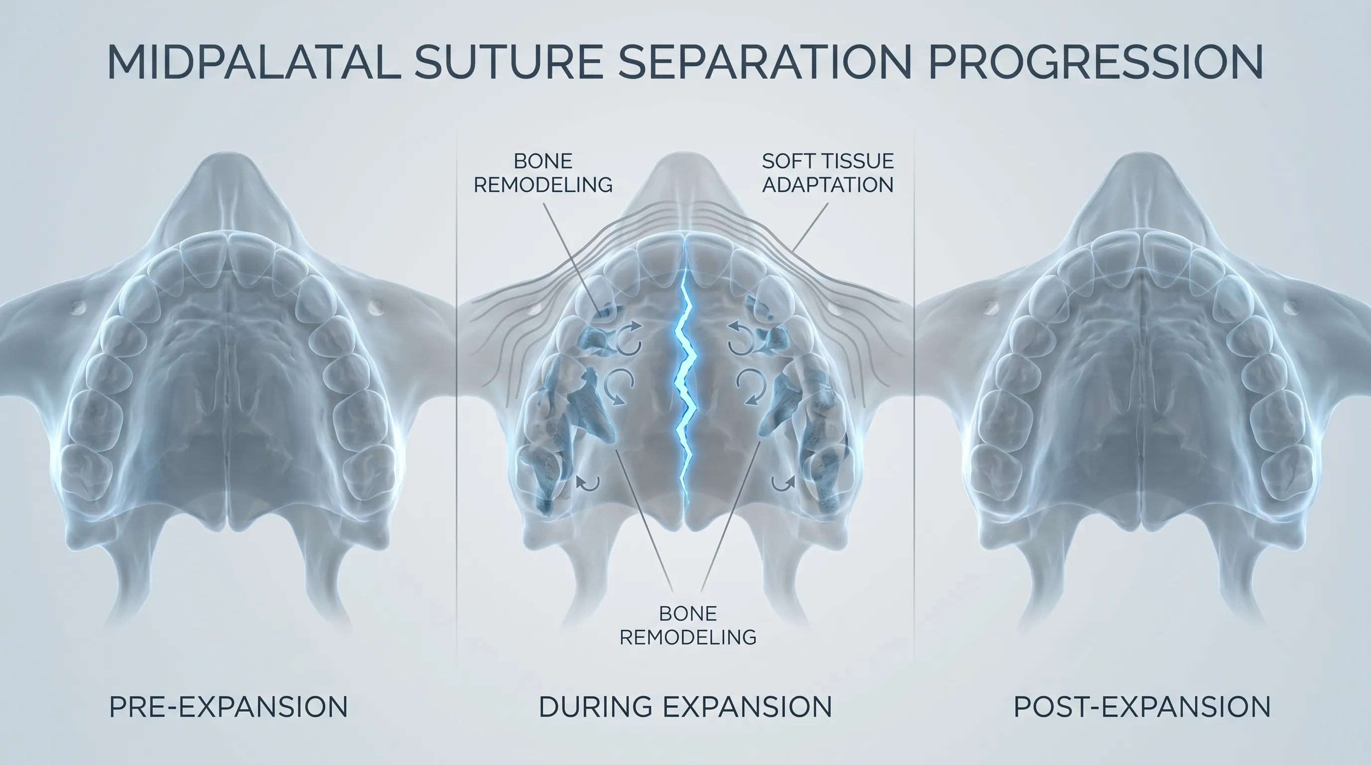
To fully appreciate why surgical intervention becomes necessary, it is essential to understand the underlying craniofacial anatomy. The human skull is not a single, solid bone; rather, it is composed of multiple cranial plates connected by fibrous joints known as sutures. The most clinically relevant of these in dentistry is the midpalatal suture, which runs longitudinally down the center of the hard palate and connects the left and right maxillary bones. During early childhood and pre-adolescence, this suture remains pliable, allowing the upper jaw to be widened using gentle, controlled orthodontic forces. However, as individuals progress into their mid-to-late teenage years, the body naturally begins a process called synostosis, where the fibrous tissue gradually ossifies into solid bone. Once this biological fusion occurs, non-surgical appliances can no longer generate enough force to safely separate the maxillary halves. Attempting to force expansion after fusion often results in undesirable dental tipping, periodontal damage, or root resorption rather than true skeletal widening.
Free calculatorRPE CalculatorRate of Perceived ExertionOpen the calculatorThe Role of the Midpalatal Suture
The midpalatal suture serves as a primary growth center for facial development. It allows the maxilla to widen in coordination with the developing dentition and the expanding nasal cavity. When transverse maxillary deficiency occurs, the suture remains closed, restricting lateral growth and forcing the body to compensate in other structural areas. This compensation frequently manifests as posterior crossbites, compromised airway dimensions, and altered facial proportions. Orthodontic expansion devices work by applying targeted lateral pressure to the molars and premolars, transmitting force directly to the suture. In a fused adult maxilla, this pressure is absorbed entirely by the periodontal ligaments and alveolar bone rather than the suture itself. Surgical intervention essentially resets the biological environment by creating controlled micro-fractures, temporarily restoring the suture's capacity for skeletal separation.
Why Age Matters in Palatal Expansion
Age is the single most critical factor in determining treatment feasibility. Children between the ages of seven and fourteen typically respond exceptionally well to conventional appliances like the Hyrax or Haas expander. During this window, the suture is still predominantly cartilaginous, requiring minimal force to achieve rapid, stable expansion. As patients approach fifteen years of age, the interdigitation of the sutural margins becomes increasingly complex, and the bone density rises significantly. For older adolescents and adults, the biological threshold shifts dramatically. Research consistently demonstrates that attempting non-surgical expansion beyond this age yields high relapse rates and compromised periodontal health. This is precisely why palate expander surgery has become the gold standard for mature patients. By strategically addressing the anatomical reality of suture fusion, clinicians can achieve true skeletal modification rather than merely shifting teeth into unstable positions. Understanding this biological timeline ensures patients pursue the correct modality for their developmental stage, preventing unnecessary discomfort and suboptimal clinical outcomes.
Types of Maxillary Expansion Procedures
Modern orthodontics and oral surgery offer multiple pathways to achieve transverse maxillary widening, each tailored to specific anatomical presentations, age groups, and clinical objectives. Selecting the appropriate technique requires careful evaluation of skeletal maturity, dental health, and patient-specific functional goals. The three primary modalities represent a spectrum of invasiveness and mechanical approach, ranging from entirely conservative to fully surgical intervention.
Non-Surgical Rapid Palatal Expansion (RPE)
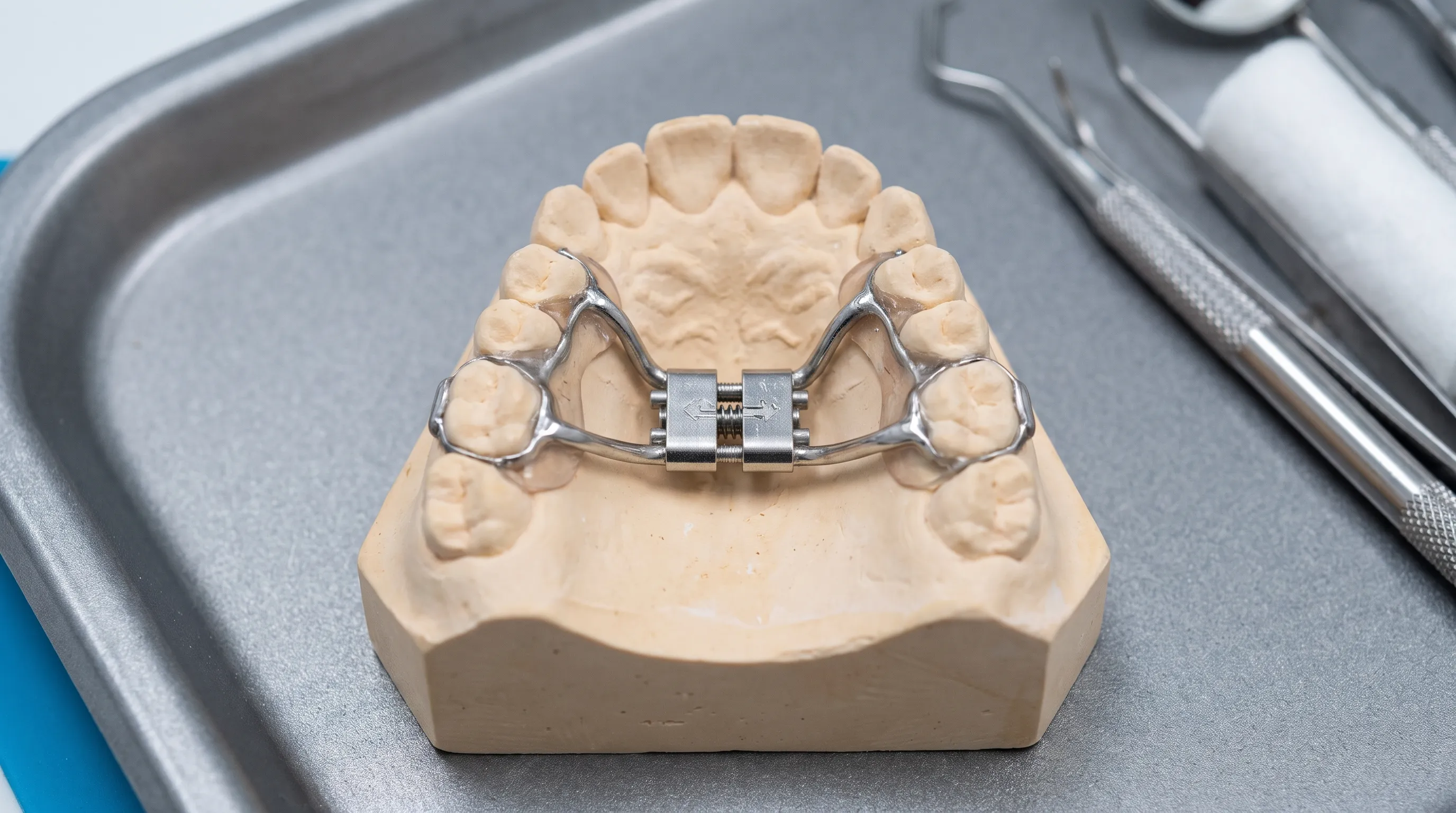
Rapid palatal expansion remains the cornerstone of pediatric orthodontic care. This procedure utilizes a fixed metal appliance cemented to the upper molars, featuring a central screw mechanism that is activated periodically using a specialized orthodontic key. Each turn advances the screw by a fraction of a millimeter, typically targeting 0.25 mm of daily expansion. The accumulated force gradually opens the midpalatal suture, encouraging rapid osteogenesis as new bone fills the widened gap. Treatment duration usually spans two to six months of active activation, followed by a prolonged retention period to stabilize the newly formed bone. While highly effective in younger populations, RPE relies entirely on suture pliability and is contraindicated in fully mature adults due to the risk of periodontal compromise and structural failure.
Surgically-Assisted Rapid Palatal Expansion (SARPE)
When skeletal maturity has eliminated the feasibility of conservative treatment, surgically-assisted rapid palatal expansion becomes necessary. SARPE is a collaborative procedure requiring precise coordination between an orthodontist and an oral and maxillofacial surgeon. The protocol begins with the placement of a customized expander device, followed by an outpatient surgical procedure under general anesthesia. The surgeon performs a controlled osteotomy, carefully severing the maxilla from adjacent skeletal structures, including the zygomatic buttress, pterygoid plates, and nasal septum. These strategic bone cuts effectively release the maxillary complex, eliminating the resistance caused by a fused suture. Following surgery, the patient activates the expander at prescribed intervals, typically twice daily, over several weeks. The orthopedic force now translates into true skeletal separation rather than dental tipping. American Association of Orthodontists recognizes SARPE as the most reliable method for achieving substantial transverse correction in adults, with documented success rates exceeding ninety percent when executed by experienced multidisciplinary teams.
Miniscrew-Assisted Rapid Palatal Expansion (MARPE)
Miniscrew-assisted rapid palatal expansion represents a groundbreaking middle ground between traditional RPE and full SARPE protocols. Developed over the past decade, this technique utilizes temporary anchorage devices, commonly referred to as mini-screws or bone screws, to bypass dental anchorage entirely. Instead of relying on the teeth to transmit expansion forces, the appliance is secured directly into the palatal bone using titanium micro-implants. This direct skeletal anchorage allows clinicians to apply higher, more targeted forces directly to the midpalatal region. In many late adolescents and young adults, MARPE successfully achieves suture separation without requiring osteotomies or hospitalization. The procedure significantly reduces surgical trauma, shortens recovery time, and minimizes soft tissue complications. While not universally applicable to all adults, MARPE has rapidly gained clinical traction as a conservative first-line option for patients who possess adequate bone density and favorable suture anatomy.
The Surgical Process Explained Step-by-Step
Navigating a maxillofacial procedure requires transparency and thorough preparation. Understanding each clinical phase helps patients set realistic expectations, adhere strictly to pre- and post-operative instructions, and maintain psychological readiness throughout the treatment journey. The process is highly structured, progressing through meticulous planning, precise execution, and disciplined activation protocols.
Pre-Operative Evaluation and 3D Imaging
Before any surgical intervention, comprehensive diagnostic imaging establishes the exact anatomical parameters required for safe expansion. Cone-beam computed tomography (CBCT) provides three-dimensional visualization of the maxilla, allowing clinicians to measure palatal width, assess suture fusion levels, and map critical neurovascular structures. Digital dental scans and cephalometric radiographs are integrated to evaluate occlusal relationships and plan the final orthodontic outcome. During this consultation phase, the surgical and orthodontic teams collaborate to fabricate a patient-specific expander. Customization ensures optimal fit, precise force distribution, and minimal soft tissue irritation. Pre-surgical blood work, medical clearance, and anesthesia consultations are completed to mitigate systemic risks. Patients are advised to discontinue certain medications, optimize nutritional intake, and arrange post-operative support well in advance of the procedure date.
The Osteotomy and Expander Placement
On the day of the procedure, patients receive local anesthesia with intravenous sedation or undergo general anesthesia, depending on surgical complexity and personal preference. The oral surgeon accesses the maxilla through intraoral incisions, carefully preserving the mucosal lining and periodontal attachments. Using specialized piezoelectric instruments and surgical burs, controlled osteotomies are performed along predetermined anatomical planes. These bone cuts release the maxilla from the pterygoid plates, zygomatic arches, and nasal floor, effectively isolating the central maxillary segments. Once the surgical release is complete, the pre-fabricated expander is cemented or bonded to the dentition, or in the case of MARPE, anchored via mini-screws. The incisions are closed with resorbable sutures, and the surgical site is packed with sterile gauze or bone graft material if necessary. The entire procedure typically lasts between one to two hours, after which patients are monitored in a recovery bay before being discharged with detailed care instructions.
The Post-Operative Activation Protocol
The activation phase begins approximately twenty-four to forty-eight hours after surgery, once initial swelling has stabilized and anesthesia has completely worn off. Patients are instructed to use a specialized activation key to turn the central screw according to a highly specific schedule established by the treating clinicians. Each turn produces an immediate audible click and a distinct sensation of pressure or mild tension across the midface. Most protocols require one to two turns daily, continuing until the targeted skeletal width is achieved. Patients often notice the development of a temporary diastema, or gap, between the upper front teeth, which serves as a positive clinical indicator that the midpalatal suture is successfully separating. This diastema typically closes spontaneously once active expansion concludes. The National Institute of Dental and Craniofacial Research (NIDCR) emphasizes strict adherence to activation schedules, as inconsistent turning can lead to asymmetrical expansion, increased discomfort, or delayed bone mineralization.
Health Benefits and Clinical Applications
The clinical rationale for palate expander surgery extends far beyond cosmetic alignment. Modern orthodontic philosophy views the maxilla as a central structural pillar that directly influences respiratory function, neuromuscular balance, and long-term dentofacial harmony. Addressing transverse deficiency yields multidimensional health improvements that profoundly impact daily quality of life.
Functional Improvements and Airway Enhancement
A narrow maxilla directly constricts the nasal floor, reducing overall nasal cavity volume and increasing airway resistance. Many patients with severe transverse deficiency become chronic mouth breathers, which compromises oxygenation, disrupts sleep architecture, and increases susceptibility to respiratory infections. Surgical maxillary expansion widens the nasal floor, improves mucosal airflow dynamics, and significantly reduces the effort required for nasal breathing. These structural changes are particularly impactful for individuals diagnosed with obstructive sleep apnea (OSA) or sleep-disordered breathing. By expanding the upper jaw, the tongue gains additional lateral space, reducing posterior collapse during sleep. Clinical trials consistently demonstrate measurable improvements in apnea-hypopnea indices, sleep continuity, and daytime alertness following successful expansion therapy.
Aesthetic and Structural Corrections
While functional benefits remain paramount, the aesthetic transformations associated with maxillary expansion are substantial. A constricted maxilla often produces a narrow dental arch, inadequate buccal corridors, and compromised lip support, which can create an aged or disproportionately long facial appearance. Expanding the upper jaw restores ideal arch form, improves smile width, and enhances midfacial projection. The improved skeletal foundation also facilitates optimal incisor inclination and posterior tooth alignment, creating a harmonious occlusal relationship. Additionally, correcting posterior crossbites eliminates asymmetric mandibular positioning, reducing uneven wear on the dentition and decreasing strain on the temporomandibular joints. These structural corrections contribute to enhanced facial symmetry, improved phonetic articulation, and a more balanced facial profile.
Comprehensive Conditions Treated with Palatal Expansion
Maxillary expansion serves as a foundational component in treating numerous complex craniofacial and dental conditions. Beyond crowding and crossbites, it plays a crucial role in comprehensive cleft lip and palate rehabilitation, where segmental expansion precedes bone grafting and dental alignment. Patients with severe Class III skeletal discrepancies often require transverse widening to create adequate space for orthognathic surgery. Additionally, individuals suffering from chronic sinus congestion, recurrent otitis media, or persistent headaches linked to cranial tension may experience symptom relief following structural optimization. The procedure also benefits orthodontic patients who have previously been advised to undergo premolar extractions due to space deficiency. By creating genuine skeletal space, palate expander surgery preserves healthy tooth structure while enabling comprehensive alignment without extractions. The multidisciplinary versatility of this intervention underscores its status as a cornerstone procedure in modern dentofacial orthopedics.
Risks, Complications, and How to Minimize Them
No medical intervention exists without inherent risks, and maxillary expansion is no exception. Understanding potential complications allows patients to recognize early warning signs, communicate effectively with their care team, and implement evidence-based prevention strategies. Most adverse outcomes are mild, transient, and highly manageable when proper protocols are followed.
Common Post-Operative Side Effects
During the initial recovery phase, patients routinely experience facial swelling, mild discomfort, and temporary speech alterations. The presence of the appliance alters tongue positioning, occasionally causing a lisp or difficulty pronouncing sibilant sounds. These articulation challenges typically resolve within two to three weeks as the neuromuscular system adapts. Dietary restrictions during the active phase often lead to temporary weight loss or altered nutritional intake. Chewing discomfort is expected, particularly when biting into harder foods or when activation tension is at its peak. Nasal congestion and occasional minor epistaxis are also common, resulting from the proximity of the surgical site to the nasal mucosa. These symptoms generally subside within the first ten to fourteen days as tissue inflammation decreases and initial bone bridging begins.
Serious Surgical Risks and Anatomical Considerations
While rare, significant complications can arise if surgical precision is compromised or if underlying anatomical anomalies are not adequately mapped. These include infection at the osteotomy site, delayed wound healing, nerve paresthesia affecting the infraorbital or nasopalatine regions, and asymmetrical maxillary separation. Inadequate surgical release can result in incomplete expansion, requiring revision osteotomies. Root resorption, though uncommon with controlled activation, may occur if forces are applied too aggressively. Periodontal complications such as gingival recession or alveolar bone loss can develop if oral hygiene deteriorates or if the appliance impinges on soft tissues without proper monitoring. The American Association of Oral and Maxillofacial Surgeons stresses that selecting a board-certified surgeon with extensive SARPE experience dramatically reduces the incidence of severe complications.
Evidence-Based Strategies for Complication Prevention
Mitigating risks begins with meticulous pre-operative planning and continues through disciplined post-operative compliance. Patients should utilize prescribed antimicrobial rinses, maintain impeccable oral hygiene, and avoid manipulating the appliance with unsterile objects. Strict adherence to activation schedules prevents sudden force spikes that could damage periodontal ligaments or induce root trauma. Following dietary guidelines, consuming nutrient-dense soft foods, and staying hydrated support optimal tissue regeneration and bone mineralization. Attending all scheduled follow-up appointments allows clinicians to monitor expansion symmetry, assess soft tissue adaptation, and adjust the treatment trajectory if deviations occur. Proactive communication regarding unusual pain, prolonged bleeding, or persistent numbness ensures timely intervention before minor issues escalate into serious complications.

Recovery, Aftercare, and Long-Term Success
The success of maxillary expansion depends not only on surgical precision but equally on post-operative diligence. The recovery timeline is divided into distinct phases, each requiring specific behavioral adjustments and clinical monitoring. Understanding these phases ensures patients maintain realistic expectations while optimizing structural stability and functional outcomes.
Immediate Post-Surgical Care and Dietary Adjustments
The first two weeks following surgery constitute the acute healing phase. Swelling typically peaks between forty-eight and seventy-two hours before gradually subsiding. Ice packs applied to the midface for twenty-minute intervals help minimize edema. Patients are prescribed analgesics and, occasionally, antibiotics to manage discomfort and prevent infection. Dietary modifications are essential; a strict soft-food regimen consisting of blended soups, yogurt, mashed vegetables, protein shakes, and smoothies prevents mechanical stress on the surgical site and the expander appliance. Avoiding hot, spicy, or crunchy foods during this period reduces mucosal irritation. Patients should refrain from strenuous physical activity, heavy lifting, and bending over for at least seven days to prevent increased blood pressure that could trigger post-operative bleeding.
The Critical Retention Phase Explained
Once the targeted skeletal width is achieved, active expansion ceases, but treatment is far from complete. The newly separated maxillary segments are held in place solely by the expander appliance and an immature, woven bone matrix. This retention phase is absolutely critical for clinical success. The expander remains in place without activation for a minimum of six months, allowing trabecular bone to gradually mineralize and consolidate into stable lamellar bone. Premature removal during this fragile period virtually guarantees skeletal relapse, as the immature bone cannot withstand masticatory forces and natural muscular tension. Clinicians monitor consolidation progress through periodic radiographs, ensuring adequate calcification before transitioning to fixed orthodontic appliances or removable retainers. Patience during this stabilization window is the single greatest predictor of long-term expansion maintenance.
Daily Maintenance, Oral Hygiene, and Lifestyle Tips
Maintaining oral hygiene with an expander in place presents unique challenges. The appliance traps food particles and plaque, significantly increasing the risk of decalcification, gingivitis, and halitosis. Patients must utilize specialized orthodontic brushes, interdental cleaners, and water flossers to navigate around expansion bands and wires. Daily irrigation with warm saline solution or alcohol-free chlorhexidine rinse reduces microbial load and soothes inflamed tissues. Avoiding sticky, sugary, or hard foods prevents mechanical damage to the appliance and reduces acid production that accelerates enamel demineralization. Wearing a protective mouthguard during athletic activities minimizes the risk of traumatic appliance displacement. Consistent hydration, balanced nutrition rich in calcium and vitamin D, and avoiding tobacco use all accelerate bone remodeling and enhance overall treatment outcomes.
Comparing Expansion Techniques: Making an Informed Decision
Selecting the optimal expansion modality requires weighing anatomical feasibility, patient preferences, recovery expectations, and long-term stability. The table below provides a direct clinical comparison to guide discussions with your orthodontic and surgical team.
| Feature | Non-Surgical RPE | Surgically-Assisted RPE (SARPE) | Miniscrew-Assisted RPE (MARPE) |
|---|---|---|---|
| Ideal Age Range | 7–14 years | 16+ years (fully fused suture) | 15–30 years (partially fused suture) |
| Invasiveness Level | Low (outpatient, non-invasive) | Moderate to High (requires osteotomy) | Low to Moderate (micro-screws only) |
| Anesthesia Required | None | General or IV Sedation | Local Anesthesia |
| Expansion Mechanism | Dental anchorage, transmits force to teeth and suture | Direct skeletal release via surgical cuts | Direct skeletal anchorage via mini-implants |
| Recovery Timeline | 1–2 weeks discomfort | 2–3 weeks acute recovery, 1–2 months total stabilization | 1 week discomfort, rapid return to routine |
| Relapse Risk | Low in compliant pediatric patients | Very low with proper retention | Low to moderate, depends on bone density |
| Cost & Insurance | Generally lower, often covered | Higher, varies by insurance plan | Moderate, emerging coverage |
Patients with severe skeletal deficiency, advanced age, or complex craniofacial anatomy typically achieve the most predictable results with SARPE. Young adults with adequate palatal bone thickness and favorable suture morphology may successfully undergo MARPE, avoiding the hospitalization and extended healing associated with osteotomies. The decision should never be based solely on convenience or cost, but rather on comprehensive diagnostic imaging and professional clinical assessment.
Frequently Asked Questions
How long does palate expander surgery take to perform?
The surgical portion of the procedure typically lasts between one and two hours, depending on anatomical complexity and whether concurrent procedures are performed. Including anesthesia administration, surgical preparation, and post-operative monitoring, patients usually remain at the surgical facility for four to six hours total on the day of surgery. The subsequent activation phase spans several weeks, followed by a multi-month retention period.
Can palate expander surgery change the shape of my face?
Yes, maxillary expansion can produce noticeable, positive changes in facial structure. By widening the upper jaw, patients often experience improved midfacial projection, broader smile dimensions, enhanced lip support, and reduced nasolabial fold prominence. These changes are particularly evident in individuals who previously suffered from severe transverse deficiency or prolonged mouth breathing, as the procedure restores natural skeletal proportions.
Is palate expander surgery covered by dental or medical insurance?
Coverage varies significantly based on the specific diagnosis, insurance provider, and regional policies. When expansion is deemed medically necessary to treat severe malocclusion, obstructive sleep apnea, or craniofacial anomalies, medical insurance often covers a substantial portion of the surgical and anesthesia fees. Dental insurance may cover the orthodontic appliance and retention phases. Pre-authorization and detailed clinical documentation from your treating clinicians are essential for successful reimbursement.
What foods should I eat and avoid during the activation phase?
Patients should prioritize soft, nutrient-dense foods that require minimal chewing and reduce mechanical stress on the appliance and surgical sites. Ideal options include scrambled eggs, oatmeal, smoothies, mashed sweet potatoes, Greek yogurt, and well-cooked pasta. Foods to strictly avoid include hard candies, nuts, whole apples, chewy meats, crusty bread, and sticky confections. Cutting foods into small, manageable pieces and chewing slowly on the posterior teeth minimizes discomfort and prevents appliance damage.
Will I need braces after palate expander surgery is completed?
In the majority of clinical cases, comprehensive orthodontic treatment with fixed braces or clear aligners follows the completion of skeletal expansion. While the surgery successfully corrects the underlying transverse skeletal deficiency, the teeth themselves still require precise alignment, leveling, and occlusal detailing. The expanded maxilla provides the necessary space for optimal tooth positioning, making the subsequent orthodontic phase more efficient and stable than it would have been prior to expansion.
Conclusion
Palate expander surgery represents a sophisticated, highly effective solution for individuals whose upper jaw development has plateaued before reaching optimal skeletal dimensions. By bridging the gap between orthodontic science and maxillofacial surgery, modern expansion techniques deliver transformative benefits that extend far beyond dental alignment. Patients experience measurable improvements in nasal breathing, sleep architecture, facial symmetry, and long-term occlusal stability. While the procedure requires careful planning, disciplined post-operative adherence, and patience during the bone consolidation phase, the clinical outcomes consistently justify the investment of time and effort. Advances in miniscrew-assisted techniques continue to make skeletal widening more accessible and less invasive than ever before. If you suspect transverse maxillary deficiency is contributing to your dental or respiratory concerns, consulting with a board-certified orthodontist and oral surgeon is the essential first step. Through comprehensive imaging, personalized treatment planning, and multidisciplinary expertise, you can safely achieve a wider, healthier, and more functionally balanced facial structure that supports optimal wellness for decades to come.
How we work
Researched and drafted with AI assistance, then reviewed by a human editor against the cited sources.
This article is general health information, not medical advice. Consult a qualified healthcare professional about your own situation.