Can You Taste a Cavity? Symptoms, Science & Expert Dental Guidance

Many patients walk into dental clinics with the same persistent question: can you taste a cavity? It is a completely reasonable concern, especially when you notice a sudden shift in oral flavor, persistent bad breath, or a subtle metallic aftertaste that toothbrushes and mouthwashes simply cannot eliminate. Understanding the relationship between tooth decay and taste perception requires a clear grasp of oral anatomy, microbial activity, and the progressive nature of dental disease. While pop culture and internet searches often suggest that cavities announce themselves immediately through distinct flavors, the clinical reality is far more nuanced. Early-stage decay operates silently beneath the surface, completely bypassing your sensory nerves and taste buds. However, once the structural breakdown reaches advanced stages, the biological byproducts of bacterial metabolism, trapped food debris, and potential abscess formation can absolutely generate unpleasant taste sensations. This comprehensive guide examines the science behind tooth decay, explains exactly when and why taste changes occur, outlines the clinical progression of cavities, and provides evidence-based prevention strategies to protect your oral health long before symptoms ever manifest.
Understanding the Science Behind Tooth Decay
To fully appreciate whether you can taste a cavity, it is essential to understand the biological and chemical processes that drive enamel demineralization. Tooth decay is not a sudden event but rather a dynamic, progressive condition driven by a complex ecosystem of oral microorganisms, dietary habits, and individual susceptibility factors. The human mouth houses over 700 distinct bacterial species, collectively forming the oral microbiome. While many of these organisms are harmless or even beneficial, certain acidogenic and aciduric strains, particularly Streptococcus mutans and Lactobacillus species, thrive in sugar-rich environments. When you consume carbohydrates, these bacteria rapidly ferment sugars and starches into organic acids, primarily lactic acid. This metabolic process drops the local pH around your teeth below the critical threshold of 5.5, triggering the dissolution of calcium and phosphate crystals from the hydroxyapatite matrix of your enamel. As noted by the World Health Organization (WHO), this acid-driven demineralization is the foundational mechanism behind global dental caries.
Free calculatorHeadache Location ChartIdentify headache types by locationOpen the calculatorHow Cavities Actually Form
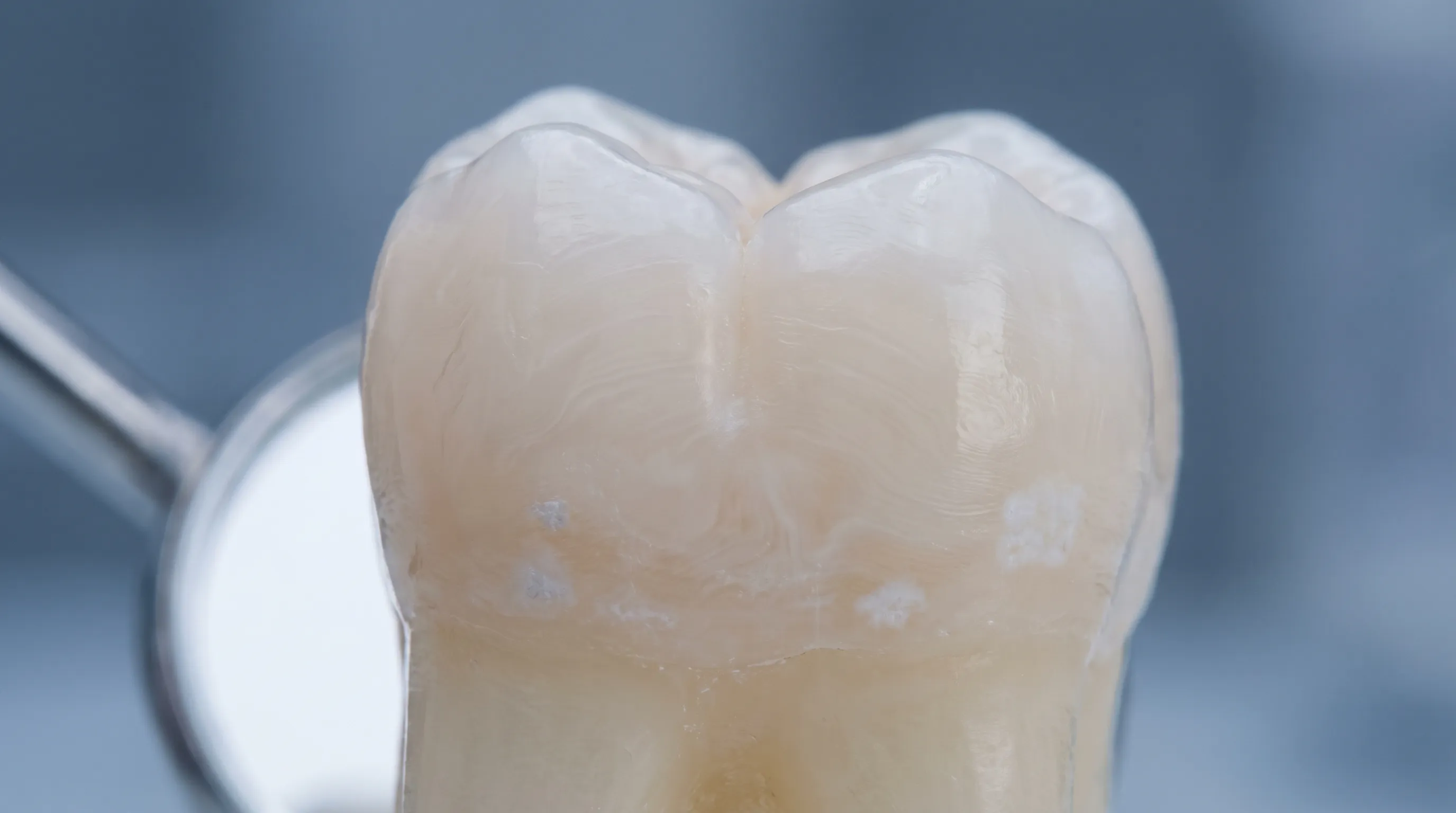
The National Institute of Dental and Craniofacial Research (NIH/NIDCR) explains that tooth decay begins when plaque-forming bacteria continuously bathe your teeth in acid. Initially, this acid exposure causes subsurface demineralization without creating a physical hole. The enamel loses minerals but remains structurally intact, appearing clinically as a chalky white spot lesion. This early stage represents a crucial window of opportunity. As detailed by Mayo Clinic, cavities are essentially decayed areas that become tiny openings or holes only after the mineral loss overwhelms the natural repair mechanisms of your teeth. If dietary sugars are consumed frequently, or if oral hygiene practices fail to disrupt the plaque biofilm, the acid attacks continue uninterrupted. Eventually, the enamel surface collapses, creating a permanent cavity that can no longer remineralize naturally without professional intervention.
The Role of Oral Microbiome and Acids
The bacterial ecosystem in your mouth operates as a living biofilm. Plaque is not merely a passive accumulation of food particles; it is a structured, multi-species bacterial community protected by an extracellular polymeric substance (EPS) matrix. This matrix acts as a shield, making the bacteria highly resistant to mechanical removal and allowing them to maintain an acidic microenvironment directly against the enamel surface. As decay progresses deeper into the tooth, the bacterial composition shifts. Aerobic bacteria are gradually replaced by anaerobic, putrefactive organisms that break down proteins and produce volatile sulfur compounds (VSCs). These compounds, including hydrogen sulfide and methyl mercaptan, are directly responsible for the foul odors and unpleasant tastes associated with advanced oral disease. Research highlighted by the Cleveland Clinic notes that cavities are fundamentally caused by mouth bacteria feeding on dietary sugars and converting them into destructive acids, highlighting the direct dietary link to microbial acid production.
Enamel Demineralization vs. Cavity Formation
It is vital to distinguish between early demineralization and actual cavity formation. Demineralization is a reversible process. Saliva plays a heroic role here by buffering acids, washing away food particles, and delivering calcium, phosphate, and fluoride ions back to weakened enamel areas. Fluoride enhances this process by forming fluorapatite, a crystalline structure significantly more acid-resistant than natural hydroxyapatite. When the rate of demineralization outpaces remineralization over weeks or months, the structural integrity fails. The surface layer caves in, forming a physical defect that traps food, harbors aggressive bacteria, and exposes the underlying dentin. Dentin contains microscopic tubules that communicate directly with the dental pulp, making the tooth highly susceptible to thermal, chemical, and osmotic sensitivity once the protective enamel shield is compromised.
Can You Taste a Cavity? The Direct Answer
Addressing the core question directly, the answer depends entirely on the stage of disease progression. Early-stage cavities produce zero taste, zero pain, and zero visible symptoms that a layperson can reliably identify. The enamel contains no living cells, blood vessels, or nerve endings. It is a highly mineralized, acellular tissue designed purely for mechanical protection. Therefore, mineral loss occurring within this outer layer generates no sensory feedback. You cannot taste a cavity when it exists only as subsurface demineralization or a microscopic white spot. The taste receptors on your tongue and the olfactory receptors in your nasal cavity require chemical stimuli to register flavor. Intact enamel, even when softened by acid, does not release flavorful compounds into the oral environment.
Why Early Decay Has No Taste
MedlinePlus and the Centers for Disease Control and Prevention (CDC) consistently emphasize that early oral diseases rarely cause noticeable signs until they reach a more advanced, often painful stage. During the initial phase of decay, the chemical environment remains localized to the microscopic space between plaque and enamel. The acids dissolve minerals but do not break down proteins or create volatile byproducts. Without nerve innervation or chemical diffusion into the saliva, your gustatory system remains completely unaware of the damage. This silent progression is precisely why routine dental screenings are non-negotiable for preventive care. Relying on taste, smell, or pain as early warning mechanisms guarantees that decay will be caught too late, often after it has penetrated the dentin and potentially reached the pulp chamber.
When a Bad Taste Actually Appears
Once decay breaches the enamel and invades the dentin, the sensory and chemical landscape changes dramatically. The cavity becomes a physical reservoir that traps food debris, particularly carbohydrates and fibrous materials. As these particles sit in the warm, moist, oxygen-poor environment of a dental defect, they begin to decompose. Oral bacteria metabolize the decaying matter, releasing metabolic waste products that diffuse into your saliva. Additionally, if the infection reaches the dental pulp and triggers an immune response, neutrophils migrate to the site, creating pus. An abscess, as defined by NIH/NIDCR, is a localized collection of pus caused by infection, frequently resulting in pain, swelling, and fever. When this purulent fluid drains through a sinus tract or the cavity itself, it produces a distinctively foul, salty, or bitter taste. This is when patients often report that their mouth tastes metallic, rotten, or persistently unpleasant despite thorough brushing.
Differentiating Cavity-Related Tastes from Other Oral Issues
A bad taste does not automatically equal a cavity. Periodontal disease (gum inflammation and bone loss), tonsil stones, gastroesophageal reflux disease (GERD), dry mouth syndrome (xerostomia), and certain medications can all alter oral flavor. Gum disease produces taste changes due to bleeding tissue and anaerobic bacterial activity in deep periodontal pockets. Tonsil stones calcify in the crypts and emit intense sulfuric odors that translate directly to taste. Acid reflux bathes the mouth in gastric contents, leaving a sour or bitter aftertaste. Dry mouth reduces saliva flow, preventing the natural clearance of debris and bacteria, which allows flavors to linger and intensify. Dentists use clinical examination, radiographs, and diagnostic testing to isolate the exact etiology. Assuming a bad taste is solely a cavity without professional evaluation risks missing other serious underlying conditions.
Symptoms and Warning Signs of Progressive Decay
While early decay remains silent, moderate and severe cavities generate a predictable cascade of symptoms. Recognizing these signs early can prevent emergency situations, complex treatments, and permanent tooth loss. The progression typically follows a timeline of months to years, though aggressive decay in children or immunocompromised individuals can advance within weeks.
Early-Stage Indicators (Often Overlooked)
The earliest detectable signs usually require professional equipment. Dentists may note chalky white opacities near the gumline or between teeth. These areas feel slightly rough to a dental explorer but do not catch. Patients rarely notice these at home. Occasionally, individuals report a vague sensation of texture irregularity when running their tongue over the tooth surface. Mild discoloration might appear as faint yellow or brown tinges, often dismissed as normal staining. At this point, the pulp is completely unaffected, and thermal stimuli cause no discomfort.
Mid-Stage Progression (Sensitivity and Staining)
Once enamel breaches and dentin exposure occurs, symptoms become unmistakable. Dentinal tubules transmit thermal changes, osmotic shifts, and chemical stimuli directly to the pulp. Patients experience sharp, transient pain when consuming sugary, cold, hot, or acidic foods. Visual changes include obvious brown, black, or gray discoloration localized to specific spots. Tiny pits or holes become visible in mirrors or feel distinct to the tongue. Pain when biting down may occur if the structural integrity of the cusp is compromised. Food impaction becomes frequent, leading to localized gum inflammation and intermittent bad breath. The Mayo Clinic highlights that untreated cavities grow larger and affect deeper layers, emphasizing the importance of addressing sensitivity immediately rather than waiting for it to resolve spontaneously, which it rarely does.
Advanced Decay and Infection (Abscess and Severe Pain)
When decay reaches the pulp, bacterial invasion triggers irreversible pulpitis and eventually necrosis. The dying nerve tissue releases inflammatory mediators, causing spontaneous, throbbing pain that often worsens at night or when lying down. Pain may radiate to the jaw, ear, or temple. Swelling develops as pus accumulates at the root apex. A dental fistula (gum boil) may appear as a small pimple on the gums, draining pus and temporarily relieving pressure while causing a persistent foul taste. Systemic signs include fever, lymphadenopathy (swollen glands), facial asymmetry, and difficulty swallowing or breathing. This stage constitutes a dental emergency requiring immediate intervention to prevent the infection from spreading into fascial spaces of the neck or entering the bloodstream, which can lead to life-threatening complications.
| Stage | Structural Damage | Sensory Symptoms | Taste Changes | Treatment Approach |
|---|---|---|---|---|
| Early (Enamel) | Subsurface demineralization, intact surface | None | None | Fluoride therapy, remineralization protocols, improved hygiene |
| Moderate (Dentin) | Cavitation, exposed dentin tubules | Thermal/sweet sensitivity, visible pits, mild bite discomfort | Mild bad taste from trapped food debris | Conservative composite or amalgam filling, sealants for adjacent surfaces |
| Advanced (Pulp) | Bacterial penetration, inflamed or necrotic nerve | Spontaneous throbbing pain, radiating discomfort, swelling | Distinctly foul, salty, or metallic taste from pus/abscess | Root canal therapy, crown restoration, or extraction if unrestorable |
| Post-Treatment | Restored structure, sealed margins | Resolution of acute symptoms, possible mild post-op sensitivity | Normalized taste, temporary metallic taste from new materials | Maintenance care, occlusal adjustment, night guard if indicated |

How Dentists Detect Cavities Before Symptoms Arise
Professional dental examinations are specifically designed to intercept decay during the asymptomatic, reversible phase. Relying on taste, pain, or mirror checks is clinically inadequate for early detection. Modern dentistry employs a multi-modal diagnostic approach to identify lesions years before they cause sensory changes.
Clinical Visual Examinations
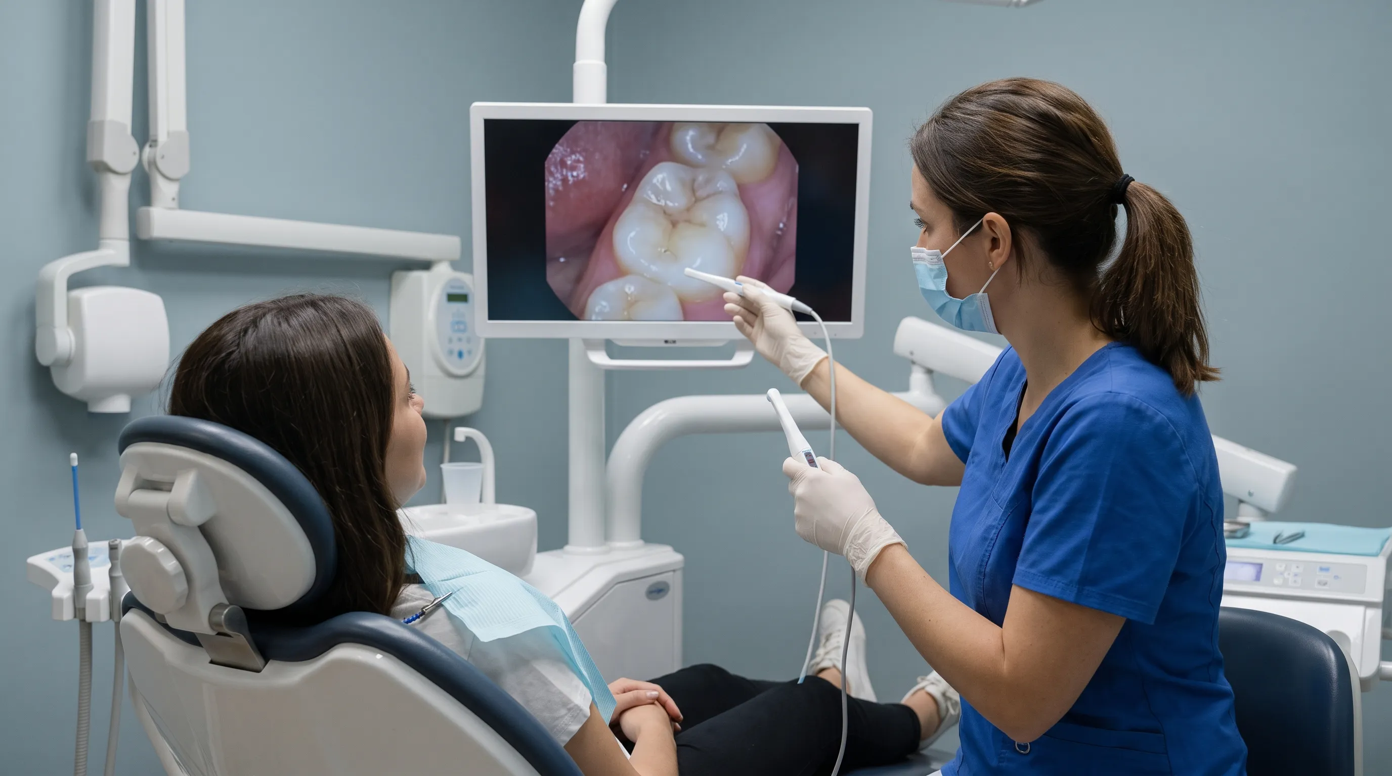
Dentists use bright overhead lighting, mouth mirrors, and gentle air drying to desiccate enamel surfaces. Drying reveals subsurface demineralization as opaque white spots that disappear when wet. This simple technique allows clinicians to spot early acid damage that patients would never notice. Tactile examination with a dental explorer helps determine if the enamel surface has actually collapsed into a physical cavity or remains intact despite demineralization. Dentists also assess gum health, plaque accumulation, and occlusal wear patterns that contribute to decay risk.
Dental Radiographs (X-Rays)
Bitewing radiographs are the gold standard for detecting interproximal cavities (between teeth) and recurrent decay under existing restorations. Enamel and dentin appear radiolucent (dark) on X-rays, while healthy tooth structure and bone appear radiopaque (light). Early decay shows as a subtle triangular shadow in the enamel. Advanced decay extends into dentin and approaches the pulp chamber. Panoramic and periapical radiographs evaluate root health, abscess formation, and bone loss. The American Dental Association (ADA) recommends radiographic frequency based on individual caries risk, ensuring patients receive appropriate diagnostic coverage without unnecessary radiation exposure.
Advanced Diagnostic Tools
Laser fluorescence devices, such as DIAGNOdent, use specific wavelengths of light to quantify bacterial metabolic byproducts within demineralized areas. The device emits a fluorescent reading that correlates with lesion depth, providing objective, numeric data to track progression or regression over time. Quantitative light-induced fluorescence (QLF) imaging captures real-time mineral loss percentages. Transillumination shines intense light through the tooth, making cracks and early decay appear as dark shadows. These technologies complement traditional methods, reducing subjective guesswork and enabling minimally invasive, precision dentistry that preserves maximum healthy tooth structure.
Prevention Strategies to Stop Decay in Its Tracks
Preventing cavities requires a multifaceted approach that addresses diet, hygiene, microbiome balance, and professional intervention. The CDC reports that drinking fluoridated water reduces cavities by approximately 25% in both children and adults, demonstrating the profound impact of public health initiatives. Individual prevention protocols must be consistent, scientifically grounded, and tailored to personal risk factors.
Daily Oral Hygiene Routines
Brushing twice daily with fluoride toothpaste remains the cornerstone of decay prevention. Use a soft-bristled brush with proper angulation at the gumline, spending at least two minutes to ensure complete surface coverage. Electric toothbrushes with pressure sensors and timers often improve compliance and reduce aggressive scrubbing that damages enamel and gums. Flossing daily is non-negotiable for removing interproximal plaque where brushes cannot reach. Water flossers serve as effective adjuncts for individuals with braces, bridges, or dexterity limitations, but traditional string floss remains superior for direct plaque removal. Antimicrobial or fluoride mouth rinses provide additional chemical protection, especially when used at separate times from brushing to avoid washing away concentrated toothpaste fluoride.
Dietary Modifications for Oral Health
Decay is driven by the frequency of acid attacks, not solely the total sugar volume. Grazing on sugary snacks, sipping soda throughout the day, or constantly consuming acidic beverages maintains a low pH environment that prevents remineralization. Limit carbohydrate intake to meal times, drink water between meals, and avoid prolonged bottle feeding in infants. Incorporate xylitol-containing products; this natural sugar alcohol inhibits S. mutans growth, reduces plaque acidity, and stimulates saliva flow. Foods rich in calcium, phosphorus, and fat-soluble vitamins (A, D, E, K) support enamel integrity and salivary buffering capacity. Chewing fibrous vegetables like carrots and celery naturally cleanses teeth and promotes salivation. Reducing tobacco use eliminates a major risk factor for periodontal disease, oral cancer, and impaired healing.
Professional Preventive Treatments
Biannual dental visits enable early interception, professional plaque removal, and application of prescription-strength fluoride varnishes. Dental sealants, recommended heavily by the CDC given that less than 42% of U.S. children utilize them, provide a physical plastic barrier over deep molar grooves, preventing plaque accumulation in vulnerable fissures. Silver diamine fluoride (SDF) offers non-invasive arrest of active decay in children and elderly patients who cannot undergo immediate drilling. Periodontal therapy addresses gum inflammation that accelerates root surface decay. Individualized risk assessments consider medical conditions (diabetes, autoimmune disorders, Sjögren syndrome), medication side effects (antihistamines, antidepressants causing dry mouth), and genetic enamel defects to customize prevention plans.
What to Do If You Notice a Bad Taste or Suspect a Cavity
Recognizing the need for professional intervention is critical. If you experience persistent unpleasant tastes, sensitivity, or visible changes, waiting is the worst possible strategy. Delayed care transforms minor, affordable treatments into complex, costly procedures.
Immediate Steps to Manage Discomfort
Until your dental appointment, maintain rigorous but gentle hygiene. Rinse with warm salt water to reduce bacterial load, soothe inflamed tissue, and draw out minor exudate. Avoid placing aspirin directly on the gums, as this causes chemical burns. Use over-the-counter analgesics like ibuprofen or acetaminophen for pain management, following package dosing instructions. Apply dental wax or temporary filling material from pharmacies to cover sharp edges or large holes that catch food, but understand this is strictly a short-term barrier, not a treatment. Limit extreme temperatures and sugary exposures to reduce pulpal inflammation.
When to Seek Emergency Dental Care
Certain symptoms demand immediate professional attention. Severe, uncontrolled pain, facial or neck swelling, difficulty breathing or swallowing, high fever, and pus drainage indicate spreading infection. These signs suggest the bacteria have breached the tooth apex and entered fascial spaces. Abscesses in the maxillary region can spread to the cavernous sinus or orbital area, while mandibular infections can compromise the airway through Ludwig angina. Emergency dental or medical evaluation is mandatory. Antibiotics alone do not cure dental abscesses; the source tooth must be treated with root canal therapy or extraction to eliminate the bacterial reservoir.
Treatment Options Based on Severity
Modern restorative dentistry prioritizes tissue conservation. Small cavities receive composite resin fillings bonded directly to tooth structure, restoring function and aesthetics. Moderate decay may require inlays or onlays when standard fillings would compromise structural strength. Large cavities with remaining sound tooth structure are covered by dental crowns for long-term durability. If the pulp is infected, root canal therapy removes the necrotic tissue, disinfects the canal system, and seals the space, preserving the natural tooth. Unrestorable teeth require extraction, followed by implants, bridges, or dentures to restore mastication and prevent shifting of adjacent teeth. Early intervention consistently yields simpler, less invasive, and more cost-effective outcomes.
Frequently Asked Questions
Can you taste a cavity in its early stages?
No, early-stage cavities are completely asymptomatic and do not produce any noticeable taste or pain. At this point, enamel is only beginning to demineralize, forming microscopic white spots that lack nerve endings or bacterial waste byproducts strong enough to alter your sense of taste. The disease operates silently, which is why professional screenings are essential for early detection.
What does a bad taste from tooth decay actually mean?
A persistent foul, metallic, or salty taste usually indicates advanced decay, trapped food particles, or an active infection like a dental abscess. The unpleasant flavor comes from pus, volatile sulfur compounds produced by anaerobic bacteria, and decomposing organic matter lodged inside the cavity. It signals that the disease has progressed beyond the enamel and requires immediate clinical attention.
How can I stop a cavity before it causes symptoms?
Early decay can often be reversed through consistent use of fluoride toothpaste, dietary modifications to reduce sugar frequency, drinking fluoridated water, and biannual dental cleanings. Fluoride promotes remineralization by attracting calcium and phosphate ions back to weakened enamel. Dental sealants provide a physical barrier over deep grooves, preventing bacteria from colonizing vulnerable areas before decay begins.
When should I see a dentist for a strange taste in my mouth?
Schedule a dental appointment immediately if a bad taste persists beyond a week, especially when accompanied by tooth sensitivity, visible pits or discoloration, localized swelling, or throbbing pain. Delaying care can allow minor decay to progress into severe infection, abscess formation, or irreversible pulp damage. Professional diagnosis ensures accurate treatment rather than temporary masking of symptoms.
Can a dental abscess cause a persistent bad taste?
Yes, a dental abscess is one of the most common causes of sudden, intense bad taste. When infection reaches the tooth pulp, the body produces pus as an immune response. This purulent fluid leaks into the oral cavity, creating a distinctly foul, salty, or bitter flavor that often requires antibiotics and definitive treatment like root canal therapy or extraction to resolve completely.
Conclusion
Understanding whether you can taste a cavity requires separating myth from clinical reality. While early decay remains completely undetectable by taste buds or sensory nerves, advanced structural breakdown, bacterial metabolism, trapped food debris, and abscess formation can absolutely generate persistent, unpleasant flavors in the mouth. This unpleasant taste is not a reliable early warning system; rather, it is a late-stage alarm indicating significant tissue destruction and potential infection. The most effective strategy for lifelong oral health centers on consistent, evidence-based prevention: brushing with fluoride, flossing daily, limiting dietary sugar frequency, utilizing professional sealants and fluoride treatments, and committing to regular dental examinations that include radiographic screening. By intercepting decay during the silent, reversible phase, patients avoid pain, preserve natural tooth structure, and maintain systemic health. If you notice any unusual taste, sensitivity, or visual changes, do not wait for symptoms to intensify. Contact a licensed dental professional promptly for accurate diagnosis and minimally invasive treatment tailored to your specific clinical needs. Your teeth deserve proactive protection, not reactive rescue.
How we work
Researched and drafted with AI assistance, then reviewed by a human editor against the cited sources.
This article is general health information, not medical advice. Consult a qualified healthcare professional about your own situation.