Can You Get a Zit on Your Tongue? Medical Facts, Causes, and Treatments
Have you ever woken up to discover an uncomfortable bump on your tongue and immediately wondered, can you get a zit on your tongue? You are certainly not alone. Millions of people search for answers regarding unexpected oral bumps, assuming they might be experiencing a form of acne. However, the answer is both anatomically complex and clinically straightforward. True acne requires hair follicles and sebaceous glands to form, structures that are entirely absent in the specialized mucosal lining of your mouth. What you are actually noticing is likely a benign inflammatory response, a minor glandular blockage, or a common oral ulcer masquerading as a pimple. Understanding the difference between actual dermatological acne and oral lesions is crucial for appropriate care, peace of mind, and preventing complications. In this comprehensive guide, we will dissect the medical realities of tongue bumps, explore their underlying causes, examine evidence-based treatment protocols, and provide actionable strategies to maintain optimal oral mucosal health.
Understanding Tongue Bumps: Why They Look Like Pimples
The oral cavity is a highly dynamic environment that serves as the entry point for digestion, respiration, and speech. When unexpected bumps appear, the visual similarity to facial acne can understandably trigger concern. However, recognizing the fundamental biological differences between cutaneous skin and oral mucosa is the first step toward accurate self-assessment and effective management, as detailed by the Cleveland Clinic.
Free calculatorHeadache Location ChartIdentify headache types by locationOpen the calculatorThe Complex Anatomy of the Tongue
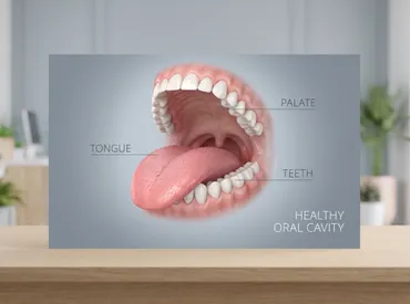
The tongue is a muscular hydrostat covered in specialized epithelial tissue known as stratified squamous epithelium. Unlike the skin on your face, arms, or back, this mucosal layer contains zero sebaceous glands, hair follicles, or sweat glands. Instead, it features thousands of microscopic projections called papillae, which house your taste buds and provide structural integrity for mastication and swallowing. The most prominent are filiform papillae (which give the tongue its rough texture), fungiform papillae (visible as tiny red dots), circumvallate papillae (located at the back of the tongue), and foliate papillae (along the lateral borders). When any of these structures become irritated, inflamed, or obstructed, they swell rapidly, creating the appearance of a raised, white, or red bump. This physiological response is entirely distinct from comedone formation, yet it shares enough visual overlap with acne to confuse laypersons asking whether they can get a zit on their tongue.
Transient Lingual Papillitis: The Most Common Culprit
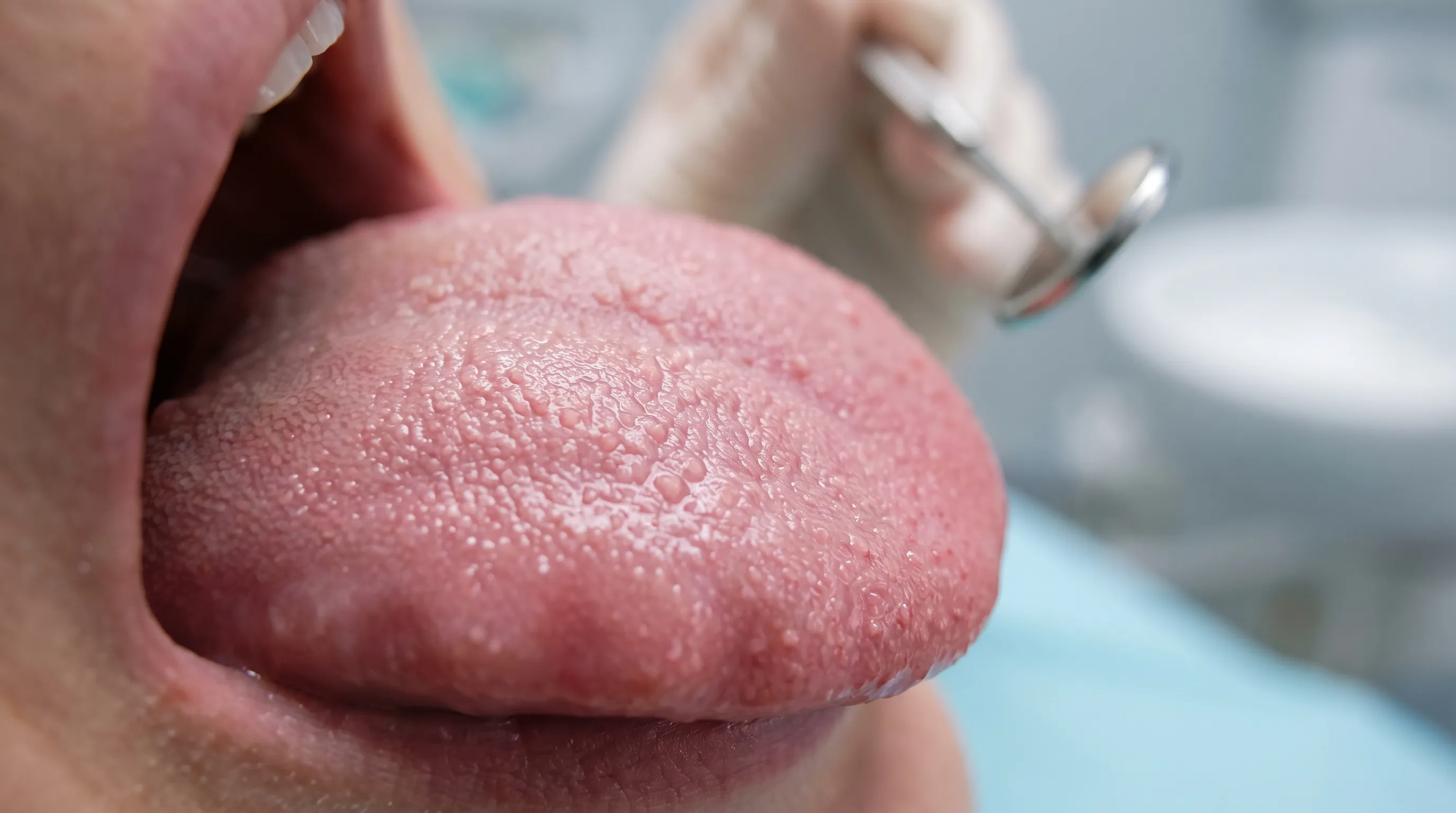
Also known as lie bumps or inflamed papillae, transient lingual papillitis is the primary condition responsible for isolated tongue bumps. Research published in the International Journal of Dermatology indicates that this condition affects a significant portion of the population at least once in their lifetime. The pathogenesis involves localized inflammation of one or more fungiform or filiform papillae, typically triggered by mechanical trauma, spicy or acidic foods, stress, or minor allergic reactions. As noted by the Mayo Clinic, these lesions present as tender, slightly raised nodules that may range from flesh-colored to pale white or erythematous. Unlike true acne, they do not contain keratin plugs or purulent exudate unless secondarily infected. The discomfort is usually short-lived, resolving spontaneously within three to seven days as the epithelial cells regenerate and inflammation subsides.
How Inflammation Mimics Acne
The confusion surrounding the question of whether you can get a zit on your tongue largely stems from shared inflammatory pathways. When tissue experiences microtrauma, the body initiates a localized immune cascade. Mast cells release histamine, blood vessels dilate, and fluid accumulates in the extracellular matrix, causing swelling. In the oral cavity, this manifests as a discrete, dome-shaped elevation. Additionally, dead epithelial cells, salivary proteins, and oral microbiome components can accumulate on the surface of an inflamed papilla, forming a whitish cap that closely resembles a pustule. However, microscopic analysis reveals that these caps are fibrinous exudates and keratinized debris rather than sebum-filled comedones. Recognizing this distinction prevents unnecessary manipulation and guides patients toward appropriate soothing interventions rather than acne-targeted topicals that may further irritate sensitive mucosa.
Common Oral Lesions That Mimic Tongue Acne
Beyond transient papillitis, several other benign conditions frequently masquerade as pimple-like protrusions on or near the tongue. Differentiating these requires understanding their etiology, duration, and clinical characteristics.
Mucoceles and Blocked Minor Salivary Glands
The floor of the mouth, underside of the tongue, and lateral borders contain hundreds of minor salivary glands. These glands continuously secrete mucous to lubricate the oral cavity and initiate digestion. When a duct becomes obstructed due to accidental biting, dental appliance friction, or chronic irritation, saliva backs up into the surrounding connective tissue. This creates a mucocele: a painless, translucent, or bluish fluid-filled cyst that often feels firm to the touch. While technically not a zit, mucoceles share the raised, nodular appearance that confuses patients. According to the Cleveland Clinic, these lesions typically resolve spontaneously within two to six weeks as the trapped fluid is gradually reabsorbed or drains naturally. Persistent mucoceles may require minor surgical excision to prevent recurrence.
Aphthous Ulcers (Canker Sores)
Aphthous stomatitis presents as shallow, crater-like sores with a yellowish-white fibrin center surrounded by an erythematous halo. In their early stages, these lesions often begin as slightly raised, tender bumps before ulcerating. The exact etiology remains multifactorial, involving immune dysregulation, genetic predisposition, stress, hormonal fluctuations, and nutritional gaps. Unlike acne, which is driven by Propionibacterium acnes and hormonal sebum production, canker sores are sterile inflammatory responses mediated by T-lymphocytes and cytokine release. They are notoriously painful, especially during eating or speaking, and typically heal within 7 to 14 days. Patients frequently mistake the initial swelling phase for a tongue zit, prompting premature topical treatments that can delay mucosal repair.
Traumatic Fibromas and Irritation Bumps
Chronic mechanical irritation from sharp teeth, ill-fitting dentures, or habitual chewing can trigger localized fibrous tissue hyperplasia. This results in an irritation fibroma: a firm, painless, pinkish nodule that develops as a protective response to repeated trauma. Fibromas are entirely benign and composed of dense collagen bundles. They do not fluctuate in size like inflammatory bumps and will not resolve spontaneously. While they pose no health threat, their persistent presence often leads patients to question if a zit on their tongue might actually be a structural change. Professional evaluation confirms diagnosis, and elective excision is recommended only if the lesion interferes with function or causes persistent discomfort.
Geographic Tongue and Inflammatory Patches
Benign migratory glossitis, commonly called geographic tongue, involves irregular, map-like patches on the dorsal surface where filiform papillae are temporarily shed. These depapillated areas appear as smooth, red zones surrounded by slightly raised, whitish or yellowish borders that migrate across the tongue over time. During active phases, the raised borders can feel slightly thickened and bumpy, leading to confusion about whether they can get a zit on their tongue when experiencing this condition. Geographic tongue is a benign inflammatory process with no known malignant potential, though it can cause transient sensitivity to acidic or spicy foods. Management focuses on symptom relief rather than lesion eradication.
| Condition | Appearance | Typical Duration | Primary Cause | Recommended Action |
|---|---|---|---|---|
| Transient Lingual Papillitis | Small, tender white/red bump | 3-7 days | Irritation, stress, acidic foods | Gentle rinses, avoid irritants |
| Mucocele | Smooth, translucent/bluish cyst | 2-6 weeks | Blocked salivary duct | Monitor, surgical excision if persistent |
| Canker Sore | Shallow ulcer with white center | 7-14 days | Immune response, stress, deficiency | Topical analgesics, B-vitamin support |
| Irritation Fibroma | Firm, pink, painless nodule | Permanent until removed | Chronic friction/biting | Dental adjustment or minor surgery |
| Geographic Tongue | Map-like patches with raised borders | Fluctuates/migrates | Inflammatory epithelial shedding | Avoid triggers, monitor symptoms |
Underlying Medical Conditions and Systemic Connections
While isolated tongue bumps are overwhelmingly benign, recurrent or persistent lesions can sometimes signal broader physiological imbalances or systemic health conditions. Recognizing these patterns enables proactive management and timely professional consultation.
Nutritional Deficiencies and Oral Manifestations
The oral mucosa is highly sensitive to micronutrient availability. Clinical studies consistently demonstrate that deficiencies in vitamin B12, folate, iron, and zinc compromise epithelial integrity and delay cellular turnover. Patients with recurrent aphthous ulcers or persistent papillary inflammation frequently test positive for subclinical deficiencies. Vitamin B12, in particular, plays a critical role in DNA synthesis and myelin maintenance; its absence triggers mucosal fragility and inflammatory cascades. Iron deficiency reduces oxygen delivery to rapidly regenerating tissues, while zinc deficiency impairs immune modulation and wound healing. Addressing these gaps through targeted supplementation and nutrient-dense dietary choices often reduces lesion frequency by up to 60 percent in clinically monitored populations, as supported by research from the National Institutes of Health Office of Dietary Supplements.
Viral Infections and Their Oral Signs
Certain viral pathogens establish latency in oral tissues and periodically reactivate, producing lesion clusters that mimic localized bumps. Herpes simplex virus type 1 (HSV-1) can cause intraoral ulcers, though these typically present as grouped vesicles on keratinized tissue rather than isolated tongue bumps. Human papillomavirus (HPV) infections occasionally manifest as small, cauliflower-like papillomas on the tongue or lateral borders. Coxsackievirus, responsible for hand, foot, and mouth disease, produces multiple painful vesicular eruptions across the oral mucosa, particularly in pediatric populations. Understanding viral etiology prevents misdiagnosis and ensures appropriate antiviral or supportive care rather than treating a non-existent zit. For comprehensive guidance on viral oral presentations, visit the Centers for Disease Control and Prevention (CDC) oral health resources.
Autoimmune Disorders and Chronic Inflammation
Systemic autoimmune conditions such as Behçet’s disease, Crohn’s disease, and celiac disease frequently present with oral manifestations. Behçet’s disease is characterized by recurrent, painful aphthous ulcers that may appear on the tongue alongside genital and cutaneous lesions, as documented by the Cleveland Clinic. Crohn’s disease can induce linear ulcerations or mucosal tags within the oral cavity, while celiac disease often correlates with enamel hypoplasia and recurrent stomatitis. In these scenarios, isolated tongue bumps are rarely the sole symptom. Patients experiencing frequent, atypical, or treatment-resistant oral lesions alongside gastrointestinal, dermatological, or systemic fatigue should pursue comprehensive rheumatological or gastroenterological screening.
Diagnosis and When to Consult a Medical Professional
Accurate diagnosis of tongue bumps requires systematic evaluation. While most cases resolve independently, establishing clear self-assessment parameters and recognizing warning signs prevents delayed care for serious conditions.
Self-Assessment Guidelines
Begin by documenting lesion characteristics using a consistent daily log. Note the exact location, color, size in millimeters, pain level on a scale of one to ten, and any associated symptoms such as fever, lymphadenopathy, or difficulty swallowing. Photograph the area under consistent lighting every 24 hours to track morphological changes. Avoid touching, biting, or applying harsh chemicals. Record recent dietary changes, new oral care products, stress levels, and sleep patterns. This data proves invaluable during clinical consultations, as healthcare providers rely on temporal progression patterns to differentiate inflammatory, infectious, and neoplastic processes.
Clinical Evaluation and Diagnostic Procedures
During an office visit, dentists, oral medicine specialists, or otolaryngologists perform a thorough intraoral examination using high-intensity illumination and palpation. They assess lesion mobility, tenderness, border regularity, and surrounding tissue architecture. If the diagnosis remains unclear, they may perform a viral swab, bacterial culture, or punch biopsy. Biopsy involves removing a small tissue sample for histopathological analysis, which definitively rules out dysplasia or carcinoma. Advanced imaging, such as ultrasound or cone-beam computed tomography, may be utilized for deep-seated mucoceles or suspected salivary gland pathology. Professional evaluation ensures that benign inflammatory responses receive appropriate conservative care while potentially serious conditions are intercepted early.
Red Flags Requiring Immediate Attention
While asking can you get a zit on your tongue usually leads to benign conclusions, certain clinical features warrant urgent evaluation. Seek immediate professional care if a bump persists beyond fourteen days without improvement, progressively enlarges, becomes hard or fixed to underlying structures, bleeds spontaneously, exhibits irregular or rolled edges, or displays mixed red and white coloration. Additional warning signs include unexplained weight loss, persistent numbness, difficulty moving the tongue, ear pain on one side, or cervical lymphadenopathy. These indicators may suggest leukoplakia, erythema, or early-stage squamous cell carcinoma, as outlined by the National Cancer Institute. Early detection dramatically improves treatment outcomes and survival rates for oral malignancies.

Evidence-Based Treatment Options and Self-Care Strategies
Effective management of tongue bumps hinges on addressing the underlying etiology while providing symptomatic relief and promoting mucosal regeneration. Evidence-based approaches prioritize gentle intervention over aggressive manipulation.
Over-the-Counter Topical Remedies
Pharmacological management focuses on pain reduction, inflammation modulation, and infection prevention. Benzocaine and lidocaine gels provide rapid localized analgesia by temporarily blocking sodium channels in sensory nerves. Use sparingly and according to package directions to avoid tissue desensitization or allergic reactions. Antiseptic mouthwashes containing cetylpyridinium chloride or hydrogen peroxide reduce bacterial load and promote a cleaner healing environment. However, alcohol-based rinses can exacerbate mucosal dryness and delay epithelial closure; opt for alcohol-free formulations instead. Anti-inflammatory gels containing hyaluronic acid create a protective barrier over ulcerated surfaces, accelerating tissue repair and reducing discomfort during meals. For detailed treatment protocols, refer to the Mayo Clinic guidelines on managing oral ulcers.
Clinically Supported Home Care
Natural and accessible home remedies remain foundational in oral lesion management. Warm saline rinses (one-half teaspoon of non-iodized salt dissolved in eight ounces of warm water) administered three to four times daily reduce edema, neutralize pH, and stimulate local circulation. Ice chips applied gently to the affected area induce vasoconstriction, numbing discomfort and limiting inflammatory mediator release. Aloe vera gel, specifically inner leaf preparations free of aloin, demonstrates clinically significant anti-inflammatory and antimicrobial properties. Honey, particularly medical-grade Manuka varieties, promotes moist wound healing and suppresses bacterial colonization through hydrogen peroxide and osmotic action. Consistent hydration maintains salivary flow, preventing xerostomia-related mucosal vulnerability.
Medical and Prescription Interventions
When conservative measures prove insufficient, healthcare providers may prescribe targeted therapies. Topical corticosteroids such as triamcinolone acetonide dental paste suppress localized immune responses in severe aphthous ulcers, reducing healing time by up to 50 percent. Antiviral medications like acyclovir or valacyclovir are indicated for confirmed herpetic or coxsackie virus involvement. For recurrent, debilitating mucoceles or fibromas, minor surgical excision under local anesthesia removes the lesion and ligates the offending duct. Laser ablation offers a minimally invasive alternative, sealing blood vessels and nerve endings while reducing postoperative pain. In cases linked to nutritional deficits, targeted intramuscular or high-dose oral supplementation corrects underlying gaps and prevents recurrence.
Lifestyle Modifications and Long-Term Prevention
Sustainable oral mucosal health requires proactive lifestyle adjustments that minimize trauma, optimize systemic wellness, and support epithelial resilience. Preventive strategies address the root triggers rather than merely treating symptomatic flares.
Dietary Adjustments for Mucosal Health
Nutritional intake directly influences mucosal integrity. Limit highly acidic foods such as citrus fruits, tomatoes, and vinegar-based dressings during active inflammation periods. Reduce consumption of spicy, crunchy, or thermally extreme foods that mechanically abrade delicate tissue. Incorporate omega-3 fatty acids from fatty fish, flaxseeds, and walnuts to modulate systemic inflammation and support cellular membrane repair. Prioritize probiotic-rich foods like yogurt, kefir, and fermented vegetables to maintain a balanced oral microbiome, preventing pathogenic overgrowth that can exacerbate mucosal irritation. Ensure adequate daily hydration by consuming eight to ten glasses of filtered water, as chronic dehydration thickens saliva and reduces its protective buffering capacity.
Optimizing Your Oral Hygiene Routine
Proper dental care extends beyond teeth to encompass the entire oral environment. Use an ultra-soft bristle toothbrush to prevent inadvertent papillary trauma. Replace toothbrushes every three months or immediately following an illness. Switch to sodium lauryl sulfate-free toothpaste, as SLS disrupts mucosal lipid barriers and correlates with increased canker sore frequency in sensitive individuals. Floss daily using gentle, gliding motions to prevent plaque accumulation near gingival margins, which can harbor inflammatory mediators. Consider incorporating a tongue scraper to remove superficial biofilm without applying excessive pressure to inflamed areas. Routine preventive care is further emphasized by the CDC.
Stress Management and Hormonal Regulation
Chronic stress elevates cortisol production, which suppresses local immune surveillance and delays mucosal healing. Implement evidence-based stress reduction techniques such as mindfulness meditation, diaphragmatic breathing exercises, and structured sleep routines. Aim for seven to nine hours of quality sleep nightly, as growth hormone and cytokine regulation peak during deep restorative phases. Women may notice cyclical lesion flares correlating with menstrual phases due to progesterone and estrogen fluctuations affecting vascular permeability. Tracking these patterns alongside oral symptom logs enables preemptive care adjustments. Regular physical activity further modulates inflammatory markers, improves circulation, and enhances stress resilience.
Frequently Asked Questions
Can you get a zit on your tongue or is it always a different type of bump?
True acne requires hair follicles and oil-producing glands, neither of which exist in the mouth. When patients ask if they can get a zit on their tongue, they are actually observing inflamed papillae, minor salivary cysts, or early ulcerative sores. These respond to different treatments than dermatological acne and should be managed with gentle oral care rather than topical acne medications.
Why does my tongue bump hurt more when I eat spicy or salty foods?
Inflamed oral mucosa experiences heightened nerve sensitivity due to compromised epithelial barriers. Spicy compounds like capsaicin and hypertonic saline directly stimulate exposed nociceptors, triggering sharp pain. This is a normal physiological response indicating active inflammation. Avoiding irritants during the healing phase allows the protective keratin layer to regenerate without repeated chemical disruption.
Is it safe to use mouthwash containing alcohol on an active tongue lesion?
Alcohol-based mouthwashes are contraindicated during active mucosal inflammation. Ethanol acts as a desiccant, stripping the oral lining of protective moisture and delaying cellular repair. Opt for alcohol-free formulations with soothing ingredients like xylitol, aloe vera, or mild antiseptics that clean without compromising the healing microenvironment.
How long should I wait before seeing a doctor if the bump does not disappear?
Clinical guidelines recommend professional evaluation for any oral lesion persisting beyond 14 days without improvement. Benign inflammatory bumps typically resolve within a week. Extended duration warrants examination to rule out chronic irritation, persistent infection, nutritional deficiency, or atypical tissue changes requiring diagnostic testing.
Can stress directly cause bumps on my tongue?
Yes. Psychological stress triggers systemic cortisol release, which alters local immune function and reduces mucosal blood flow. This environment increases susceptibility to transient lingual papillitis, aphthous ulcers, and viral reactivation. Incorporating daily stress-management practices significantly reduces recurrence rates and supports faster tissue recovery.
Conclusion
Understanding whether you can get a zit on your tongue requires shifting away from dermatological assumptions and embracing oral medicine fundamentals. The mouth lacks the anatomical structures necessary for true acne formation, yet it remains highly susceptible to inflammatory, mechanical, and systemic disruptions that produce remarkably similar-looking bumps. Transient lingual papillitis, mucoceles, canker sores, and irritation fibromas represent the most common explanations, all of which respond well to conservative care, proper hygiene, and targeted nutritional support. By recognizing the distinct clinical features of each condition, applying evidence-based soothing strategies, and maintaining vigilant monitoring for persistent or atypical changes, patients can effectively manage oral discomfort and safeguard long-term mucosal health. Remember that consistent prevention, stress reduction, and professional guidance form the foundation of optimal oral wellness. When in doubt, consult a qualified dental or medical professional to ensure accurate diagnosis and appropriate intervention tailored to your unique physiological needs.
How we work
Researched and drafted with AI assistance, then reviewed by a human editor against the cited sources.
This article is general health information, not medical advice. Consult a qualified healthcare professional about your own situation.