Tongue Spots Black: Causes, Diagnosis, Treatment, and Prevention Guide

Discovering unexpected tongue spots black can instantly trigger alarm, leading many individuals to fear serious underlying pathology or systemic illness. While the sudden appearance of dark discoloration, elongated bumps, or unusual coating on the dorsal surface of the tongue looks remarkably alarming, it is most frequently linked to a completely benign and highly treatable oral condition. Medical professionals commonly refer to this phenomenon as black hairy tongue, though the terminology only describes the visual presentation rather than an actual infectious or malignant process. The condition arises when the microscopic filiform papillae on the tongue's surface fail to shed properly, resulting in keratin buildup that traps chromogenic bacteria, yeast, food pigments, and cellular debris. Understanding the physiological mechanisms behind these changes is the first step toward demystifying the condition and implementing effective, evidence-based management strategies.
Oral discoloration often generates significant psychological distress due to its proximity to the face and the social implications of visible symptoms. Patients frequently report heightened anxiety about halitosis, altered taste perception, or potential contagiousness. Fortunately, contemporary clinical research confirms that tongue spots black are overwhelmingly temporary, non-infectious, and highly responsive to targeted oral hygiene modifications. By examining the anatomical foundations, microbial interactions, environmental triggers, and clinical management protocols associated with this presentation, individuals can confidently navigate their symptoms, eliminate triggering factors, and restore normal oral physiology. This comprehensive guide provides medically accurate, practitioner-vetted insights into the causes, diagnostic pathways, therapeutic interventions, and long-term prevention strategies for managing tongue discoloration safely and effectively.
Understanding the Anatomy and Clinical Presentation
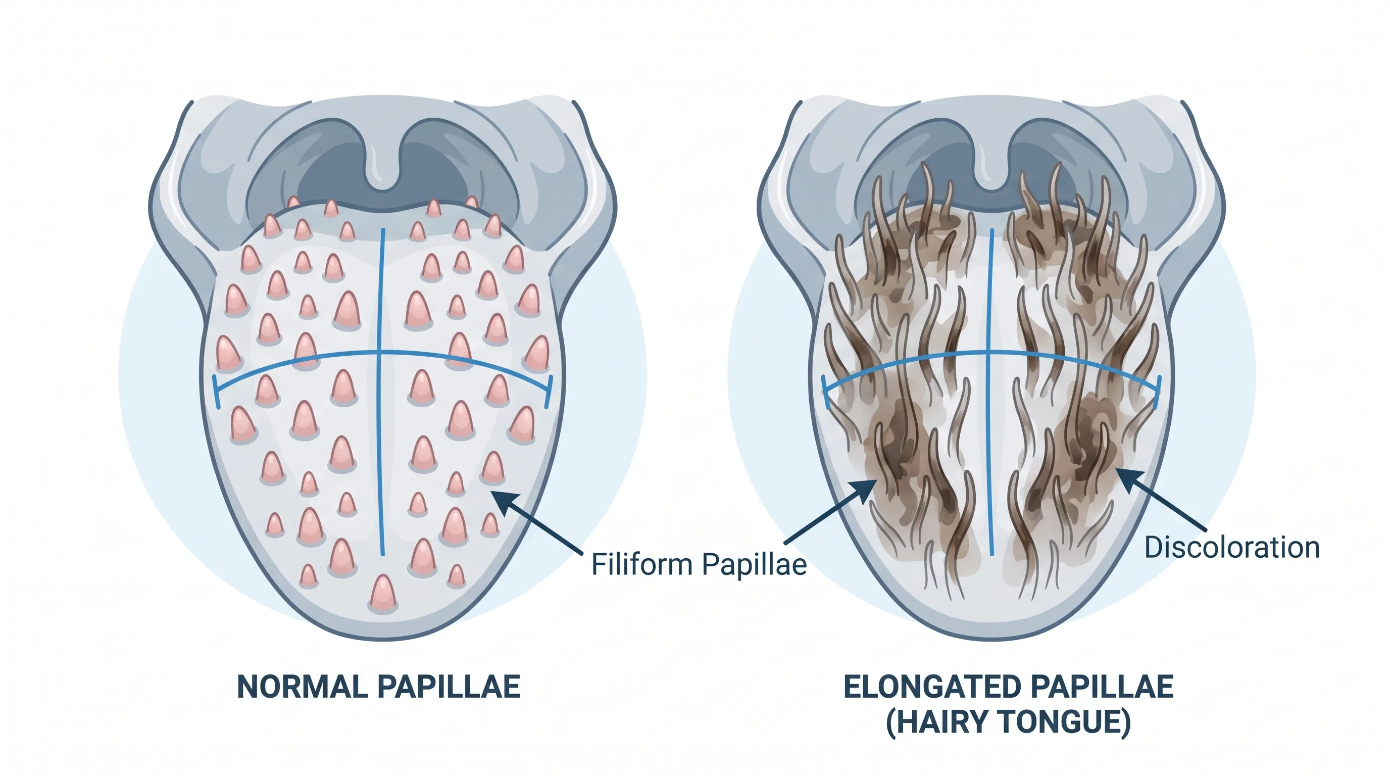
To properly address tongue spots black, it is essential to first understand the underlying structural components of the tongue and how normal physiological processes can temporarily become disrupted. The dorsal surface of the tongue is covered with thousands of microscopic projections called papillae, which serve distinct functional roles in tactile sensation, taste perception, and mechanical food manipulation. Unlike the fungiform, circumvallate, and foliate papillae that house taste buds, the filiform papillae are densely packed, keratinized structures responsible for creating friction during chewing and swallowing. In healthy oral environments, these papillae continuously undergo a natural desquamation cycle, where superficial epithelial cells shed at a rate that matches their production, maintaining a smooth, pink appearance.
Free calculatorWater Intake CalculatorCalculate daily water needsOpen the calculatorDefining Black Hairy Tongue and Its Medical Classification
The medical terminology for this condition includes Lingua Villosa Nigra, Melanoglossia, and Black Hairy Tongue Syndrome. Despite the dramatic nomenclature, clinical literature consistently classifies it as a benign, self-limiting oral disorder. The term "hairy" refers exclusively to the dramatic elongation of keratinized papillae, not to the presence of actual hair follicles or mammalian hair growth. When desquamation is delayed or halted, papillae can elongate up to fifteen times their normal length of approximately one millimeter. This hyperkeratotic state creates a dense, carpet-like environment that readily traps staining compounds from dietary sources, tobacco products, and microbial pigments, resulting in the characteristic black, brown, yellow, green, or white discoloration often associated with tongue spots black.
Epidemiology and Demographic Prevalence
Clinical surveillance and epidemiological reviews indicate that this condition predominantly affects adults rather than pediatric populations. There is a notable male-to-female predominance, which researchers attribute to higher rates of tobacco use, occupational dietary patterns, and varying oral care habits across demographics. The condition is also frequently documented among individuals experiencing systemic debilitation, those undergoing head and neck radiation therapy, HIV-positive patients with altered immune function, and individuals utilizing certain intravenous medications. Smokers and heavy consumers of dark beverages such as black tea and coffee consistently demonstrate elevated prevalence rates, highlighting the significant role of exogenous chromogenic exposure and microenvironmental changes in triggering papillary hyperplasia and discoloration.
The Pathophysiology Behind Papillary Elongation
The development of visible tongue spots black is fundamentally a disorder of epithelial turnover and microbial ecology. Under normal circumstances, the tongue undergoes continuous mechanical abrasion from food intake, swallowing, and mucosal movement. This physical friction, combined with healthy salivary flow, naturally exfoliates superficial keratin layers and prevents microbial colonization. When this delicate balance is disrupted, a cascade of physiological changes initiates the characteristic appearance.
Disrupted Desquamation and Keratin Accumulation
Hyperkeratosis and parakeratosis of the filiform papillae occur when cellular shedding mechanisms are impaired. Without consistent exfoliation, dead epithelial squames accumulate and compact, forming a dense keratin matrix that extends outward from the papillary base. This accumulation acts as a biological scaffold that retains moisture, cellular debris, and exogenous pigments. The thickened epithelial layer also creates a low-oxygen microenvironment that favors the proliferation of anaerobic and facultative anaerobic organisms, further accelerating the discoloration process and potentially contributing to altered taste perception and mild oral malodor.
Microbial Shifts and Chromogenic Colonization
The oral cavity hosts a highly complex microbiome consisting of hundreds of bacterial and fungal species. A balanced microbiome maintains neutral pH levels, supports epithelial integrity, and prevents pathogenic overgrowth. When salivary flow decreases or oral hygiene practices diminish, chromogenic microorganisms rapidly colonize the elongated papillary network. Species such as Porphyromonas, Prevotella, and certain Candida strains produce pigmented metabolic byproducts including melanin-like compounds, porphyrins, and hemoglobin-derived chromophores. These pigments bind tightly to keratin matrices, transforming the tongue's surface into a visually striking canvas of dark tones. The presence of tongue spots black in this context is essentially a visible biomarker of localized microbial dysbiosis rather than a primary infection.
Primary Causes and Modifiable Risk Factors
Identifying and eliminating triggering factors remains the cornerstone of effective management. Multiple clinical, lifestyle, and pharmacological variables converge to disrupt normal papillary turnover and promote chromogenic colonization.
Tobacco Use, Dietary Habits, and Hydration Levels
Smoking tobacco, whether through cigarettes, cigars, or smokeless alternatives, introduces intense heat, chemical irritants, and tannins directly into the oral cavity. These compounds desiccate mucosal tissues, reduce protective salivary clearance, and deposit persistent staining agents. Similarly, diets predominantly composed of soft, highly processed foods lack the necessary mechanical friction to naturally exfoliate papillae. Chronic low-grade dehydration or xerostomia further compounds the issue by diminishing salivary enzymes and buffering capacity. Individuals experiencing persistent mouth breathing due to nasal congestion or sleep apnea often exhibit compounded papillary changes due to prolonged mucosal drying.
Medications That Trigger Discoloration
Pharmacological agents frequently alter the oral ecosystem without causing direct tissue damage. Broad-spectrum antibiotics disrupt commensal bacterial populations, allowing opportunistic chromogenic species to flourish. Bismuth-containing medications, widely recognized for their antidiarrheal and gastroprotective properties, react with oral sulfur compounds to form bismuth sulfide, a harmless but visibly dark precipitate that adheres to papillary surfaces. Antipsychotics, antihypertensives, and anticholinergics often induce hyposalivation as a secondary effect, indirectly promoting the conditions necessary for tongue spots black to develop. Additionally, oxidizing mouthwashes containing hydrogen peroxide, witch hazel, or high concentrations of menthol can paradoxically exacerbate hyperkeratosis by irritating the epithelial lining and accelerating abnormal cellular proliferation.
Environmental and Occupational Exposure
Individuals working in environments with high chemical vapor exposure, poor hydration practices, or irregular meal schedules may experience accelerated oral desquamation delays. Occupational stressors that increase sympathetic nervous system activity can reduce parasympathetic-mediated salivation, subtly altering the oral microenvironment over time. Chronic betel nut chewing, common in certain geographic regions, introduces intense staining alkaloids and mechanical friction that profoundly alter papillary architecture and pigmentation.
| Risk Factor Category | Primary Mechanism of Action | Common Examples |
|---|---|---|
| Pharmacological | Microbiome disruption or salivary reduction | Broad-spectrum antibiotics, anticholinergics, bismuth subsalicylate |
| Behavioral/Tobacco | Mucosal desiccation and pigment deposition | Cigarette smoking, chewing tobacco, betel quid |
| Dietary/Habitual | Reduced mechanical abrasion and low hydration | Soft food diets, excessive coffee/tea, inadequate water intake |
| Product-Induced | Epithelial irritation and abnormal keratinization | Peroxide-based mouthwashes, alcohol-heavy rinses, mentholated gels |
| Systemic/Medical | Immune alteration and tissue repair delay | Head/neck radiation, HIV, advanced age, general debilitation |
Recognizing Symptoms and Clinical Presentation
While visual changes dominate patient concerns, associated sensory symptoms frequently impact daily comfort and nutritional habits. Most individuals remain largely asymptomatic beyond the striking visual presentation, yet a subset experiences functional disruptions that warrant clinical attention.
Visual Characteristics and Anatomical Location
The discoloration typically manifests as patchy, diffuse, or confluent areas of dark pigmentation concentrated along the central and posterior dorsal tongue, immediately anterior to the circumvallate papillae. The lateral borders and ventral surface usually remain unaffected, preserving normal mucosal coloration. The appearance ranges from velvety brown to jet black, occasionally presenting with yellowish, greenish, or grayish hues depending on the specific microbial species and dietary pigments involved. When tongue spots black first appear, they may begin as isolated, small macules that gradually coalesce into broader coated regions if initiating factors persist.
Associated Sensory and Functional Symptoms
Elongated papillae frequently trap volatile organic compounds produced by anaerobic bacteria, resulting in noticeable intraoral halitosis. Patients often report a persistent metallic or bitter taste known clinically as dysgeusia, which interferes with food enjoyment and appetite. A mild tickling or scratching sensation against the soft palate may occur during swallowing or speech due to the extended papillary tips making unintended mucosal contact. In cases where Candida albicans or other fungal organisms overgrow within the keratinized matrix, patients may experience glossopyrosis, characterized by a burning or stinging sensation that intensifies with spicy or acidic food consumption. Severe cases occasionally trigger exaggerated gag reflexes or mild nausea due to altered tactile stimulation of the oropharyngeal mucosa.
Psychological and Social Considerations
The visible nature of this condition frequently generates disproportionate anxiety relative to its clinical severity. Patients often fear contagious transmission, malignant transformation, or permanent tissue damage. Dental and medical professionals emphasize that reassurance and clear patient education significantly reduce distress. Normalizing the condition as a temporary physiological response rather than a disease state allows individuals to focus on practical hygiene interventions without unnecessary fear. Addressing the emotional component of oral discoloration remains an integral part of comprehensive care, particularly for patients with preexisting health anxiety or body image concerns.

Diagnostic Approaches and Rule-Out Conditions
Clinical evaluation primarily relies on visual assessment and patient history rather than invasive testing. Experienced clinicians can typically identify the condition based on classic morphological features and reported lifestyle factors, though careful differential evaluation remains essential to exclude more serious pathology.
Clinical Examination and Visual Assessment
Diagnosis occurs through direct intraoral inspection under adequate lighting. Practitioners examine the dorsal surface for hyperkeratotic, elongated papillae with characteristic dark pigmentation, verifying the absence of ulceration, induration, or fixed nodular formations. Because the condition is entirely superficial, tissue remains soft and pliable to palpation. Patient history focusing on medication use, tobacco habits, oral hygiene routines, and recent dietary changes provides crucial contextual information. In straightforward presentations, no additional laboratory testing or biopsies are required.
Differential Diagnosis: Distinguishing from Serious Conditions
Clinicians must carefully differentiate this benign presentation from several conditions with overlapping visual characteristics. Hairy leukoplakia, caused by Epstein-Barr virus replication in immunocompromised individuals, typically appears as corrugated white plaques on the lateral tongue margins rather than central dorsal discoloration. Oral thrush presents as creamy white, easily wipeable patches that reveal erythematous underlying mucosa, often accompanied by significant discomfort. Geographic tongue exhibits migrating erythematous patches with serpiginous white borders and lacks the hyperkeratotic "hairy" morphology. Melanotic macules appear as isolated, flat, brownish spots without papillary elongation. Most critically, oral mucosal melanoma is exceptionally rare but presents as irregularly pigmented, rapidly expanding, or nodular lesions that may bleed or ulcerate, necessitating immediate biopsy and specialist referral.
When Histology or Microbiology Is Necessary
Biopsy is rarely indicated but may be pursued if lesions demonstrate atypical features such as asymmetry, rapid progression, fixed induration, or failure to respond to aggressive hygiene modifications. Histologic examination typically reveals marked filiform papillae hyperplasia with hyperparakeratosis and superficial bacterial colonization without dysplasia or malignant transformation. Microbial cultures or fungal smears may be utilized when concurrent thrush symptoms, severe burning sensations, or antibiotic history suggest secondary yeast overgrowth, guiding targeted antifungal therapy when standard hygiene proves insufficient.
Evidence-Based Treatment and Management
Management prioritizes gentle mechanical removal of accumulated keratin, restoration of normal salivary flow, and elimination of precipitating factors. The condition responds exceptionally well to conservative, patient-driven interventions, making invasive procedures largely unnecessary.
First-Line Oral Hygiene Protocols
The foundational treatment involves twice-daily gentle mechanical cleaning of the dorsal tongue using a soft-bristled toothbrush or medical-grade tongue scraper. Patients should apply light, backward-moving strokes starting near the tip and progressing toward the posterior tongue, avoiding aggressive pressure that could cause epithelial microtrauma or trigger gag reflexes. Combining this practice with standard twice-daily tooth brushing establishes a consistent routine that prevents further accumulation. Increased fluid intake throughout the day maintains mucosal hydration, supporting natural enzymatic clearance and reducing keratin compaction.
Introducing dietary roughage such as raw vegetables, fibrous fruits, and whole grains provides natural abrasive action during mastication, mechanically assisting papillary exfoliation without requiring additional instrumentation. Avoiding excessive reliance on blended or pureed foods allows normal physiological abrasion to resume, accelerating resolution. For additional guidance on evidence-based hygiene routines, comprehensive resources are available at WebMD - Black Hairy Tongue: Causes and Treatments.
Pharmacological Interventions
When conservative measures prove inadequate after two to three weeks, clinicians may consider targeted pharmacotherapy. Short-course systemic or topical antifungals such as nystatin oral suspension or fluconazole effectively manage secondary Candida overgrowth contributing to burning sensations and persistent discoloration. In select cases where bacterial dysbiosis severely disrupts normal healing, brief targeted antibiotic therapy may be prescribed, though this approach requires careful monitoring to prevent further microbiome imbalance. Topical retinoids such as tretinoin have demonstrated efficacy in promoting normalized epithelial turnover by accelerating cellular differentiation and reducing hyperkeratosis, though their use remains off-label and should be supervised by a qualified practitioner.
Surgical intervention is reserved exclusively for refractory cases unresponsive to extended conservative and medical management. Laser ablation or electrosurgical clipping of severely hypertrophied papillae provides immediate mechanical reduction but carries risks of temporary mucosal sensitivity, altered taste, and potential recurrence if underlying triggering factors remain unaddressed. Most patients achieve complete resolution long before surgical options become relevant.
Lifestyle and Dietary Modifications
Successful long-term management requires sustained behavioral adjustments. Smoking cessation represents the single most impactful intervention, eliminating thermal irritation, chemical deposition, and mucosal desiccation. Reducing daily consumption of dark pigmented beverages such as black tea, strong coffee, and red wine minimizes exogenous staining agents that readily adhere to keratinized surfaces. Limiting alcohol intake preserves salivary gland function and prevents chronic dehydration of oral tissues.
Patients should carefully evaluate their oral hygiene products, temporarily discontinuing mouthwashes containing peroxide, alcohol, witch hazel, or high menthol concentrations until papillary architecture normalizes. Switching to neutral pH, alcohol-free rinses or simple warm saline solutions maintains oral cleanliness without disrupting epithelial turnover. Incorporating these changes aligns with clinical recommendations detailed by specialists at Healthgrades - Black Hairy Tongue: Causes, Diagnosis, and Treatment and ensures sustainable mucosal health.
Prevention and Long-Term Oral Care
Maintaining optimal tongue physiology requires consistent, proactive habits that support natural desquamation, microbial balance, and adequate hydration. Prevention strategies seamlessly integrate into daily routines without demanding significant lifestyle disruption.
Daily Maintenance Routines
Establishing a structured morning and evening oral care sequence that includes gentle tongue cleaning forms the cornerstone of prevention. Using light, deliberate strokes removes accumulating debris before it compacts into hyperkeratotic layers. Hydration goals should prioritize consistent water intake throughout waking hours, aiming for clear or pale yellow urine output as a practical hydration marker. When traveling, working long shifts, or consuming caffeine-heavy beverages, individuals should consciously increase water consumption and briefly rinse their mouth with water to dilute potential staining agents.
Monitoring and Professional Dental Checkups
Routine dental examinations every six months allow practitioners to assess papillary health, identify early signs of microbial imbalance, and provide personalized hygiene feedback. During cleanings, dental professionals can safely perform professional tongue debridement if minor buildup occurs between visits, particularly for patients with chronic dry mouth or medication-induced xerostomia. Individuals managing systemic conditions like diabetes, autoimmune disorders, or those receiving radiation therapy should maintain closer monitoring intervals to preemptively address mucosal changes before they progress.
When to Seek Professional Medical Evaluation
While most cases resolve independently with hygiene modifications, certain clinical scenarios warrant prompt professional assessment. Consult a dentist, primary care physician, or oral medicine specialist if the condition persists beyond three to four weeks despite strict adherence to hygiene protocols. Seek immediate evaluation if you develop localized pain, ulceration, unexplained bleeding, rapidly expanding lesions, or significant difficulty swallowing. Persistent dysgeusia, severe halitosis, or gagging that interferes with adequate nutrition requires clinical intervention to rule out secondary infections or structural abnormalities. Individuals with immunosuppression, recent head and neck radiation, or unexplained systemic weight loss should prioritize early professional evaluation to ensure comprehensive diagnostic accuracy and appropriate care coordination.
Frequently Asked Questions
Are black spots on the tongue a sign of oral cancer?
In the vast majority of cases, tongue spots black are caused by benign, temporary conditions like black hairy tongue or transient surface staining from food, beverages, or medications. However, any persistent, irregularly shaped, or growing dark lesion that does not respond to improved oral hygiene should be evaluated by a dentist or oral medicine specialist to rule out rare but serious conditions like oral melanoma.
How long does it take for black hairy tongue to go away?
With consistent oral hygiene practices like gentle tongue brushing, hydration, and dietary adjustments, most cases resolve within one to two days up to a few weeks. Discontinuing triggering factors like certain mouthwashes, smoking, or soft diets typically accelerates the shedding of elongated papillae and restores normal coloration.
Can antibiotics cause tongue spots black?
Yes. Broad-spectrum antibiotics can disrupt the natural balance of the oral microbiome by killing beneficial bacteria while allowing chromogenic organisms and yeasts like Candida albicans to overgrow. This microbial imbalance, combined with reduced desquamation of papillae, is a well-documented trigger for temporary black or dark discoloration on the tongue surface.
Should I use a tongue scraper or a toothbrush for treatment?
Both are effective when used correctly. A soft-bristled toothbrush or a dedicated tongue scraper can gently remove accumulated keratin, bacteria, and debris. The key is to use light pressure and avoid vigorous scraping, which can cause microtrauma. Consistency with gentle mechanical removal twice daily is more important than the specific tool chosen.
Why does the tongue sometimes feel hairy or ticklish with this condition?
The "hairy" sensation and tickling feeling occur because the filiform papillae elongate dramatically instead of shedding normally. As they grow up to fifteen times their typical length, they can brush against the palate during swallowing or speaking, creating a mild tickling or gagging sensation. This physical elongation, not actual hair growth, is responsible for the tactile and visual symptoms.
Conclusion
The sudden appearance of tongue spots black often triggers unnecessary fear, yet clinical evidence consistently demonstrates that this presentation represents a benign, highly responsive disruption of normal papillary turnover. By understanding the anatomical basis of filiform papillae, recognizing the role of microbial colonization, and identifying modifiable risk factors such as tobacco use, medication effects, and oral hygiene habits, individuals can confidently implement targeted management strategies. Gentle mechanical cleaning, adequate hydration, dietary fiber incorporation, and temporary avoidance of oxidizing oral products typically restore normal mucosal architecture within days to weeks. Maintaining consistent preventive practices and attending routine dental evaluations ensures long-term oral ecosystem balance and early identification of any atypical changes. For those seeking deeper clinical insights or professional treatment guidance, authoritative resources such as Wikipedia - Black Hairy Tongue provide comprehensive medical literature references. With informed action and consistent care, patients can seamlessly resolve symptoms and maintain optimal oral health without prolonged distress.
How we work
Researched and drafted with AI assistance, then reviewed by a human editor against the cited sources.
This article is general health information, not medical advice. Consult a qualified healthcare professional about your own situation.