Understanding Gum Abscess Stages: A Complete Guide to Symptoms, Treatment & Prevention

A sudden, throbbing sensation in your gums that disrupts your sleep, eating, and daily routine is rarely something you can ignore. When a localized dental infection takes hold, it doesn't happen overnight. Instead, it follows a predictable biological progression that dental professionals carefully monitor and categorize. Understanding the underlying progression of a gum abscess is crucial for anyone who values long-term oral health. Many patients delay seeking professional help because early signs mimic mild irritation or routine sensitivity, but waiting can transform a manageable condition into a complex medical emergency. By familiarizing yourself with the warning signs at each phase of infection, you empower yourself to act quickly, minimize tissue damage, and avoid invasive surgical interventions later on.
The human mouth is home to hundreds of bacterial species that exist in delicate balance. When that balance is disrupted by poor oral hygiene, untreated decay, or compromised immune defenses, harmful microorganisms multiply rapidly. They produce acidic byproducts and toxins that trigger an inflammatory immune response. This inflammatory cascade is what ultimately leads to tissue breakdown, pus accumulation, and the characteristic symptoms that bring patients to the dental chair. Navigating the gum abscess stages successfully requires both awareness of early indicators and a clear understanding of how clinical treatment aligns with the infection's progression. In this comprehensive guide, we will explore the biological mechanisms, clinical presentations, evidence-based treatment protocols, and long-term prevention strategies that define modern periodontal and endodontic care.
Understanding the Biological Basis of a Gum Abscess
To properly grasp how dental infections develop and evolve, we must first examine the anatomical and microbial foundations of the periodontium. The gums, alveolar bone, periodontal ligament, and cementum work together as a highly integrated support system for your teeth. When plaque biofilm accumulates along and beneath the gumline, mineralized deposits called calculus form rapidly. This hardened matrix acts as a porous fortress, sheltering anaerobic bacteria like Porphyromonas gingivalis, Prevotella intermedia, Tannerella forsythia, and Fusobacterium nucleatum. These pathogens secrete enzymes and lipopolysaccharides that trigger a localized immune response.
Free calculatorHeadache Location ChartIdentify headache types by locationOpen the calculatorThe body sends white blood cells, particularly neutrophils and macrophages, to combat the invading microbes. As this immune battle intensifies, dead bacteria, immune cells, and tissue debris accumulate, forming purulent exudate known as pus. Because the pus is trapped within a confined anatomical space, pressure builds rapidly, causing the characteristic swelling and pain. Differentiating between the types of infections is essential when evaluating the gum abscess stages, as treatment protocols vary significantly based on the origin point.
The Microbiology Behind Dental Infections
Dental infections are rarely caused by a single organism. Instead, they involve polymicrobial communities that exhibit complex synergistic behaviors. Gram-negative anaerobic bacteria dominate advanced periodontal pockets because these oxygen-deprived environments favor their survival. These bacteria produce collagenases, hyaluronidases, and prostaglandins that actively degrade connective tissue and stimulate bone resorption. As the infection progresses, the local pH drops, creating an even more favorable environment for acid-tolerant pathogens while damaging healthy epithelial cells. The host immune response, while attempting to protect the area, often contributes to collateral tissue damage. Matrix metalloproteinases (MMPs) released by activated neutrophils break down the periodontal ligament and alveolar bone matrix. This self-perpetuating cycle of bacterial proliferation and immune-mediated destruction forms the clinical foundation of periodontal disease.
Differentiating Abscess Types and Their Clinical Relevance
Periodontal abscesses originate in the gingival sulcus or periodontal pocket and typically affect the supporting structures around a vital tooth. They are commonly triggered by food impaction, incomplete calculus removal, or systemic conditions like uncontrolled diabetes. Conversely, periapical abscesses develop at the apex of a non-vital tooth following untreated caries, traumatic pulp exposure, or failed root canal therapy. The infection travels through the root canal system, exits the apical foramen, and establishes itself in the periapical region. Both types present with overlapping symptoms, but diagnostic testing like pulp vitality assessments and radiographic imaging helps clinicians pinpoint the exact source. Accurate identification directly influences whether treatment focuses on pocket debridement or endodontic intervention.
The Early Phase: Initial Inflammation and Gingival Swelling
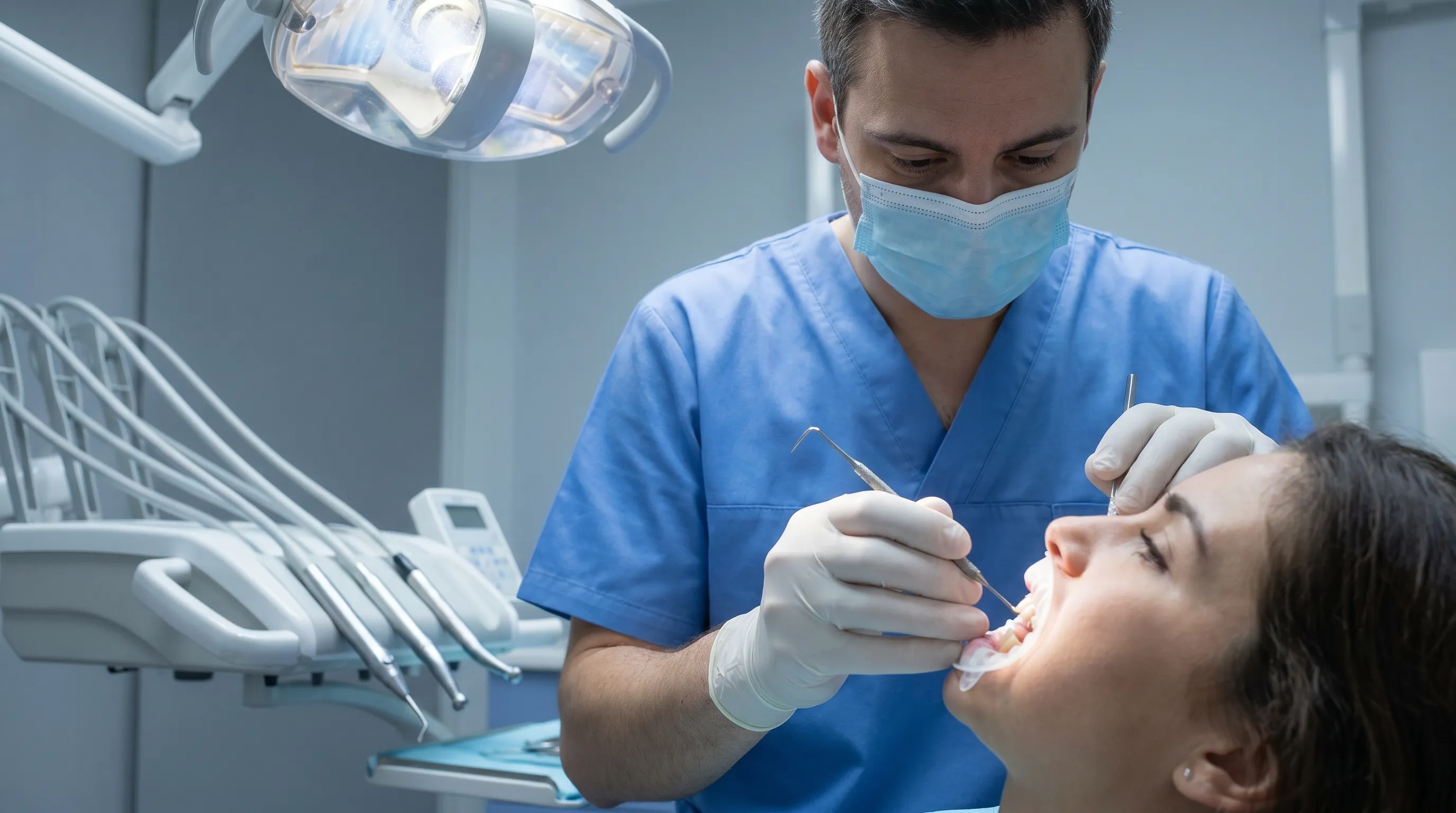
The initial phase represents the body's first active response to bacterial accumulation beneath the gumline. During this period, the gingival margin becomes erythematous, edematous, and highly sensitive to mechanical stimulation. Patients often notice mild bleeding during brushing or flossing, along with a subtle sense of pressure or fullness around one specific tooth. The tissue may appear slightly bulbous or roll outward, indicating early inflammatory infiltration. Recognizing these early signals is critical, as prompt intervention during this phase can often halt disease progression and prevent irreversible structural damage.
Recognizing Early Warning Signs and Symptoms
In the earliest manifestations, the gingiva exhibits a bright red or purplish discoloration rather than the healthy coral-pink hue of normal tissue. Probing depths may increase slightly from the baseline 1 to 3 millimeters to 4 or 5 millimeters, signaling early pocket formation. The area might feel tender to touch but does not yet cause spontaneous throbbing pain. Patients frequently report a localized "heavy" sensation or mild discomfort when chewing on that side. A slight foul taste may occasionally appear, particularly in the morning, as overnight bacterial metabolism produces volatile sulfur compounds. At this point, the gum abscess stages have not yet reached acute severity, making conservative management highly effective.
Immediate Actionable Steps for Patients
If you suspect early inflammation, begin with a warm saltwater rinse. Dissolve half a teaspoon of pharmaceutical-grade sodium chloride in eight ounces of warm water and gently swish for thirty seconds, repeating three to four times daily. This hyperosmotic solution draws out excess fluid, reduces edema, and temporarily shifts the local bacterial environment. Switch to a soft-bristled toothbrush and employ gentle, circular motions rather than aggressive horizontal scrubbing. Avoid flossing directly through the most inflamed area if it causes excessive bleeding, but continue interproximal cleaning around neighboring teeth to reduce overall biofilm load. Over-the-counter nonsteroidal anti-inflammatory drugs (NSAIDs) like ibuprofen can manage discomfort and reduce inflammation, provided you follow package directions and consult a pharmacist regarding contraindications. Schedule a dental evaluation within forty-eight to seventy-two hours to receive professional cleaning and clinical assessment.
The Development Phase: Pus Formation and Pocket Expansion
As bacterial proliferation overwhelms local immune defenses, the infection transitions into a purulent phase. The periodontal pocket deepens, creating an anaerobic sanctuary where bacteria multiply exponentially. Pus begins to accumulate within the confined tissue space, generating measurable hydrostatic pressure. This phase is characterized by visible swelling, pronounced tenderness, and the emergence of a localized "gum pimple" or fluctuant mass. The infection is no longer superficial; it has established a structured pathological environment that requires professional intervention to resolve. Understanding how to navigate these intermediate gum abscess stages helps patients avoid dangerous home remedies and seek appropriate clinical care.
Clinical Indicators of Progression
The gingiva becomes visibly swollen and may take on a shiny, stretched appearance due to underlying fluid accumulation. The affected tooth may exhibit mild mobility as the supporting bone matrix undergoes early inflammatory resorption. Chewing generates a sharp, localized pain, and hot or cold stimuli may cause lingering sensitivity if the pulp remains vital. A parulis, commonly referred to as a gum boil, may form as the infection creates a drainage pathway through the gingival tissue. When this tract opens, patients often notice a sudden salty or metallic taste accompanied by immediate, temporary pain relief as pressure dissipates. However, the tract rarely heals permanently without professional debridement, as the underlying bacterial colony remains intact.
Diagnostic Procedures Dentists Use to Confirm Severity
Clinical evaluation begins with a comprehensive visual and tactile examination. Dentists utilize a calibrated periodontal probe to measure pocket depths around each tooth, documenting any readings exceeding four millimeters as pathological. Pulp vitality testing employs thermal or electric stimuli to determine whether the tooth nerve remains responsive. Bitewing and periapical radiographs reveal early bone loss patterns, root morphology anomalies, and periapical radiolucencies. In complex cases, cone-beam computed tomography (CBCT) provides three-dimensional visualization of bone architecture and infection spread. Microbial sampling is rarely necessary for routine cases but may be indicated for patients with recurrent, antibiotic-resistant infections. These diagnostic tools collectively inform the clinical treatment pathway and ensure targeted, effective intervention.
The Advanced Phase: Acute Pain, Systemic Symptoms, and Fistula Formation
When left untreated, the infection breaches local tissue boundaries and triggers widespread inflammatory responses. This represents the most critical juncture in the progression of a dental abscess. The hydrostatic pressure within the abscess cavity reaches extreme levels, causing intense, throbbing pain that frequently disrupts sleep and radiates to the jaw, ear, or neck region. Swelling extends beyond the gingival margin into adjacent facial tissues, and systemic symptoms indicate that the immune response has escalated to a whole-body level. Recognizing the red flags of advanced infection is essential for preventing life-threatening complications.
When to Seek Emergency Dental Care
Immediate professional intervention is required if you experience rapid facial swelling, difficulty swallowing, limited mouth opening (trismus), fever exceeding 100.4°F (38°C), or swollen lymph nodes beneath the jaw. These symptoms suggest that the infection is spreading into fascial planes and potentially compromising airway patency. A spreading dental infection can evolve into cellulitis, Ludwig's angina, or cavernous sinus thrombosis, all of which constitute medical emergencies. Do not wait for a routine dental appointment; contact an emergency dental clinic or visit the nearest hospital emergency department. Intravenous antibiotics, surgical drainage, and airway monitoring may be necessary to stabilize your condition.
Potential Complications of Delayed Treatment
Chronic dental infections exert significant strain on systemic health. Persistent periodontal pathogens have been linked to increased cardiovascular inflammation, poorly controlled blood glucose in diabetic patients, adverse pregnancy outcomes, and rheumatoid arthritis exacerbations. Locally, untreated abscesses can cause irreversible alveolar bone destruction, leading to tooth mobility and eventual loss. The infection may migrate into the maxillary sinus, causing sinusitis, or erode into adjacent anatomical structures. Additionally, chronic pain and infection trigger sustained cortisol release, disrupting sleep architecture, weakening immune function, and negatively impacting mental well-being. Prompt treatment not only preserves teeth but also safeguards overall physiological health.
The Resolution and Healing Phase: Treatment and Recovery
Once professional intervention begins, the focus shifts from infection control to tissue regeneration and functional restoration. Modern dental protocols prioritize minimally invasive techniques whenever possible, preserving healthy tissue while thoroughly eliminating pathogenic biofilm. Successful navigation of the resolution phase requires strict adherence to post-operative instructions, consistent home care, and scheduled follow-up evaluations. Patients who actively participate in their recovery typically experience excellent long-term outcomes.
Professional Interventions and Surgical Options
Initial management focuses on establishing drainage. A dentist or periodontist makes a precise incision into the fluctuant area to release purulent exudate, immediately relieving pressure and pain. The abscess cavity is then thoroughly irrigated with sterile saline or antimicrobial solutions. If periodontal pockets are the source, scaling and root planing (SRP) removes calculus and bacterial endotoxins from root surfaces below the gumline. Localized antibiotic delivery systems may be placed directly into deep pockets. For periapical infections, root canal therapy removes necrotic pulp tissue, disinfects the canal system, and seals the space to prevent recontamination. In cases of severe bone loss or non-restorable teeth, extraction followed by socket preservation or implant planning may be recommended.
Post-Treatment Care Guidelines and Timeline
Recovery follows a predictable biological timeline. The first forty-eight hours focus on inflammation control and initial wound stabilization. Apply cold compresses externally for fifteen-minute intervals, consume soft lukewarm foods, and avoid rinsing vigorously to protect the forming blood clot. Days three through seven typically involve noticeable reduction in swelling and gradual return of normal function. Continue saltwater rinses twice daily and transition to gentle brushing with an extra-soft brush. Avoid smoking, alcohol, and strenuous exercise, as these impair microcirculation and delay tissue repair. By week two, epithelialization is usually complete, and connective tissue remodeling begins. Week four marks the transition into long-term maintenance, requiring a comprehensive periodontal reevaluation to confirm pocket reduction and bone stability.

| Stage | Duration | Primary Symptoms | Treatment Urgency | Key Clinical Signs |
|---|---|---|---|---|
| Early Inflammation | Days 1–3 | Mild bleeding, redness, localized tenderness, slight swelling | Routine visit within 48–72 hrs | Erythematous gingiva, probing depths 4–5mm, intact tooth vitality |
| Purulent Development | Days 3–7 | Throbbing pain, visible swelling, bad taste, mild sensitivity to pressure | Prompt evaluation within 24–48 hrs | Parulis formation, pocket depths ≥5mm, purulent exudate, mild tooth mobility |
| Advanced Infection | Days 7+ | Severe radiating pain, facial swelling, fever, difficulty chewing, lymphadenopathy | Emergency care immediately | Facial cellulitis, systemic fever, trismus, bone loss on radiographs |
| Resolution & Healing | Weeks 1–4 | Decreasing swelling, reduced pain, returning normal function | Scheduled follow-ups | Epithelial closure, pocket reduction to ≤4mm, stable probing depths |
Prevention and Long-Term Gum Health Maintenance
Preventing recurrence requires transforming daily habits into evidence-based oral hygiene protocols. Gum health is not achieved through occasional intervention but through consistent, scientifically validated maintenance routines. By understanding how bacteria colonize and how the host environment influences tissue resilience, patients can implement targeted strategies that significantly reduce the likelihood of future infections.
Daily Oral Hygiene Protocols and Tools
Effective plaque removal relies on mechanical disruption combined with chemical control. Brush twice daily using a fluoride toothpaste and an oscillating-rotating or sonic electric toothbrush, angling the bristles at forty-five degrees toward the gumline to clean the sulcus effectively. Supplement brushing with daily interdental cleaning using size-appropriate interdental brushes or soft dental floss, as approximately forty percent of tooth surfaces remain inaccessible to bristles alone. For patients with deeper pockets or bridgework, a water flosser set to a medium pressure effectively disrupts subgingival biofilm. Incorporate a therapeutic mouthwash containing cetylpyridinium chloride or essential oils, but avoid using it immediately after brushing to preserve the concentrated fluoride layer left on the teeth.
Lifestyle Factors That Influence Periodontal Health
Systemic health and gum integrity are deeply interconnected. Smoking and tobacco use severely compromise gingival microcirculation, mask early bleeding symptoms, and impair neutrophil function, making smokers three to five times more likely to develop advanced periodontal disease. Managing systemic conditions like diabetes mellitus is equally critical, as hyperglycemia creates an inflammatory state that accelerates tissue destruction and impairs wound healing. A nutrient-dense diet rich in vitamin C, vitamin D, omega-3 fatty acids, and antioxidants supports collagen synthesis and modulates inflammatory pathways. Chronic stress elevates cortisol levels, which can suppress immune surveillance and trigger bruxism, further traumatizing vulnerable periodontal tissues. Addressing these modifiable risk factors alongside professional maintenance creates a resilient oral ecosystem capable of resisting pathogenic colonization.
Frequently Asked Questions
Can a gum abscess heal on its own without dental intervention?
While the immune system may temporarily contain the infection or allow spontaneous drainage through a fistula, the underlying bacterial colony and necrotic tissue remain intact. Without professional debridement and targeted therapy, the infection will inevitably recur and progress. Attempting to self-treat often leads to delayed care, increased bone loss, and higher treatment complexity.
What is the difference between a periodontal and a periapical gum abscess?
Periodontal abscesses originate in the supporting gum tissues and bone, typically driven by deep periodontal pockets and plaque accumulation. Periapical abscesses begin within the tooth pulp following decay or trauma, spreading infection through the root apex into surrounding tissues. Diagnostic testing distinguishes the two, guiding appropriate treatment pathways.
Is it safe to pop or squeeze a gum abscess at home?
No. Self-draining with unsterile instruments or fingers can force bacteria deeper into facial fascial planes, significantly increasing the risk of severe cellulitis or systemic sepsis. Proper incision and drainage must be performed by a licensed clinician under sterile conditions with appropriate local anesthesia and postoperative antimicrobial management.
How long does it take for a gum abscess to heal after professional treatment?
Initial symptom relief typically occurs within forty-eight to seventy-two hours following drainage and medication initiation. Complete soft tissue healing and periodontal stabilization generally require two to four weeks, depending on infection severity and surgical extent. Follow-up evaluations ensure proper tissue integration and prevent disease recurrence.
Are antibiotics always required to treat a gum abscess?
Not necessarily. If the abscess is localized, effectively drained, and the patient has a healthy immune system, mechanical treatment alone may suffice. Antibiotics are prescribed when systemic symptoms appear, facial swelling spreads rapidly, or the patient has underlying medical conditions like uncontrolled diabetes. Always adhere to your clinician's specific prescribing protocol.
Conclusion
The progression of a dental infection is never random; it follows a predictable biological pathway that demands informed recognition and timely intervention. By understanding the distinct clinical presentations across each phase, patients can move beyond fear-based reactions and make proactive, evidence-based decisions about their oral health. Early-stage inflammation responds exceptionally well to conservative hygiene modifications and prompt professional cleaning. As the infection advances into purulent formation, structured clinical intervention becomes essential to preserve supporting bone and prevent tissue loss. When systemic symptoms emerge, emergency care protects not just your teeth, but your overall physiological stability.
Long-term gum health is built on consistency, not perfection. Implementing scientifically validated daily protocols, attending regular periodontal maintenance appointments, and managing modifiable systemic risk factors create an environment where pathogenic bacteria struggle to thrive. Whether you are navigating early swelling, managing post-treatment recovery, or establishing a preventive routine, professional guidance remains the cornerstone of sustainable oral wellness. Prioritize your gum health today, recognize the warning signs before they escalate, and maintain a lifelong partnership with dental professionals to protect your smile and your systemic well-being.
For comprehensive clinical guidelines on periodontal management, visit the American Dental Association. For patient education on dental infections and emergency care protocols, reference the Mayo Clinic and NHS. Always consult a licensed dental professional for personalized diagnosis and treatment planning tailored to your unique clinical presentation. For detailed home care recommendations, explore resources from the Cleveland Clinic and evidence-based oral hygiene guides from Colgate.
How we work
Researched and drafted with AI assistance, then reviewed by a human editor against the cited sources.
This article is general health information, not medical advice. Consult a qualified healthcare professional about your own situation.