Teeth Removal for Braces: A Complete Guide to Extraction, Recovery, and Alignment

Making the decision to undergo dental extractions as part of your orthodontic journey can feel overwhelming, especially when you are already adjusting to the idea of wearing braces. The concept of removing perfectly healthy teeth to improve alignment raises valid concerns about long-term oral function, facial aesthetics, and overall treatment comfort. However, teeth removal for braces remains one of the most extensively studied, clinically validated, and predictably successful interventions in modern orthodontics. When strategically indicated, this procedure creates the essential physical space required to correct severe crowding, resolve protrusive bites, and establish a harmonious, stable occlusion. Understanding the scientific rationale, procedural steps, and evidence-based aftercare protocols can transform uncertainty into confidence, empowering you to actively participate in your orthodontic success story.
The human jaw does not always evolve in proportion with tooth size or number. Evolutionary shifts toward softer diets and smaller mandibular structures have left many individuals with insufficient arch length to accommodate all permanent dentition comfortably. This discrepancy often manifests as overlapping teeth, rotated canines, and compromised periodontal health. Rather than forcing teeth into an anatomically restricted space, contemporary orthodontic science prioritizes creating equilibrium between dental mass and available bone. Teeth removal for braces is never performed arbitrarily. It is the culmination of comprehensive diagnostic imaging, cephalometric analysis, and individualized biomechanical planning designed to optimize both function and facial harmony.
Understanding the Clinical Rationale Behind Orthodontic Extractions
Orthodontic treatment fundamentally revolves around the principle of controlled dental movement within living alveolar bone. Teeth shift gradually through the application of sustained, light forces that stimulate osteoclastic activity on the compression side and osteoblastic activity on the tension side. This biological remodeling process requires physical space to operate effectively. When arch length deficiency exceeds two to three millimeters per quadrant, simple expansion or interproximal enamel stripping becomes biomechanically impractical and periodontally risky. At this threshold, strategic extraction emerges as the most conservative and sustainable pathway to achieving stable, healthy occlusion.
Free calculatorHeadache Location ChartIdentify headache types by locationOpen the calculatorWhy Do Specialists Recommend Removing Permanent Teeth?
The primary indication for teeth removal for braces is severe dental crowding, a condition where the available jawbone cannot accommodate the full permanent dentition without significant overlap. When teeth are forced to align within a restricted space, root parallelism is compromised, and roots may tilt dangerously toward the cortical plate of the jawbone, increasing the risk of gingival recession and alveolar bone loss. Extraction alleviates this anatomical constraint, allowing orthodontic mechanics to guide roots into upright, parallel positions that distribute occlusal forces evenly across the dental arches. Additionally, extractions are frequently indicated in Class II and Class III malocclusions where anterior protrusion or mandibular deficiency creates functional and aesthetic imbalances. By selectively removing specific teeth, clinicians can retract protruding incisors, correct deep overbites, and establish proper anterior guidance without compromising posterior occlusion.
Which Teeth Are Typically Targeted for Extraction?
The selection of teeth for extraction follows established clinical protocols based on anatomical position, periodontal status, restorability, and impact on treatment mechanics. First premolars, located just distal to the canine teeth, are the most frequently extracted teeth in comprehensive orthodontic therapy. Their strategic removal creates uniform space distribution, facilitates anterior retraction, and maintains essential posterior occlusal support. Second premolars may be chosen when minimal crowding exists or when preserving first premolars enhances facial profile stability. In select cases involving severe decay, non-restorable caries, or compromised root structure, affected teeth are prioritized for removal regardless of their standard extraction ranking. Third molars, or wisdom teeth, are rarely extracted solely for braces but may be recommended concurrently to prevent future impaction, pericoronitis, or distal tipping of second molars. The decision matrix always prioritizes periodontal health, long-term stability, and biomechanical efficiency over arbitrary removal.
The Biomechanics and Long-Term Stability of Space Management
Orthodontic space closure is a precisely calibrated process governed by Newtonian physics and cellular biology. When brackets and archwires are engaged following extraction, orthodontic forces generate controlled tooth movement through the periodontal ligament. This ligament acts as a shock-absorbing matrix, transmitting mechanical signals to surrounding osteocytes and fibroblasts. Understanding this physiological response is essential for appreciating why teeth removal for braces yields such predictable outcomes when properly executed. The space created does not merely disappear; it is systematically occupied through sequential dental migration, root uprighting, and alveolar bone remodeling. This gradual transition ensures that the final occlusion distributes masticatory forces across all posterior teeth, minimizing premature wear, temporomandibular joint strain, and relapse potential.
How Created Space Translates to Optimal Dental Alignment
Once extraction sites have achieved initial clot stabilization and early osseous healing, typically within the first four to six weeks, orthodontic appliances begin the active phase of space closure. Modern orthodontic systems utilize sliding mechanics, sectional wires, and temporary anchorage devices to move teeth in controlled trajectories. Anterior teeth gradually retract into the posterior space while posterior segments may advance forward slightly to maintain midline symmetry. The rate of movement averages one millimeter per month, a pace optimized to prevent hyalinization and root resorption. As teeth glide through the extraction socket, the surrounding alveolar ridge remodels simultaneously, ensuring that bone volume keeps pace with dental positioning. This coordinated remodeling is why teeth removal for braces rarely results in visible gaps during the treatment phase and consistently produces arches with proper intercanine and intermolar widths. Properly executed space management also corrects transverse discrepancies, improves tongue posture, and enhances airway patency in select cases of mild skeletal constriction.
Long-Term Stability and Retention Protocols
The final phase of any orthodontic treatment involves stabilization, and extraction cases are no exception. Once spaces are fully closed, roots must undergo secondary remodeling to establish new periodontal fiber orientations. Supra-alveolar collagen fibers, particularly the gingival and transeptal fibers, exhibit remarkable memory and tend to relapse toward their original configuration. To counteract this biological tendency, comprehensive retention protocols are mandatory. Fixed lingual retainers bonded to the anterior teeth provide continuous stabilization during the first three to five years of post-treatment life, while removable vacuum-formed or Hawley retainers maintain arch integrity during sleep. Long-term studies demonstrate that extraction cases exhibit equivalent or superior stability compared to non-extraction cases when retention guidelines are strictly followed. The misconception that removal inherently causes relapse stems from inadequate retention compliance rather than the procedure itself. Consistent retainer wear, combined with periodic orthodontic evaluations, ensures that the investment in teeth removal for braces delivers lifelong functional and aesthetic benefits.
Navigating the Extraction Procedure: Step-by-Step Clinical Guidance
Patient anxiety surrounding dental surgery is entirely normal but often stems from outdated perceptions of invasive, uncomfortable procedures. Contemporary oral surgery and orthodontic extraction protocols prioritize minimally invasive techniques, advanced pain management, and rapid recovery. The entire process is meticulously coordinated between your orthodontist, oral surgeon, or general dentist to ensure seamless care. From initial diagnostics to post-operative follow-ups, every step is designed to maximize patient comfort, preserve tissue integrity, and prepare the arches for subsequent alignment therapy. Understanding what occurs before, during, and after the procedure empowers you to participate actively in your treatment timeline and reduces procedural uncertainty.
Comprehensive Diagnostic Imaging and Surgical Planning
Before any tooth is removed, your clinical team conducts a thorough evaluation utilizing three-dimensional cone-beam computed tomography, digital intraoral scans, and traditional cephalometric radiographs. These imaging modalities map root angulations, assess proximity to the inferior alveolar nerve, measure alveolar bone density, and identify any anatomical variations such as fused roots or dilacerations. Digital treatment planning software then simulates the post-extraction arch form, allowing your orthodontist to preview space closure mechanics and predict final occlusal outcomes. This virtual blueprint guides the surgical team in selecting the least traumatic extraction approach. Patients are thoroughly counseled on the procedure, anesthesia options, expected recovery timeline, and any necessary pre-operative instructions such as fasting requirements or antibiotic prophylaxis for specific medical conditions.
The Clinical Extraction Process and Anesthesia Protocols
On the day of the procedure, the clinical environment is equipped with advanced monitoring systems, sterile instrumentation, and sedation capabilities tailored to patient anxiety levels. Local anesthetic is administered via targeted infiltration or nerve blocks, ensuring complete numbness of the surgical field while preserving conscious awareness. Nitrous oxide or oral sedation may be utilized for highly apprehensive patients. Once anesthesia takes full effect, the clinician employs a controlled luxation technique to gently expand the periodontal ligament space using specialized elevators and forceps. Modern atraumatic extraction techniques minimize bone trauma and preserve the buccal plate, which is critical for subsequent orthodontic tooth movement. The entire surgical sequence typically lasts twenty to forty minutes, depending on root complexity and surgical difficulty. Patients remain pain-free throughout, with vital signs continuously monitored by trained clinical staff.
Immediate Post-Extraction Physiology and Clot Formation
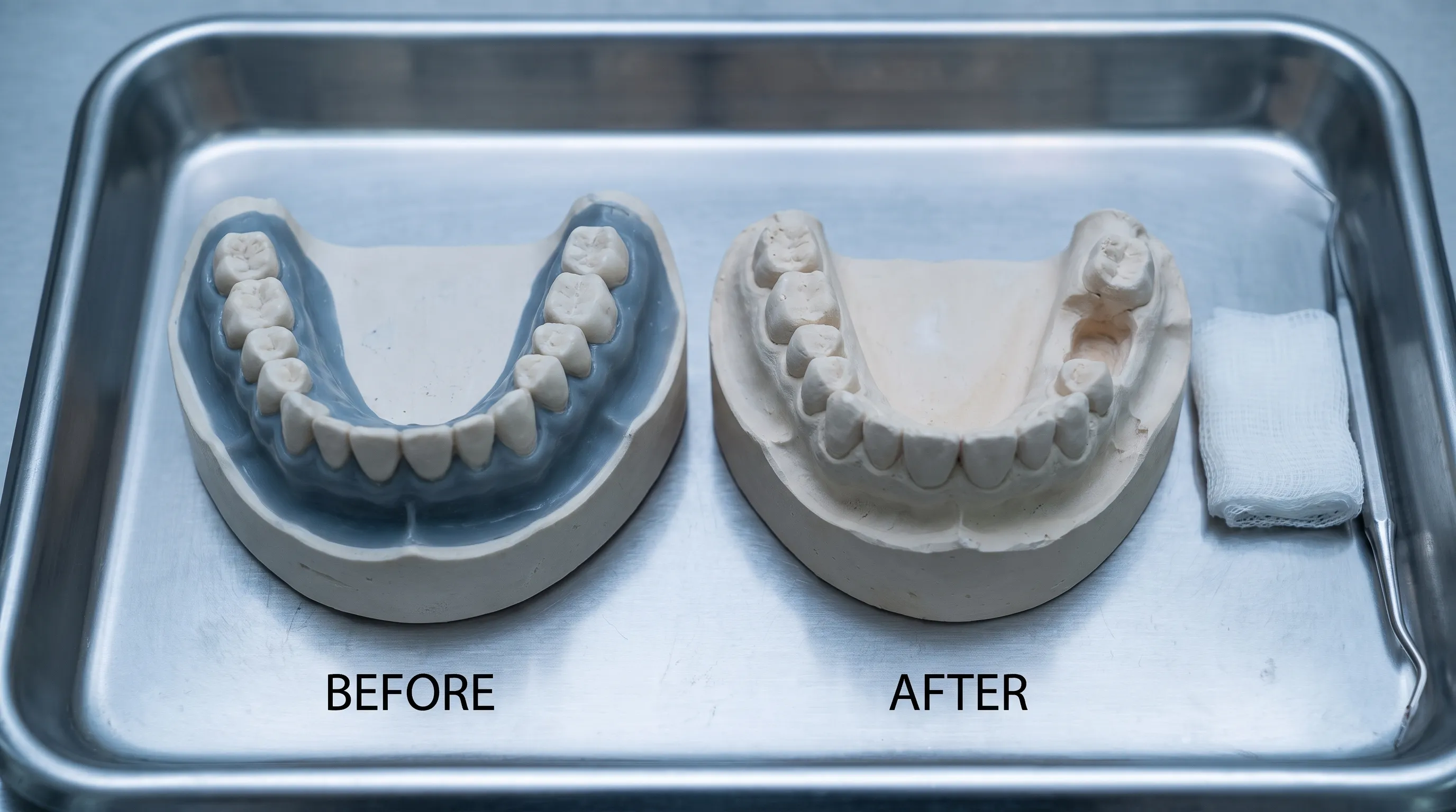
Following tooth removal, the extraction socket immediately begins the natural healing cascade. Hemostasis is achieved through gauze pressure, and a stable fibrin clot forms within minutes, serving as the biological scaffold for tissue regeneration. Protecting this clot is paramount during the first seventy-two hours, as its dislodgment can lead to alveolar osteitis, commonly referred to as dry socket. Your clinician will provide detailed post-operative instructions, including bite pressure management, cold compress application schedules, and activity restrictions. Swelling peaks around day two, gradually subsiding by day five. Mild oozing and discomfort are normal physiological responses. Medications prescribed or recommended focus on inflammation reduction and analgesia. Within ten to fourteen days, granulation tissue fills the socket, and by four to six weeks, immature woven bone begins to mineralize, preparing the site for orthodontic force application.
Evidence-Based Alternatives to Orthodontic Extractions
While teeth removal for braces remains the gold standard for severe crowding and skeletal discrepancies, contemporary orthodontics offers several non-extraction modalities for select cases. These alternatives leverage technological advancements, growth modification, and enamel conservation techniques to create space without permanent tooth loss. However, they are not universally applicable and require strict case selection criteria to avoid periodontal compromise, unstable occlusion, or prolonged treatment times. Understanding the indications, limitations, and biomechanical trade-offs of these approaches helps patients and clinicians make informed, individualized decisions.
Palatal Expansion and Skeletal Modification
In growing children and early adolescents, rapid palatal expansion (RPE) or mini-screw assisted expansion (MSE) can widen the maxillary arch by separating the midpalatal suture. This technique creates significant transverse space, alleviates crowding, and improves nasal airflow by opening the palatal vault. MSE extends expansion capabilities to late adolescents and young adults by anchoring expansion forces directly to the maxillary bone rather than dentition. While highly effective for specific transverse deficiencies, skeletal expansion does not address anteroposterior crowding or anterior protrusion. It also requires strict compliance, periodic activation adjustments, and a consolidation phase to prevent relapse. Patients with fused sutures or mature craniofacial structures typically cannot achieve meaningful skeletal widening, making extraction therapy more predictable in adult populations.
Interproximal Reduction and Enamel Stripping
Interproximal reduction (IPR) involves the precise removal of 0.1 to 0.3 millimeters of enamel from the contact surfaces of adjacent teeth. This conservative approach generates localized space without extractions, making it suitable for mild to moderate crowding, triangular tooth shapes, or black triangle reduction. IPR is performed using calibrated strips, diamond discs, or oscillating devices under direct visualization. Studies confirm that enamel reduction up to fifty percent of total thickness does not increase caries risk or compromise tooth vitality when followed by fluoride therapy and proper polishing. However, IPR cannot address severe crowding exceeding three millimeters per arch, and excessive reduction may lead to increased sensitivity, altered proximal contours, or periodontal vulnerability. It works best when combined with clear aligner therapy or minimal anchorage requirements.
Weighing the Pros and Cons of Non-Extraction Therapy
The decision between extraction and non-extraction approaches hinges on comprehensive cephalometric analysis, patient age, growth potential, periodontal health, and aesthetic goals. Non-extraction protocols preserve all natural teeth and may appeal to patients hesitant about surgical procedures. They often require less aggressive anchorage and shorter treatment times in mild cases. Conversely, non-extraction treatment in severely crowded patients frequently results in dental proclination, where teeth are flared forward to create arch space. This proclination increases the risk of gingival recession, compromises long-term stability, and may create an unbalanced facial profile with excessive lip protrusion. Extraction therapy, when clinically indicated, typically yields superior occlusal contacts, better root parallelism, and more harmonious soft-tissue relationships. The following comparison table illustrates the clinical trade-offs to guide informed decision-making.
| Treatment Approach | Ideal Indications | Primary Advantages | Potential Limitations | Long-Term Stability Outlook |
|---|---|---|---|---|
| Teeth Removal for Braces | Severe crowding (>3mm), Class II division 1, significant protrusion | Predictable space creation, optimal root alignment, improved facial profile, stable occlusion | Requires surgical recovery, temporary aesthetic gaps, retention compliance mandatory | Excellent with proper retainer wear and follow-up care |
| Non-Extraction Expansion | Transverse discrepancies, growing patients, mild crowding | Preserves dentition, improves nasal breathing, enhances arch width | Limited anteroposterior correction, relapse risk without stabilization, suture-dependent | Moderate; depends on growth phase and retention protocol |
| Interproximal Reduction | Mild crowding, triangular teeth, clear aligner candidates | Minimally invasive, no surgical recovery, preserves root structure | Limited space generation, potential sensitivity, requires enamel polishing | Good for selected cases; not suitable for severe discrepancies |
Comprehensive Recovery Guidelines and Pain Management
Successful orthodontic outcomes extend beyond the clinic chair and into the patient's daily habits during the recovery phase. Proper post-operative care accelerates tissue healing, minimizes complications, and ensures that the extraction sites are fully prepared for subsequent orthodontic forces. Recovery from teeth removal for braces follows a predictable biological timeline, but adherence to clinical guidance significantly influences comfort levels and healing efficiency. Integrating practical, evidence-based self-care strategies into your daily routine transforms recovery from a passive waiting period into an active phase of treatment success.
First 72 Hours: Managing Discomfort and Inflammation
The initial seventy-two hours represent the most critical healing window. A stable blood clot must form and remain undisturbed to serve as the foundation for tissue regeneration. Apply cold compresses intermittently for twenty minutes on and twenty minutes off during the first day to constrict blood vessels, reduce swelling, and provide localized analgesia. Avoid strenuous physical activity, heavy lifting, and hot liquids, as elevated blood pressure can dislodge the clot or trigger renewed bleeding. Keep your head elevated above heart level while resting to minimize vascular pressure in the maxillofacial region. Over-the-counter analgesics such as ibuprofen or acetaminophen, taken as directed by your provider, effectively manage pain and suppress inflammatory mediators. Do not rinse vigorously, spit forcefully, or use straws during this period, as negative pressure disrupts the delicate fibrin matrix. Gentle saltwater rinses may begin after twenty-four hours to maintain oral cleanliness without mechanical trauma.
Dietary Modifications and Oral Hygiene Optimization
Nutrition plays a pivotal role in cellular regeneration and tissue repair. Consume soft, nutrient-dense foods for the first three to five days, prioritizing protein, vitamin C, zinc, and calcium to accelerate collagen synthesis and bone remodeling. Smoothies, mashed vegetables, scrambled eggs, yogurt, and lukewarm soups provide essential micronutrients without requiring aggressive mastication. Avoid crunchy, spicy, acidic, or extremely hot foods that can irritate surgical sites or introduce particulate debris into healing sockets. Oral hygiene requires strategic modification. Continue brushing non-surgical teeth with a soft-bristled brush while carefully avoiding the extraction sites. After forty-eight hours, gentle warm saltwater rinses or alcohol-free antimicrobial rinses help reduce bacterial colonization without disrupting granulation tissue. Replace your toothbrush with a new one to prevent reintroduction of oral bacteria. Flossing can resume around surgical areas once soft tissue closure is complete, typically after ten to fourteen days.
Identifying Complications and When to Contact Your Provider
While most extractions heal uneventfully, recognizing warning signs enables prompt intervention and prevents secondary complications. Normal post-operative symptoms include mild oozing, localized tenderness, swelling that peaks on day two, and low-grade elevation in body temperature during the first twenty-four hours. Abnormal symptoms requiring immediate clinical evaluation include persistent bright red bleeding that does not respond to gauze pressure after thirty minutes, severe throbbing pain radiating to the ear or temple that intensifies after day three (indicative of dry socket), visible pus or foul-tasting discharge from the socket, fever exceeding one hundred one degrees Fahrenheit, or difficulty swallowing and breathing. These signs suggest infection, alveolar osteitis, or allergic reaction and warrant urgent professional assessment. Early intervention prevents prolonged recovery delays and ensures orthodontic treatment resumes on schedule. Always maintain open communication with your dental team and report concerns promptly.
Psychological Impact and Lifestyle Adaptation
Undergoing dental extractions, even as part of a planned orthodontic sequence, can evoke emotional responses ranging from mild apprehension to significant anxiety. The permanence of tooth removal, combined with temporary aesthetic changes and dietary restrictions, often triggers body image concerns and treatment fatigue. Acknowledging these psychological dimensions is as important as mastering the clinical aspects. Modern orthodontic practice increasingly integrates patient-centered counseling, visual treatment simulations, and transparent milestone tracking to build trust and reduce emotional strain. Preparing mentally for the temporary phases of treatment ensures that short-term adjustments translate into long-term satisfaction with your final smile.
Addressing Patient Anxiety and Body Image Concerns
Many patients worry that teeth removal for braces will alter their facial profile permanently or create an unnatural appearance. In reality, when executed according to evidence-based protocols, extraction therapy typically refines facial proportions by reducing lip protrusion, enhancing chin definition, and establishing proper lip competence. Temporary extraction gaps are completely normal and become invisible as teeth gradually close the spaces during the alignment phase. Utilizing digital smile design software, orthodontists can preview your post-treatment profile, transforming uncertainty into visual confidence. If anxiety becomes overwhelming, discuss sedation options, relaxation techniques, or brief counseling support with your provider. Peer support groups and treatment journey documentation also normalize the experience, helping you recognize that extraction therapy is a temporary bridge to a healthier, more functional occlusion.
Maintaining Treatment Compliance and Routine Adjustments
Orthodontic success depends heavily on consistent appliance wear, meticulous oral hygiene, and adherence to follow-up appointments. Extraction recovery introduces temporary dietary and activity modifications that must be integrated into your existing routine without disrupting treatment progress. Plan meals ahead, keep oral care supplies accessible, and establish a medication schedule to avoid missed doses. Attend all adjustment appointments, as archwire changes and force recalibration are timed precisely to match tissue healing rates. If you use clear aligners, coordinate their placement with socket closure timelines to prevent food trapping or discomfort. Tracking your progress through treatment photos, symptom journals, and milestone celebrations reinforces motivation and provides tangible evidence that your commitment is yielding visible results. Patience during the healing phase, combined with disciplined compliance, ensures that teeth removal for braces delivers the transformative, lifelong outcomes you envisioned.

Frequently Asked Questions
Does teeth removal for braces permanently change your facial structure?
When performed by a licensed orthodontist using modern cephalometric planning, extraction therapy is designed to maintain or enhance facial harmony. The procedure primarily adjusts dental positioning rather than skeletal architecture. By correcting anterior protrusion and aligning teeth within the natural arch boundaries, extraction often improves lip closure, soft-tissue balance, and overall facial proportion. Permanent facial alteration typically only occurs in extreme skeletal cases requiring orthognathic surgery, not in routine extraction therapy.
How long does it take for extraction gaps to close completely?
Space closure progresses at a biologically optimized rate of approximately one to one and a half millimeters per month. Complete closure usually requires six to twelve months, depending on patient age, bone density, orthodontic mechanics, and compliance with appliance wear. Your orthodontist will monitor root parallelism and tissue health throughout this phase, adjusting forces as needed to prevent hyalinization or delayed movement.
Can I refuse extraction and still achieve successful alignment results?
Mild to moderate crowding may respond well to interproximal reduction, arch expansion, or molar distalization. However, for severe crowding, significant protrusion, or transverse discrepancies, non-extraction approaches often compromise periodontal health, increase relapse risk, or produce flared teeth with unstable occlusion. Your orthodontist will present both options with clinical data, but in cases where extraction is indicated, it remains the most predictable pathway to long-term stability.
Is the extraction procedure painful during or after surgery?
The surgical phase is completely painless due to targeted local anesthesia, with optional sedation for anxious patients. Post-operative discomfort is normal but highly manageable. Peak tenderness occurs within forty-eight hours and subsides significantly by day four. Following your clinician's medication protocol, utilizing cold therapy, and adhering to dietary restrictions ensures comfortable recovery without disrupting daily life or orthodontic progress.
Do extracted teeth need to be replaced with implants or bridges?
In orthodontic extraction cases, replacement is unnecessary and contraindicated. The primary goal is to close the spaces permanently by moving adjacent teeth into the vacated positions. This controlled migration restores a full dental arch, maintains proper occlusal contacts, and eliminates the need for prosthetic replacement. Retention protocols ensure the closed spaces remain stable indefinitely.
Conclusion
Teeth removal for braces represents a meticulously planned, scientifically validated intervention that prioritizes long-term occlusal stability, periodontal health, and facial harmony. While the decision to undergo extraction requires careful consideration, contemporary diagnostic protocols, minimally invasive techniques, and comprehensive aftercare guidelines have transformed the experience into a predictable, manageable phase of orthodontic treatment. By creating necessary anatomical space, extraction therapy enables teeth to align in their natural positions, distribute biting forces evenly, and resist relapse through proper retention. Understanding the biomechanical principles, adhering to evidence-based recovery practices, and maintaining open communication with your dental team ensures that temporary discomfort yields to lasting functional and aesthetic improvements. Whether you are navigating the initial consultation, preparing for surgery, or actively managing the alignment phase, trusting in clinically proven protocols and prioritizing consistent compliance will ultimately deliver the healthy, confident smile you deserve. For additional clinical guidance, explore resources from the American Association of Orthodontists, review patient care standards from the American Dental Association, and consult evidence-based orthodontic literature through the Mayo Clinic.
How we work
Researched and drafted with AI assistance, then reviewed by a human editor against the cited sources.
This article is general health information, not medical advice. Consult a qualified healthcare professional about your own situation.