Sprained Toe vs Fractured Toe: How to Tell the Difference and Recover Safely

Stubbing your foot against a doorframe, dropping a heavy object on your foot, or misstepping during a run can send sharp, sudden pain radiating through your toes within seconds. In those initial agonizing moments, it is nearly impossible to know whether you are dealing with a mild soft tissue injury or a structural bone problem. The confusion between these two common injuries leads many people to self-diagnose incorrectly, which often results in inadequate protection, prolonged discomfort, and in some cases, permanent joint dysfunction. Understanding the critical distinctions when evaluating a sprained toe vs fractured toe is not merely academic; it directly influences your recovery timeline, your treatment choices, and your ability to safely return to daily activities. This comprehensive guide breaks down the anatomical differences, clinical presentation patterns, diagnostic approaches, and evidence-based treatment protocols so you can navigate your injury with confidence. For verified clinical guidance on lower extremity trauma, refer to Mayo Clinic’s overview on toe fractures and sprains. Whether you are an athlete managing training loads or a busy professional trying to stay mobile, learning how to properly assess and care for toe trauma will empower you to make informed health decisions and avoid costly medical complications down the line.
Understanding Toe Anatomy and the Mechanics of Injury
To fully grasp the differences between soft tissue damage and structural bone trauma, we must first examine the intricate architecture of the foot's distal segments. The human foot contains twenty-six bones, with fourteen located entirely within the toes. Each toe consists of three small bones called phalanges, except for the great toe, which contains only two. These bones articulate at the metatarsophalangeal joints and interphalangeal joints, creating a highly mobile yet remarkably stable lever system that absorbs ground reaction forces, propels the body forward during gait, and maintains balance on uneven terrain. For detailed anatomical references, consult the Cleveland Clinic’s guide to foot structure and conditions.
Free calculatorFoot Reflexology ChartFoot reflexology points and corresponding organsOpen the calculatorThe Complex Structure of the Phalanges and Soft Tissues
Surrounding the phalangeal bones are dense networks of ligaments, tendons, and joint capsules that stabilize articulation points. Collateral ligaments run along both sides of each toe joint, preventing excessive lateral or medial movement. Plantar ligaments form a thick fibrous band beneath the joints, resisting hyperextension during the push-off phase of walking. These structures work in concert with the intrinsic foot muscles to maintain dynamic alignment. When forces exceed the physiological tolerance of these connective tissues, microtrauma occurs. Ligaments can stretch, partially tear, or rupture completely, while the dense cortical bone of the phalanges may develop microcracks or fracture entirely under direct impact or extreme torque. The distribution of force, the angle of impact, and the existing health of the bone and connective tissue all determine which structure fails first. The NIH MedlinePlus resource on sprains provides clinical insights into ligament failure and tissue tolerance thresholds.
Biomechanics of Trauma: How Force Translates to Injury
Most toe injuries occur through two primary mechanisms: direct axial loading and rotational trauma. Axial loading happens when a heavy object falls onto the toe or when the foot strikes a rigid object at high velocity, such as a wall, furniture edge, or sports equipment. This compressive force travels directly through the phalanges, potentially causing bone displacement. Rotational trauma occurs when the foot is planted and the body twists unexpectedly, placing extreme shear stress on the joint capsules and collateral ligaments. Athletes in sports like basketball, soccer, football, and martial arts frequently experience this mechanism. Recognizing the exact moment of injury and the vector of force applied provides clinicians with crucial diagnostic clues when differentiating between structural bone failure and ligamentous overload. For a deeper visual breakdown of toe injury mechanics, you can watch this comprehensive medical explanation:
What Exactly Is a Sprained Toe?
A toe sprain represents an injury to the ligaments surrounding a joint without any disruption to the underlying bone structure. Ligaments are composed of dense, collagen-rich connective tissue that connects bone to bone. When stretched beyond their elastic limit, the collagen fibers sustain damage. This condition is remarkably common, yet it is frequently minimized as a minor inconvenience until chronic instability sets in.
Pathophysiology of Ligament Overstretching
At the cellular level, a sprain involves the disruption of the extracellular matrix within the ligament. Grade I injuries consist of microscopic fiber tearing accompanied by localized inflammation. The inflammatory cascade brings neutrophils and macrophages to clear damaged tissue while fibroblasts begin synthesizing new collagen strands. Grade II injuries involve partial macroscopic tears that visibly elongate the ligament, causing joint laxity and significant swelling. Grade III sprains feature complete ligament rupture, resulting in gross joint instability and requiring extended immobilization to allow scar tissue formation. Understanding this grading system helps clinicians tailor treatment intensity and rehabilitation timelines.
Clinical Presentation and Symptom Progression
The hallmark symptoms of a sprained toe include localized pain at the joint line, progressive swelling that peaks within twenty-four to forty-eight hours, and stiffness during early morning movement. Bruising may appear but typically remains mild compared to bone trauma. Patients often report a feeling of joint looseness or clicking during weight-bearing activities. Pain usually increases with active range of motion or direct palpation of the ligamentous attachment points rather than along the bone shaft itself. The discomfort is often sharp during specific directional movements but dulls to a manageable ache at rest.
Sprain Grading System: From Microtears to Complete Ruptures
Medical professionals categorize toe sprains using a standardized severity scale to guide intervention strategies. Mild grade I sprains typically resolve within one to two weeks with conservative care and modified activity. Moderate grade II injuries require structured taping, possible short-term immobilization, and three to four weeks of targeted physical therapy to restore ligamentous tension. Severe grade III ruptures may need custom orthotics, extended bracing, and up to eight weeks of rehabilitation to address compensatory muscle weakness and prevent chronic joint subluxation. Proper classification ensures that rehabilitation protocols match tissue healing phases accurately.
Defining a Fractured Toe
When the cortical or trabecular bone of a phalanx sustains structural failure, the condition is classified as a fracture. Unlike sprains, which primarily affect soft tissue stabilization, fractures compromise the skeletal framework that transmits mechanical load. The healing process differs fundamentally because bone remodeling requires precise mineralization and callus formation rather than collagen matrix reorganization.
Bone Stress and Structural Failure
Phalangeal fractures result from either acute high-energy trauma or repetitive low-grade stress that exceeds bone fatigue thresholds. Stress fractures develop gradually through microdamage accumulation, often seen in runners who rapidly increase mileage without adequate recovery. Acute fractures occur instantaneously from direct impact. The fracture pattern dictates the treatment pathway. Bone integrity depends heavily on patient age, bone density, nutritional status, and underlying conditions like osteoporosis or diabetes. When the structural matrix fails, the periosteum tears, triggering significant hemorrhage into the surrounding tissue and activating a robust osteogenic healing response. Review Cleveland Clinic’s clinical overview of broken toes for detailed fracture pathology and management standards.
Classifying Toe Fractures: Hairline, Displaced, and Comminuted
Hairline fractures represent non-displaced microcracks that maintain anatomical alignment. Displaced fractures involve bone fragments shifting out of position, often requiring manual reduction. Comminuted fractures feature multiple bone fragments shattered into three or more pieces, typically from high-velocity impacts. Intra-articular fractures extend into the joint space, raising the risk of post-traumatic arthritis. The great toe fractures warrant special attention because they bear approximately forty percent of body weight during gait and provide critical push-off leverage. Improper healing in this digit can permanently alter walking mechanics and increase stress on the knees and hips.
Recognizing Fracture-Specific Warning Signs
Fracture symptoms often present more dramatically than sprains. Immediate, severe pain that does not subside with elevation is common. Swelling appears rapidly and extends across the entire toe segment. Ecchymosis, or deep purple bruising, frequently spreads to surrounding digits and the dorsal foot surface. Audible crunching or grinding sensations, known as crepitus, may be felt during attempted movement. Visible deformity, including abnormal angulation, shortening, or rotational misalignment, strongly indicates structural displacement. Pain is typically constant rather than movement-dependent and worsens significantly with any weight-bearing activity.
Key Differences: Sprained Toe vs Fractured Toe
Navigating the clinical overlap between these two injuries requires careful observation of symptom patterns, timing, and functional limitations. While both conditions share swelling, tenderness, and mobility restriction, the underlying pathology dictates distinct clinical trajectories. Learning to identify the subtle divergence when comparing a sprained toe vs fractured toe empowers patients to seek appropriate care at the optimal time.
Pain Localization and Intensity Patterns
Sprain-related pain concentrates specifically around the joint margins and ligament insertion points. It fluctuates with joint movement and typically decreases significantly at rest. Fracture pain localizes along the bone shaft and joint surfaces simultaneously, remaining intense regardless of position. Night pain that disrupts sleep is more characteristic of bone trauma due to periosteal irritation and intraosseous pressure changes from inflammation.
Visible Changes: Bruising, Deformity, and Alignment
Soft tissue sprains rarely produce dramatic deformity. Any visible changes usually involve mild swelling and superficial discoloration around the joint capsule. Bone fractures frequently generate pronounced deformities, including dorsal elevation, plantar displacement, or lateral deviation. Bruising in fractures appears darker, more extensive, and often tracks along fascial planes. A fractured toe may also exhibit skin tenting, where displaced bone presses against the dermis, creating a pale, taut appearance.
Functional Impact: Weight-Bearing and Joint Mobility
Individuals with sprained toes can often bear partial weight with minimal assistance, though discomfort limits full stride length. Joint stiffness predominates, but active motion remains possible with conscious effort. Fractured toes severely compromise load-bearing capacity. Most patients cannot support their body weight without significant pain or mechanical collapse. Active range of motion triggers sharp, radiating pain that discourages voluntary movement entirely.
| Clinical Feature | Sprained Toe | Fractured Toe |
|---|---|---|
| Primary Tissue Affected | Ligaments & Joint Capsule | Phalangeal Bone Structure |
| Pain Quality | Dull ache, sharp with joint stress | Constant throbbing, severe at rest |
| Swelling Onset | Gradual (6-24 hours) | Rapid (immediate to 4 hours) |
| Bruising Pattern | Mild, localized around joint | Extensive, dark purple, spreads |
| Deformity | Rare | Common (angulation, shortening) |
| Weight-Bearing | Possible with discomfort | Often impossible or severely limited |
| Crepitus | Uncommon | Common during movement attempts |
| Healing Timeline | 2-6 weeks | 3-8 weeks depending on severity |
Diagnosis: How Medical Professionals Tell Them Apart
Clinical differentiation between soft tissue and bone trauma relies on a systematic approach combining patient history, physical examination maneuvers, and diagnostic imaging. Emergency department physicians, orthopedic specialists, and podiatrists follow standardized protocols to ensure accurate classification and appropriate intervention planning.
Physical Examination Techniques
Clinicians begin by observing gait mechanics, noting antalgic patterns where patients rapidly transfer weight off the injured foot. Palpation follows a structured sequence, systematically compressing the metatarsals, phalanges, and joint spaces. The axial compression test involves gentle longitudinal pressure applied to the toe tip; reproduction of deep pain strongly suggests bone involvement rather than ligamentous injury. Range of motion testing evaluates active and passive movement, while stability tests assess ligamentous integrity by applying controlled varus and valgus stress to the interphalangeal joints. Specialized assessments like the plantar stress test identify hypermobility that differentiates high-grade sprains from stable fractures.
Imaging Modalities and Diagnostic Accuracy
Standard anteroposterior, lateral, and oblique X-rays remain the gold standard for confirming bone fractures. Radiographic imaging reveals fracture lines, displacement measurements, and joint surface involvement. However, stress fractures may not appear on initial X-rays due to the microscopic nature of early damage. In these cases, repeat imaging after ten to fourteen days or advanced modalities like MRI provide definitive diagnosis. MRI excels at visualizing soft tissue integrity, identifying ligament tears, bone marrow edema, and occult fractures simultaneously. Ultrasound offers dynamic assessment of tendon and ligament function in real-time, though it cannot penetrate cortical bone. The choice of imaging depends on clinical suspicion, mechanism of injury, and suspected complication risk.
Red Flags Requiring Urgent Medical Intervention
Certain clinical indicators demand immediate orthopedic evaluation. Open wounds with visible bone or joint fluid leakage indicate compound fractures with infection risk. Circulatory compromise, evidenced by cold toes, delayed capillary refill, or cyanosis, suggests vascular injury. Numbness or tingling indicates nerve compression requiring decompression. Severe rotational deformity of the great toe or multiple digit involvement mandates specialist referral. Patients with diabetes, peripheral neuropathy, or immunosuppression require prompt assessment due to elevated complication risks. Ignoring these warning signs can lead to malunion, chronic pain syndromes, or permanent mobility restrictions. Consult CDC guidelines on recognizing urgent injury symptoms for emergency care benchmarks.
Treatment Protocols and Evidence-Based Recovery
Successful recovery depends on matching treatment intensity to injury severity while adhering to tissue healing timelines. Evidence-based protocols emphasize early protection, progressive loading, and structured rehabilitation to restore biomechanical function and prevent recurrence.
Immediate First Aid: Beyond the RICE Protocol
The traditional RICE method (Rest, Ice, Compression, Elevation) remains foundational, but modern sports medicine emphasizes the PEACE and LOVE framework for soft tissue management. Protect the toe from further stress immediately. Elevate above heart level to reduce hydrostatic pressure. Avoid anti-inflammatory medications during the first forty-eight hours, as they may interfere with natural healing cascades. Compression should be gentle to avoid compromising circulation. After the acute phase, Load optimally through pain-free movement, maintain positive rehabilitation expectations, encourage cardiovascular conditioning for tissue perfusion, and incorporate progressive exercise to restore joint mechanics. For bone injuries, strict immobilization takes priority during the inflammatory phase.
Conservative Management for Ligamentous Injuries
Mild to moderate sprains respond well to buddy taping, which stabilizes the injured digit against an adjacent healthy toe using medical-grade adhesive tape with foam padding between the digits to prevent skin maceration. Taping should remain secure during daily activities but be loosened overnight. Rigid-soled shoes or post-operative walking boots limit joint flexion, reducing stress on healing ligaments. Progressive mobilization begins after seventy-two hours, starting with gentle isometric contractions and advancing to controlled range-of-motion exercises. Tactile proprioceptive training restores neuromuscular coordination, while eccentric strengthening rebuilds ligamentous resilience. 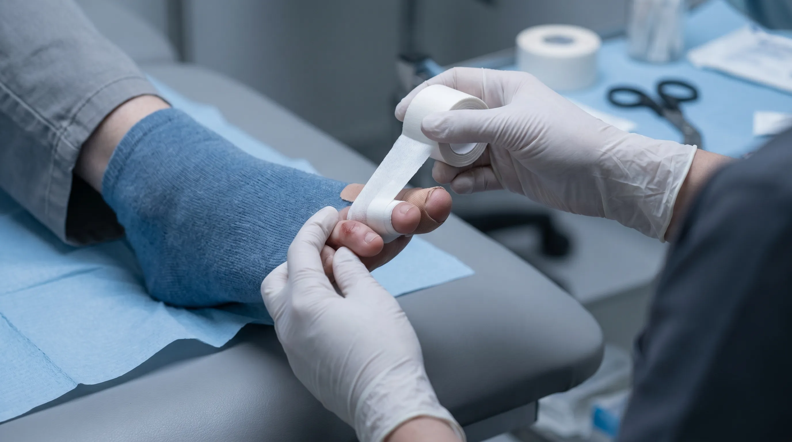
Orthopedic Interventions for Bone Fractures
Stable, non-displaced fractures require immobilization through buddy taping and protective footwear for three to four weeks. The great toe often necessitates custom orthotics or specialized post-op shoes to maintain neutral alignment. Displaced fractures require closed reduction under local anesthesia to realign bone fragments before casting or splinting. Comminuted or intra-articular fractures may demand surgical fixation using pins, screws, or plates to restore joint congruency. Post-operative care includes strict non-weight-bearing status initially, transitioning to partial loading once radiographic callus formation confirms early union. Medication management balances pain control with constipation prevention and bone health optimization through calcium and vitamin D supplementation.
Phased Rehabilitation and Physical Therapy
Rehabilitation follows distinct phases aligned with tissue healing biology. Phase one focuses on edema control, pain management, and gentle mobility restoration. Phase two introduces progressive loading, gait retraining, and intrinsic foot muscle activation. Phase three emphasizes dynamic stability, plyometric progressions, and sport-specific movement patterns. Balance training on unstable surfaces restores proprioception critical for preventing recurrent injuries. Manual therapy techniques address fascial restrictions and joint hypomobility. Home exercise programs ensure compliance and accelerate functional return. 
Long-Term Care, Prevention, and Lifestyle Adaptations
Preventing future toe injuries requires addressing biomechanical vulnerabilities, optimizing footwear selection, and implementing targeted conditioning routines that enhance structural resilience and movement efficiency. WHO recommendations on musculoskeletal injury prevention provide foundational strategies for long-term risk reduction.
Footwear Optimization and Protective Equipment
Proper footwear dramatically reduces toe trauma risk. Athletic shoes require adequate toe box width allowing digits to splay naturally without compression. Toe guards made from silicone or thermoplastic elastomer absorb impact forces during contact sports or heavy manual labor. Work boots with composite toe caps meet safety standards while preventing crush injuries from falling objects. Regular shoe replacement every three hundred to five hundred miles ensures midsole cushioning maintains shock absorption capacity. Orthotic inserts correct overpronation and supination patterns that place uneven stress on toe joints.
Preventive Strengthening and Mobility Drills
Targeted exercises build structural resilience. Towel scrunches activate intrinsic foot muscles while improving arch stability. Toe spreads enhance neuromuscular control and joint mobility resistance band exercises strengthen flexors and extensors through full range of motion. Calf stretching reduces Achilles tension that increases forefoot loading pressure. Ankle alphabet drills improve joint kinematics. Consistent daily practice of these movements maintains tissue elasticity and proprioceptive acuity. Proper warm-up protocols before physical activity increase blood flow to distal structures, preparing ligaments and tendons for dynamic loads.
Activity Modification and Safe Return to Play
Gradual progression prevents re-injury during recovery. Athletes must meet specific functional benchmarks before returning to competition, including pain-free single-leg balance, symmetric jumping capacity, and full range of motion without crepitus. Cross-training maintains cardiovascular fitness while minimizing impact stress. Surface selection matters; training on firm, level ground reduces unpredictable joint loading during early rehabilitation phases. Communication with coaches and trainers ensures appropriate practice modifications. Patience during recovery yields superior long-term outcomes compared to premature return that risks chronic joint degeneration.
Frequently Asked Questions
How can I tell if my toe is broken without going to a doctor immediately?
While definitive diagnosis requires imaging, clinical clues strongly suggest fractures. Look for rapid swelling within hours, dark bruising spreading across multiple toes, visible angulation, inability to bear weight without sharp pain, and constant throbbing that persists during sleep. If you experience crepitus or deformity after trauma, treat the injury as a fracture until medical evaluation confirms otherwise. Avoid testing mobility repeatedly, as this can worsen displacement.
Should I apply heat or ice to a freshly injured toe?
Always apply ice during the first forty-eight to seventy-two hours following acute injury. Cold therapy constricts blood vessels, reduces metabolic demand in damaged tissue, limits edema formation, and numbs nerve endings to decrease pain perception. Use a thin towel barrier between ice and skin, applying for fifteen to twenty minutes every two to three hours. Heat should never be used during the acute inflammatory phase, as it increases blood flow and exacerbates swelling. After the third day, contrast therapy or gentle warmth may promote circulation once swelling stabilizes.
Can I continue exercising or running with a mildly injured toe?
Exercise modification depends entirely on injury classification and severity. Mild sprains tolerate low-impact activities like swimming or stationary cycling within days if protected properly. Running places repetitive impact forces exceeding two times body weight on the forefoot, which can delay ligament healing or convert a stable fracture into a displaced one. If pain alters your gait pattern or causes limping, cease weight-bearing exercise immediately. Consult a sports medicine professional for alternative training strategies that maintain fitness without compromising recovery.
Why does my toe hurt weeks after the initial injury has healed?
Persistent pain beyond expected healing timelines often indicates incomplete rehabilitation, joint stiffness, or compensatory biomechanical dysfunction. Scar tissue formation can restrict gliding between ligament and bone. Muscle atrophy around the foot reduces shock absorption, transferring excessive load to healing structures. Undiagnosed intra-articular damage or stress reaction may also contribute. Targeted physical therapy addressing mobility deficits, proprioceptive training, and gradual loading progression typically resolves lingering discomfort. If pain persists beyond eight weeks, advanced imaging rules out avascular necrosis or early osteoarthritis.
When should I consider surgical intervention for a toe injury?
Surgery remains reserved for specific clinical scenarios that conservative management cannot address. Indications include intra-articular fractures disrupting joint congruency, open fractures with infection risk, multiple displaced fractures causing mechanical collapse, great toe injuries with significant malalignment, and complete ligament ruptures resulting in chronic instability. Surgical correction aims to restore anatomical alignment, stabilize joint surfaces, and enable early mobilization. Post-operative rehabilitation is mandatory to regain function. Most toe injuries respond excellently to non-surgical protocols, making surgery the exception rather than the rule.
Key Takeaways
Distinguishing between a sprained toe and a fractured toe requires careful attention to symptom onset, pain characteristics, visible structural changes, and functional limitations. While both conditions demand respect and appropriate care, their underlying pathologies dictate fundamentally different treatment pathways and recovery expectations. Ligamentous sprains benefit from early protected movement and progressive loading, whereas bone fractures require strict immobilization until radiographic evidence confirms structural union. Diagnostic imaging remains the definitive tool for eliminating uncertainty and guiding clinical decisions. Implementing immediate first aid measures, adhering to phased rehabilitation protocols, and addressing biomechanical vulnerabilities through proper footwear and targeted conditioning dramatically improve outcomes and reduce recurrence risk. When in doubt about the severity of your injury, consulting a qualified podiatrist or orthopedic specialist ensures accurate assessment and personalized intervention strategies. Prioritizing evidence-based care today preserves your mobility, prevents chronic complications, and supports lifelong foot health. 
How we work
Researched and drafted with AI assistance, then reviewed by a human editor against the cited sources.
This article is general health information, not medical advice. Consult a qualified healthcare professional about your own situation.