Retrolisthesis Grade 1: Complete Guide to Symptoms, Diagnosis, and Conservative Management

Living with chronic back discomfort or discovering an unexpected spinal alignment finding on a routine medical scan can be deeply unsettling, particularly when unfamiliar medical terminology enters your health vocabulary. When a clinician mentions a diagnosis of retrolisthesis grade 1, many patients immediately envision severe spinal deformities, surgical interventions, or permanent disability. The reality is far more manageable. Retrolisthesis grade 1 represents the mildest classification of posterior vertebral displacement, characterized by a subtle backward slippage of one vertebral body relative to its neighbor. This condition is remarkably common, particularly in aging populations and individuals with long histories of manual labor or poor postural habits, as noted in epidemiological reviews by the National Institutes of Health (NIH). Understanding the biomechanics behind this diagnosis, recognizing the subtle symptoms, and implementing evidence-based conservative management strategies can empower you to take control of your spinal health. By focusing on targeted rehabilitation, ergonomic optimization, and proactive lifestyle modifications, most individuals experience significant symptom relief and maintain excellent functional mobility for decades. This comprehensive guide will walk you through the clinical presentation, diagnostic pathways, and scientifically supported treatment protocols necessary to navigate retrolisthesis grade 1 with confidence and long-term spinal resilience.
Understanding Retrolisthesis Grade 1: The Fundamentals
To effectively manage any spinal condition, it is crucial to first understand the precise anatomical and biomechanical mechanisms at play. The human spine is a complex, dynamic structure composed of stacked vertebrae, intervertebral discs, facet joints, ligaments, and a robust muscular support system. Each component must maintain precise alignment to distribute mechanical loads evenly, protect the delicate spinal cord, and facilitate pain-free movement across multiple planes of motion. When structural integrity is compromised, even subtly, the resulting biomechanical shifts can cascade into localized discomfort, nerve irritation, or progressive degenerative changes.
Free calculatorBMI CalculatorCalculate Body Mass IndexOpen the calculatorWhat Is Retrolisthesis?
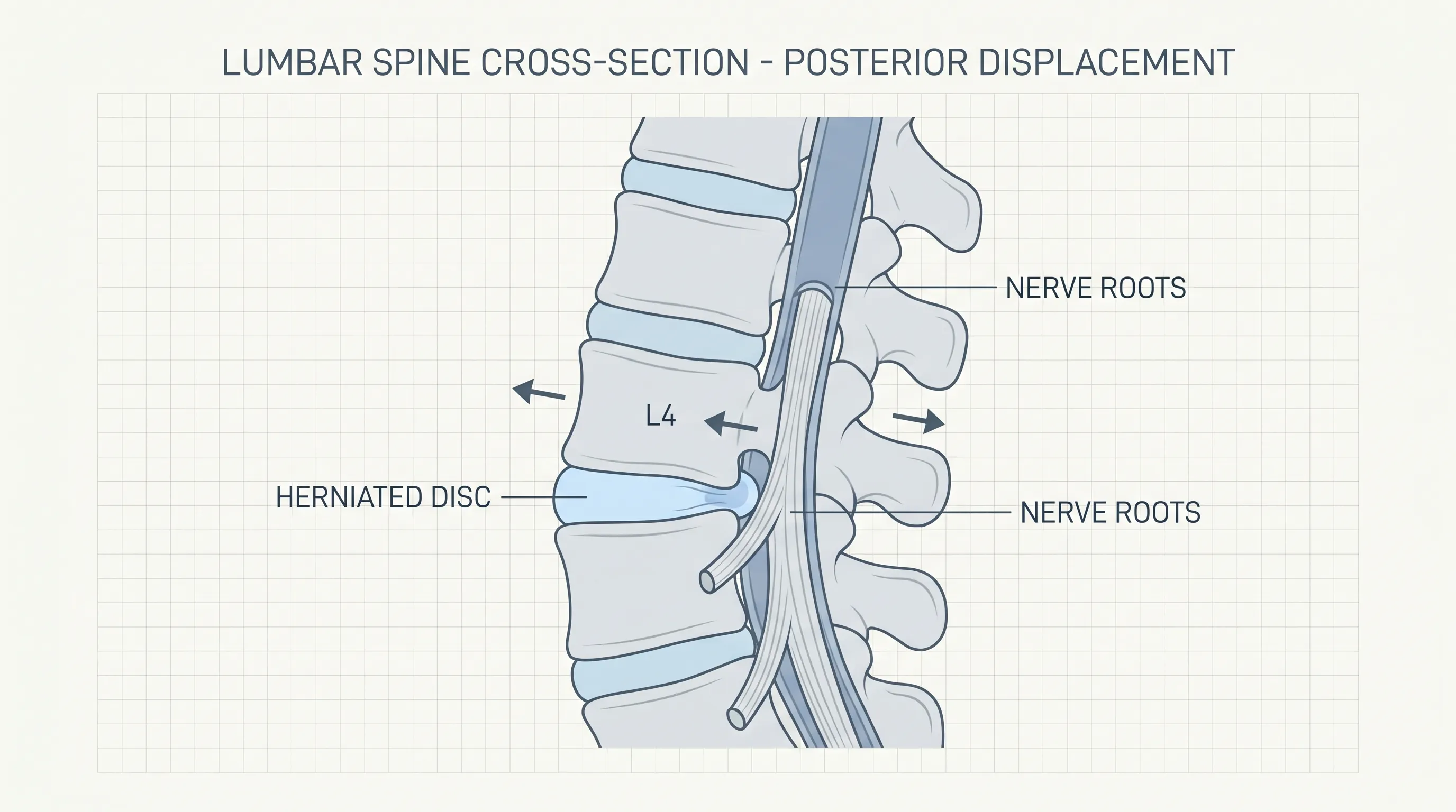
Retrolisthesis refers specifically to the posterior displacement, or backward slippage, of a vertebral body relative to the adjacent vertebra situated directly below it. Unlike a complete dislocation, which represents a severe traumatic injury requiring emergency medical intervention, retrolisthesis involves a partial misalignment where the vertebra remains largely seated within the spinal column but sits slightly out of its natural anterior-posterior position. This condition predominantly affects the highly mobile cervical and lumbar regions of the spine. The cervical spine bears the considerable weight of the head while facilitating extensive rotational and flexion-extension movements. The lumbar spine, conversely, absorbs the majority of the body's axial load during standing, walking, and lifting. Both regions are highly susceptible to mechanical stress, disc degeneration, and ligamentous laxity, creating an environment where posterior slippage can gradually develop. The underlying mechanism typically involves a reduction in intervertebral disc height, degeneration of the posterior longitudinal ligament, or hypertrophy of the facet joints, all of which alter the natural gliding mechanics of the vertebral segments, as detailed by the Cleveland Clinic.
Differentiating Retrolisthesis from Spondylolisthesis
Medical terminology can often cause unnecessary confusion for patients navigating orthopedic diagnoses. While retrolisthesis and spondylolisthesis both involve vertebral displacement, they represent fundamentally opposite directional pathologies. Spondylolisthesis describes the anterior, or forward, displacement of a vertebra relative to the segment below it. This forward slip is frequently associated with pars interarticularis defects, stress fractures, or congenital spinal anomalies. Retrolisthesis grade 1, in contrast, involves a backward shift driven primarily by disc space narrowing, degenerative joint changes, or muscular imbalances that pull the vertebra posteriorly. Recognizing this directional distinction is clinically significant because the biomechanical forces, rehabilitation protocols, and long-term prognostic expectations differ substantially between the two conditions. Treatment strategies for retrolisthesis grade 1 emphasize posterior chain stabilization, disc decompression techniques, and careful avoidance of excessive lumbar extension, whereas anterior displacement protocols often prioritize flexion-biased exercises and stabilization of the anterior spinal column.
The Clinical Grading System Explained
Spinal specialists utilize a standardized grading scale to classify the severity of vertebral displacement, ensuring consistent communication across medical disciplines and guiding appropriate treatment decisions. The grading system for retrolisthesis is primarily determined by measuring the percentage of occlusion within the intervertebral foramina (IVF), the bony canals through which spinal nerve roots exit the spinal canal. In retrolisthesis grade 1, the posterior displacement measures up to one-quarter, or 25 percent, of the intervertebral foramina's vertical height. When measured directly in millimeters on lateral imaging, this grade typically corresponds to a displacement range of approximately one to three millimeters. This minimal degree of slippage preserves the majority of neural foramen patency, meaning that nerve compression, when it occurs, is usually mild and highly responsive to conservative interventions, per diagnostic standards outlined by the Mayo Clinic. Grades two through four represent progressively severe displacements, with increasing neural compromise, spinal canal narrowing, and higher probabilities of neurological deficits requiring advanced imaging and potential surgical consultation.
Clinical Subtypes
Beyond the primary grading scale, clinicians categorize retrolisthesis into specific subtypes based on the directional relationship between multiple vertebral segments. Complete retrolisthesis occurs when a single vertebra shifts posteriorly relative to both the segments situated above and below it, creating a localized posterior convexity. Partial retrolisthesis describes a scenario where the affected vertebra displaces backward relative to either the superior or inferior segment only, often seen in early degenerative changes or unilateral facet joint pathology. Stairstepped retrolisthesis presents a more complex alignment pattern where a vertebra moves posteriorly relative to the segment above but remains anterior to the segment below, creating a zigzag spinal alignment. Understanding these subtypes allows physical therapists and chiropractors to tailor their manual therapy techniques and exercise prescriptions to address the specific mechanical imbalances present in your individual case.
Symptoms and Clinical Presentation
The clinical presentation of retrolisthesis grade 1 varies dramatically across individuals, largely influenced by the exact spinal level involved, baseline musculoskeletal fitness, disc health, and individual pain thresholds. Many patients discover their condition entirely by accident during imaging ordered for unrelated complaints, while others experience persistent, low-grade discomfort that gradually interferes with daily activities. Recognizing the subtle warning signs early enables prompt intervention and prevents the progression toward more severe structural compromise.
Localized Spinal Discomfort and Stiffness
The most common symptomatic presentation involves localized axial pain concentrated directly over the affected spinal segment. Patients frequently describe this discomfort as a deep, aching sensation that worsens with prolonged static postures, such as sitting at a desk for extended periods or standing on hard surfaces. Morning stiffness is another hallmark symptom, often resulting from nocturnal fluid redistribution within the degenerative intervertebral disc and temporary muscle guarding around the misaligned segment. This stiffness typically resolves within thirty to sixty minutes of gentle movement and dynamic stretching. As the day progresses and mechanical load accumulates, a dull throbbing may emerge, particularly in the late afternoon when postural muscles fatigue and compensatory strategies fail to adequately support the vertebral column. Palpation over the spinous processes may reveal localized tenderness, and range of motion testing often demonstrates mild restriction in flexion, extension, or lateral bending depending on the primary biomechanical fault.
Neurological Manifestations and Radiating Pain
When the mild posterior displacement narrows the neural foramen sufficiently to irritate adjacent nerve roots, patients may experience radicular symptoms that extend beyond the primary spinal region. In lumbar retrolisthesis grade 1, this commonly manifests as sharp, electric-like pain, tingling, numbness, or a heavy, aching sensation traveling through the buttocks, posterior thighs, calves, and occasionally into the feet. Cervical involvement can produce similar neurological disturbances radiating down the shoulders, arms, forearms, and into the hands, sometimes accompanied by subtle grip weakness or altered temperature sensation. These symptoms are typically position-dependent, flaring during spinal extension or rotational movements that further compress the neural exit pathway. It is important to note that neurological symptoms do not automatically indicate severe nerve damage; rather, they reflect mild, reversible nerve root irritation that responds exceptionally well to targeted decompression exercises and postural retraining.
The Reality of Asymptomatic Cases
Contrary to popular belief, a significant portion of individuals diagnosed with retrolisthesis grade 1 remain completely asymptomatic throughout their lives. Radiographic studies consistently demonstrate that minor vertebral misalignments and disc height reductions are extremely common in the general population, often correlating with normal age-related degenerative processes rather than pathological disease. In these incidental cases, the robust paraspinal musculature, healthy connective tissue, and adaptive spinal biomechanics effectively compensate for the subtle misalignment. When asymptomatic, medical professionals generally recommend no active treatment beyond maintaining optimal spinal hygiene, staying physically active, and monitoring for the onset of symptoms. However, patients who receive an incidental diagnosis should still incorporate preventative core strengthening and ergonomic practices into their routine, as proactive management significantly reduces the risk of future symptomatic progression or acute pain flare-ups.
Underlying Causes and Risk Factors
Retrolisthesis grade 1 rarely develops in isolation. Instead, it emerges from a complex interplay of genetic predisposition, cumulative mechanical stress, age-related tissue degradation, and lifestyle factors. Identifying the primary contributing drivers allows for highly targeted therapeutic interventions that address the root cause rather than merely masking symptoms.
Degenerative Disc Disease and Facet Joint Arthritis
The most prevalent etiological factor in retrolisthesis development is progressive intervertebral disc degeneration. Healthy spinal discs consist of a tough, fibrous outer annulus surrounding a gelatinous, water-rich nucleus pulposus. This hydrostatic cushion absorbs axial shock, maintains intervertebral spacing, and facilitates smooth vertebral articulation. As individuals age, discs gradually lose proteoglycan content, desiccate, and narrow, a natural aging process thoroughly documented by the Mayo Clinic. This disc height reduction concurrently stresses the facet joints, which stabilize and guide spinal movement, leading to osteoarthritic changes, including cartilage wear, joint space narrowing, and osteophyte formation. This combined degeneration alters the instantaneous axis of rotation within the spinal motion segment, predisposing the vertebra to posterior migration under load. Individuals with a history of repetitive spinal compression or occupations requiring prolonged heavy lifting face accelerated degenerative timelines, significantly elevating their risk for developing retrolisthesis grade 1 at younger ages.
Traumatic Injury and Congenital Predispositions
Acute mechanical trauma, including vehicular accidents, falls from elevation, or high-impact sports collisions, can instantly compromise spinal ligamentous integrity and disc structure, precipitating immediate or delayed vertebral displacement. Whiplash-associated injuries, for example, frequently damage the anterior longitudinal ligament while overstretching the posterior ligamentous complex, creating biomechanical conditions favorable for posterior slippage. Additionally, certain individuals inherit congenital spinal variations, such as transitional vertebrae, asymmetric facet joint orientation, or inherently thinner intervertebral discs, which predispose them to earlier-onset retrolisthesis. Congenital predispositions often remain dormant until triggered by cumulative mechanical stress or age-related tissue changes, explaining why some patients present with symptomatic retrolisthesis grade 1 in their forties while others never develop clinically relevant symptoms.
Lifestyle, Biomechanics, and Nutritional Factors
Daily movement patterns profoundly influence spinal alignment and disc health. Chronic poor posture, particularly forward head posture and excessive lumbar kyphosis from prolonged screen time, places uneven compressive loads across the intervertebral segments, gradually weakening the posterior supporting structures. Repetitive microtrauma from improper lifting techniques, where the knees remain straight and the lower back rounds, generates enormous shear forces across the lumbar vertebrae. Conversely, nutritional deficiencies significantly accelerate spinal tissue degradation. Inadequate intake of calcium, vitamin D, magnesium, and high-quality protein compromises bone mineral density, ligament tensile strength, and disc matrix synthesis. Smoking and chronic alcohol consumption further impair microvascular circulation to the already poorly vascularized intervertebral discs, hastening cellular death and structural collapse. Addressing these modifiable lifestyle and nutritional factors represents a cornerstone of comprehensive retrolisthesis management, offering patients direct control over their long-term spinal trajectory.
Diagnostic Imaging and Clinical Evaluation
Accurate diagnosis of retrolisthesis grade 1 requires precise imaging protocols and careful clinical correlation. Unlike forward displacement, which is often readily apparent on standard anteroposterior views, posterior slippage demands specific radiographic techniques to ensure accurate measurement and avoid false negatives.
The Critical Role of True Lateral X-Rays
Standard radiographic evaluation begins with true lateral X-ray views of the affected spinal region. Achieving a true lateral projection requires meticulous patient positioning to eliminate rotational artifacts that artificially alter vertebral alignment and disc space measurements. When properly positioned, lateral radiographs clearly demonstrate the anterior-posterior relationship between adjacent vertebral endplates and allow clinicians to quantify the exact millimeter displacement or calculate the percentage of intervertebral foramen occlusion. Flexion and extension lateral X-rays may also be ordered to assess dynamic spinal instability, revealing whether the retrolisthesis progresses or reduces during active movement. True lateral plain X-rays remain the gold standard for initial diagnosis due to their high sensitivity, specificity, and cost-effectiveness in detecting structural misalignment.
Advanced Imaging Modalities: CT and MRI
While plain radiographs excel at bony alignment assessment, they provide limited visualization of soft tissue structures. Computed tomography (CT) scanning offers superior cross-sectional imaging of the intervertebral discs, facet joints, and neural foramina. Clinical studies, including research by Giles et al., have demonstrated that approximately 53 percent of patients presenting with lumbar retrolisthesis exhibit concurrent disc bulging or protrusion visible on CT imaging, compared to only 47 percent in age-matched controls without posterior displacement. This correlation highlights the importance of evaluating disc integrity alongside bony alignment. Magnetic resonance imaging (MRI) is reserved for cases involving suspected nerve root compression, spinal cord involvement, or severe radicular symptoms. MRI provides unparalleled visualization of soft tissue pathology, including disc herniations, ligamentum flavum hypertrophy, neural inflammation, and spinal cord signal changes. The selection of advanced imaging follows clinical necessity, ensuring patients receive the most diagnostic information without unnecessary radiation exposure or healthcare costs.
Disc Bulging and Spinal Alignment Correlations
Recent orthopedic research has increasingly focused on the biomechanical relationship between posterior vertebral displacement and compensatory spinal alignment patterns. Studies published in peer-reviewed journals, such as those indexed in the PubMed database, indicate that retrolisthesis often functions as a biomechanical adaptation to maintain horizontal gaze and upright posture in the presence of progressive spinal kyphosis or anterior column collapse. Research by Mihara et al. demonstrated that individuals exhibiting multiple sites of three-millimeter posterior lumbar slippage displayed distinct global spinal alignment characteristics, suggesting that retrolisthesis grade 1 frequently represents a compensatory realignment rather than an isolated pathological defect. Understanding this adaptive mechanism informs clinical decision-making, emphasizing restoration of global spinal balance and postural optimization over aggressive corrective maneuvers that could disrupt compensatory equilibrium.
Evidence-Based Treatment and Management Strategies
The cornerstone of retrolisthesis grade 1 management lies in conservative, non-surgical interventions designed to restore muscular balance, reduce neural irritation, and enhance functional mobility. Surgical intervention remains exceptionally rare, reserved only for cases demonstrating progressive neurological deterioration or severe pain refractory to extended rehabilitation.
First-Line Physical Therapy and Rehabilitation
Physical therapy represents the primary therapeutic modality for retrolisthesis grade 1, focusing on three critical rehabilitation objectives: core stabilization, flexibility restoration, and neuromuscular re-education. Targeted exercises begin with deep abdominal and paraspinal muscle activation, specifically engaging the transversus abdominis and multifidus muscles that act as natural corsets supporting the vertebral column. Core strengthening protocols typically progress from isometric holds to dynamic stabilization exercises, emphasizing controlled movement patterns that avoid excessive spinal flexion or extension. Concurrent flexibility training addresses common muscular restrictions in the hamstrings, hip flexors, and thoracolumbar fascia, which frequently contribute to posterior pelvic tilt and increased disc compression, consistent with rehabilitation guidelines published by the Cleveland Clinic. Low-impact aerobic conditioning, including aquatic therapy, stationary cycling, and walking on even surfaces, enhances regional blood flow, delivers essential nutrients to the degenerative disc environment, and promotes systemic endorphin release for natural pain modulation. Therapeutic ultrasound, manual joint mobilizations, and myofascial release techniques may supplement the home exercise program during acute phases.
Pharmacological and Interventional Pain Management
While rehabilitation addresses the underlying biomechanical dysfunction, short-term pain management facilitates active participation in physical therapy and accelerates functional recovery. Nonsteroidal anti-inflammatory drugs (NSAIDs) such as ibuprofen or naproxen effectively reduce localized facet joint inflammation and nerve root edema when used judiciously under medical supervision. Muscle relaxants provide temporary relief from severe paraspinal spasms that develop as protective guarding mechanisms around the misaligned segment. Adjunctive therapies, including targeted heat application to relax hypertonic musculature or ice therapy to reduce acute inflammatory flares, offer immediate symptomatic relief. In cases where conservative medication management fails to resolve persistent radicular symptoms, fluoroscopically guided epidural steroid injections deliver potent anti-inflammatory agents directly to the affected neural foramen, creating a pain-free window that allows patients to aggressively pursue physical rehabilitation.
Ergonomic Optimization and Daily Lifestyle Modifications
Sustainable long-term management requires permanent integration of spinal hygiene principles into daily routines. Workplace ergonomics play a pivotal role, beginning with properly adjusted seating that maintains natural lumbar lordosis, supports the thoracic region, and keeps feet flat on the floor or a supportive footrest. Computer monitors should be positioned at eye level to prevent forward head posture, while keyboards and mice remain close to the body to avoid shoulder strain. For individuals engaged in physical occupations or manual labor, proper lifting mechanics are non-negotiable: bending at the knees and hips while maintaining a neutral spinal alignment distributes load across the powerful lower extremity muscles rather than the vulnerable lumbar vertebrae. Avoiding prolonged static postures through regular movement breaks, utilizing sit-stand workstations, and incorporating micro-stretching routines every sixty minutes significantly reduces cumulative disc compression. Supportive footwear with adequate arch stabilization and shock absorption further minimizes axial loading transmitted through the spinal column during weight-bearing activities.
Surgical Considerations: Rare Exceptions
Surgical intervention for retrolisthesis grade 1 is exceptionally uncommon and reserved for highly specific clinical scenarios. Indications include progressive neurological deficits such as worsening motor weakness, loss of bowel or bladder control, or severe spinal instability that advances to higher grades despite six to twelve months of dedicated conservative care, as outlined in surgical guidelines reviewed by the NIH. When surgery becomes medically necessary, spinal fusion procedures may stabilize the affected segment, decompressive laminectomy can relieve neural compression, and instrumentation restores proper vertebral alignment. However, the overwhelming majority of patients experience complete symptom resolution through dedicated rehabilitation and lifestyle optimization, never progressing toward operative intervention. Maintaining realistic expectations and committing fully to conservative management protocols ensures optimal outcomes for this mild spinal condition.

Long-Term Prognosis and Preventative Care
A diagnosis of retrolisthesis grade 1 does not signify a life sentence of chronic pain or physical limitation. On the contrary, with proactive management, consistent rehabilitation, and mindful lifestyle choices, individuals typically achieve excellent long-term prognoses, maintaining high levels of physical function and quality of life well into advanced age.
Monitoring Disease Progression
Regular clinical monitoring ensures that minor biomechanical shifts are identified early before they escalate into significant functional limitations. Patients are advised to maintain open communication with their healthcare providers, reporting any changes in pain intensity, symptom distribution, or physical capability. Follow-up lateral radiographs every six to twelve months, or as clinically indicated, track vertebral alignment and disc space preservation. Advanced imaging remains unnecessary for stable cases, preventing unnecessary healthcare expenditures and patient anxiety. By recognizing early warning signs, such as increasing morning stiffness, new onset of radiating symptoms, or declining exercise tolerance, clinicians can promptly adjust rehabilitation protocols and implement additional conservative interventions before progression occurs.
Building Long-Term Spinal Stability
The foundation of long-term spinal health rests upon sustained muscular support and movement competence. Transitioning from supervised physical therapy to independent home exercise programs requires commitment, discipline, and progressive overload principles. Incorporating Pilates, therapeutic yoga, or functional training systems that emphasize neutral spine awareness, diaphragmatic breathing, and controlled load distribution creates lasting structural resilience. Swimming, elliptical training, and recumbent cycling offer excellent cardiovascular conditioning without imposing harmful compressive or shear forces on the posteriorly displaced segment. Patients should view exercise not as a temporary treatment but as a permanent lifestyle pillar, recognizing that consistent neuromuscular activation remains the most effective defense against degenerative progression.
Dietary and Supplemental Support
Optimizing nutritional status profoundly influences spinal tissue integrity and systemic inflammation regulation. Consuming an anti-inflammatory dietary pattern rich in omega-3 fatty acids, leafy green vegetables, berries, nuts, and lean proteins supports cellular repair and reduces chronic inflammatory mediators that accelerate disc degeneration. Adequate hydration maintains intervertebral disc hydrostatic pressure and nutrient diffusion, while sufficient dietary protein provides essential amino acids for collagen synthesis and ligamentous repair. Targeted supplementation with high-quality collagen peptides, glucosamine-chondroitin complexes, magnesium, and vitamin D3 may provide additional structural support, particularly for individuals with documented deficiencies or accelerated degenerative timelines. Combining nutritional optimization with mechanical rehabilitation creates a synergistic therapeutic environment that maximizes tissue resilience and pain-free functionality.
Frequently Asked Questions
Is retrolisthesis grade 1 considered a serious spinal condition?
Retrolisthesis grade 1 represents the mildest classification of posterior vertebral displacement, typically measuring one to three millimeters. It rarely causes severe neurological compromise and responds exceptionally well to conservative management strategies including physical therapy, ergonomic modifications, and targeted pain relief. Most individuals maintain unrestricted daily activities and never require surgical intervention.
What specific exercises should I avoid if I have grade 1 retrolisthesis?
High-impact activities, heavy axial loading, deep spinal flexion or extension movements, and explosive rotational exercises should be avoided or heavily modified. Traditional deadlifts, heavy squats, overhead presses, sit-ups, toe touches, and contact sports generate excessive shear forces that can worsen disc degeneration and vertebral instability. Focus instead on low-impact conditioning, controlled core activation, and stability-focused movements.
Can retrolisthesis grade 1 be permanently reversed or cured?
While the structural misalignment rarely reverts completely to its original anatomical position once degenerative changes occur, the symptomatic presentation and functional limitations can be fully resolved. Through dedicated core strengthening, postural retraining, disc decompression, and lifestyle optimization, patients frequently achieve complete pain freedom and return to all desired physical activities without restrictions.
How often should imaging be repeated to monitor retrolisthesis grade 1?
Routine imaging is unnecessary for asymptomatic or stable cases. When monitoring is clinically warranted, lateral X-rays are typically repeated every six to twelve months or when significant symptom changes occur. Magnetic resonance imaging is reserved specifically for evaluating suspected nerve compression or when planning interventional procedures, ensuring patients receive appropriate diagnostic information without excessive radiation exposure.
Does a lumbar support brace help manage retrolisthesis symptoms?
Lumbar support braces provide valuable temporary relief during acute pain flare-ups by limiting excessive spinal motion and redistributing axial load away from the affected segment. However, prolonged daily use is strongly discouraged because it promotes paraspinal muscle atrophy and dependency. Bracing should be utilized intermittently alongside active rehabilitation exercises to facilitate movement while building long-term muscular stabilization.
Key Takeaways
Navigating a retrolisthesis grade 1 diagnosis requires understanding that this mild posterior vertebral displacement is highly manageable through evidence-based conservative interventions. The condition rarely progresses beyond initial stages when patients commit to targeted physical therapy, maintain optimal postural hygiene, and address underlying biomechanical imbalances. Diagnostic accuracy relies on true lateral imaging, while advanced modalities like CT and MRI evaluate soft tissue and neural involvement when necessary. Treatment success hinges on core strengthening, flexibility restoration, ergonomic optimization, and short-term pain management to facilitate active rehabilitation. By prioritizing spinal stability, monitoring for symptom progression, and maintaining consistent movement practices, individuals with retrolisthesis grade 1 can confidently preserve long-term mobility, eliminate chronic discomfort, and thrive without surgical intervention. Proactive self-management transforms this mild spinal alignment finding into an opportunity for lifelong musculoskeletal resilience and enhanced overall wellness.
How we work
Researched and drafted with AI assistance, then reviewed by a human editor against the cited sources.
This article is general health information, not medical advice. Consult a qualified healthcare professional about your own situation.