Understanding the Female Reproductive System Chart: Anatomy, Function & Health
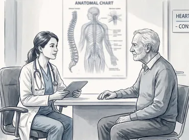
Understanding the intricate architecture of the human body begins with visual mapping, and few anatomical frameworks are as clinically vital as the female reproductive system chart. This comprehensive visual reference serves as a foundational tool for medical professionals, students, and individuals seeking to understand their own physiological health. For a detailed clinical overview, refer to the female reproductive system guidelines. The female reproductive system is a complex, highly integrated network of organs, ducts, and hormonal feedback mechanisms designed for gamete production, fertilization, gestation, and parturition. When viewed through an anatomical lens, the chart reveals not just static structures but dynamic pathways that undergo cyclical transformations, respond to neuroendocrine signals, and interact continuously with adjacent pelvic organs. Modern medicine relies heavily on accurate anatomical visualization to diagnose disorders, plan surgical interventions, counsel patients on fertility preservation, and implement preventative healthcare strategies. By learning how to interpret these diagrams, individuals can better comprehend their menstrual cycles, recognize early warning signs of reproductive pathology, and engage proactively in health maintenance. The following guide provides an evidence-based, clinically grounded exploration of this essential anatomical reference, translating complex physiological concepts into actionable insights for everyday wellness and long-term reproductive vitality.
Understanding the Anatomical Framework of the Female Reproductive Tract
The human pelvis houses a remarkably coordinated network of structures that collectively enable reproduction, urinary continence, and pelvic floor stability. A standard female reproductive system chart illustrates both the externally visible anatomy and the deeply seated internal organs, highlighting their spatial relationships and functional interdependence. The reproductive tract does not exist in isolation; it shares fascial planes, vascular supplies, and neural networks with the gastrointestinal and urinary systems, which is why clinical evaluation often requires a multidisciplinary approach. Medical education has long emphasized the importance of precise anatomical literacy, particularly because subtle structural variations can significantly influence diagnostic accuracy and treatment outcomes. Understanding these baseline structures provides the necessary context for interpreting clinical symptoms, evaluating imaging results, and engaging in informed discussions with healthcare providers about reproductive wellness.
Free calculatorOvulation CalculatorCalculate ovulation periodOpen the calculatorExternal Structures and Their Protective Functions
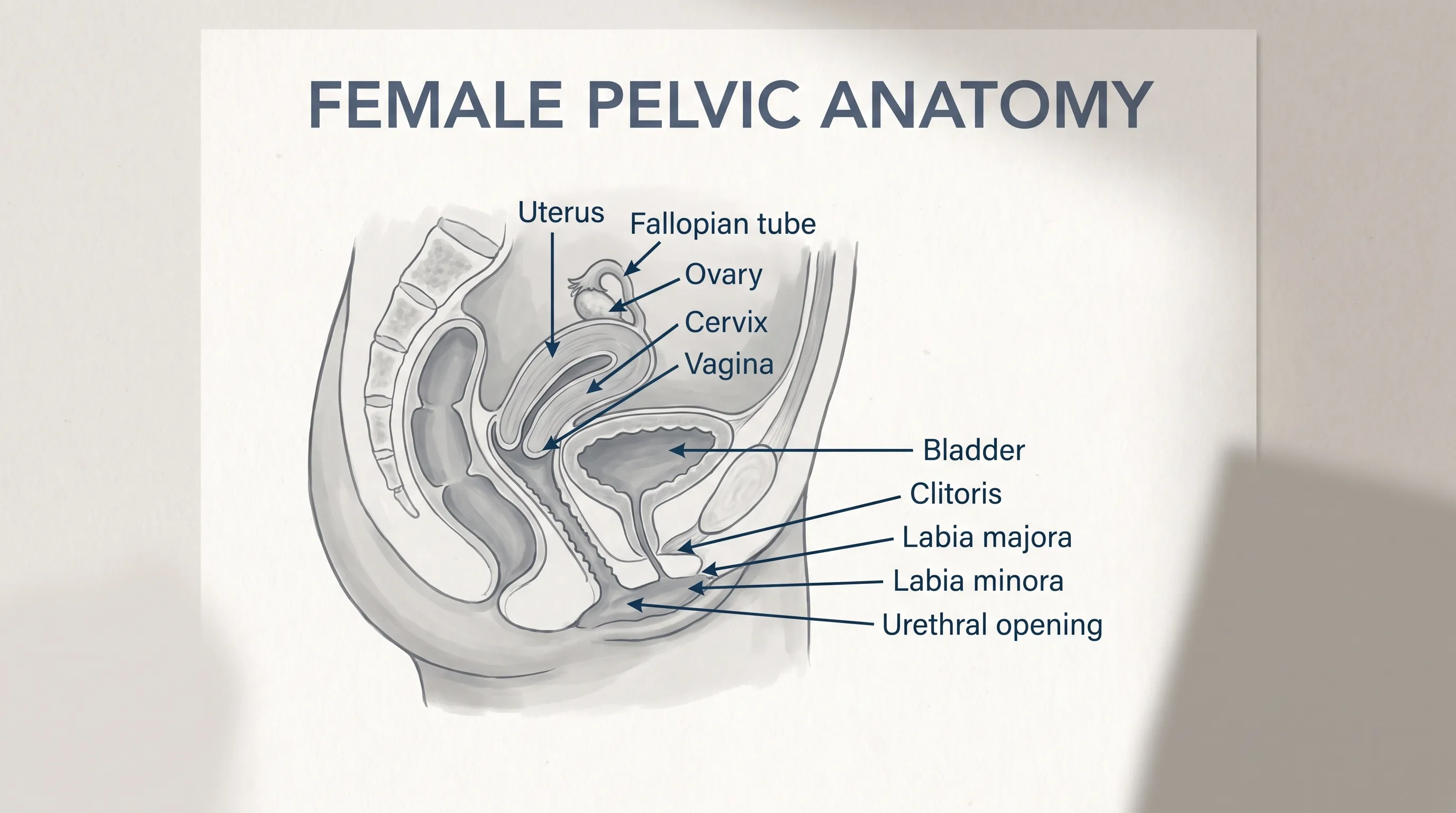
The external genitalia, collectively referred to as the vulva, serve as the primary barrier against pathogens while also playing a crucial role in sensory perception, lubrication, and urinary function. The mons pubis, a cushion of subcutaneous adipose tissue, provides mechanical protection to the underlying bony pelvis and contains numerous androgen-sensitive hair follicles. Inferiorly, the labia majora extend from the mons toward the perineum, forming protective folds that shield the more delicate inner structures. The labia minora, highly vascular and rich in nerve endings, encircle the vestibular opening and contain sebaceous glands that maintain mucosal moisture. Anteriorly, the clitoris functions as the primary neurovascular structure for sexual response, containing thousands of specialized nerve endings comparable in embryological origin to the male glans penis. The vestibule itself houses the urethral meatus, the vaginal orifice, and the openings of the Bartholin and Skene glands, which secrete mucoid fluids during sexual arousal to reduce friction and maintain an acidic vaginal environment that discourages pathogenic bacterial proliferation. Recognizing these external landmarks is essential for proper hygiene, infection prevention, and early identification of dermatological or inflammatory conditions.
Internal Organs: The Uterine and Adnexal Complex
Moving internally, the reproductive tract transitions into a muscular and mucosal network designed for transport, implantation, and gestation. The vagina is a highly distensible fibromuscular canal lined with stratified squamous epithelium that undergoes cyclical cellular turnover in response to estrogen. Learn more about vaginal health and microbiome balance. This epithelium maintains an acidic pH, typically between 3.8 and 4.5, primarily through lactobacilli metabolism of glycogen into lactic acid. The cervix, a cylindrical structure at the distal end of the uterus, contains a mucus-secreting columnar epithelium that undergoes dramatic viscosity changes throughout the menstrual cycle. During ovulation, cervical mucus becomes thin, alkaline, and highly penetrable, facilitating sperm migration. The uterus itself is a pear-shaped organ composed of three layers: the outer serosal perimetrium, the thick muscular myometrium responsible for labor contractions and structural support, and the inner endometrium, which proliferates and sheds cyclically. Extending laterally are the fallopian tubes, or uterine tubes, which feature ciliated mucosal linings and smooth muscle layers that generate peristaltic waves to transport oocytes. At the distal ends, the fimbriae sweep across the ovarian surface to capture released gametes. The ovaries, paired gonads located near the lateral pelvic walls, contain primordial follicles, corpus luteum tissue, and interstitial cells responsible for steroid hormone synthesis. Together, these structures form the core anatomical units depicted on every clinical female reproductive system chart.
Decoding the Female Reproductive System Chart
Visual references have long served as critical educational and diagnostic tools in clinical medicine. When interpreting a female reproductive system chart, it is essential to recognize that these diagrams are not merely artistic representations but clinically standardized maps used to guide patient evaluation, surgical planning, and physiological education. Medical charts typically employ anterior, sagittal, and lateral cross-sectional views to demonstrate three-dimensional spatial relationships. These visualizations help healthcare providers correlate subjective symptoms with objective anatomical locations, enabling more accurate differential diagnoses as outlined in pelvic anatomy guidelines. For instance, understanding the proximity of the ovaries to the sigmoid colon and the rectouterine pouch (pouch of Douglas) explains why certain gastrointestinal symptoms frequently overlap with reproductive complaints. Additionally, charts highlight vascular networks such as the uterine, ovarian, and internal iliac arteries, which are vital for understanding hemorrhage risks during menstruation, childbirth, or gynecological procedures.
Spatial Relationships and Anatomical Landmarks
The accuracy of a female reproductive system chart hinges on its ability to convey spatial orientation relative to fixed pelvic landmarks. The uterus typically assumes an anteverted and anteflexed position, meaning it tilts forward over the bladder rather than pressing posteriorly against the rectum. However, anatomical variation is common, with approximately twenty to thirty percent of individuals exhibiting a retroverted or retroflexed uterine position. Charts must account for these variations to avoid misdiagnosis during palpation or ultrasound evaluation. The broad ligament, a double fold of peritoneum, drapes over the uterus and fallopian tubes, anchoring them to the lateral pelvic walls while allowing physiological mobility. Suspensory ligaments house the ovarian vasculature and nerves, while the round ligaments maintain anterior uterine positioning. Neurological pathways, including the pudendal nerve, pelvic splanchnic nerves, and superior hypogastric plexus, are frequently mapped alongside these structures to illustrate pain referral patterns. Understanding these spatial relationships enables clinicians to distinguish between visceral pain originating from the uterus or ovaries and somatic pain arising from the pelvic floor musculature or perineal tissues.
How Medical Professionals Utilize Visual References
Gynecologists, reproductive endocrinologists, urogynecologists, and primary care providers routinely reference anatomical charts during patient consultations, surgical briefings, and imaging interpretation. Transvaginal ultrasound technicians rely on cross-sectional charts to identify normal follicular development, measure endometrial thickness, and detect pathological masses. During hysteroscopy or laparoscopy, surgeons use mental and physical chart references to navigate trocar placement, identify adhesions, and preserve fertility-sparing structures. Medical education programs integrate these visual tools early to ensure students develop strong spatial reasoning before entering clinical rotations. Furthermore, patient-facing charts are increasingly utilized in telehealth consultations to improve health literacy and facilitate shared decision-making. When patients can visually identify the location of a fibroid, cyst, or endometrial lesion, they are more likely to adhere to treatment plans, understand medication mechanisms, and report symptom changes accurately. The female reproductive system chart thus bridges the gap between clinical expertise and patient empowerment.
Hormonal Regulation and Cyclical Physiological Changes
The female reproductive system operates under the precise orchestration of neuroendocrine signaling, with the hypothalamic-pituitary-ovarian axis serving as the central control mechanism. Unlike most organ systems that maintain homeostasis through negative feedback loops alone, the reproductive tract exhibits a complex interplay of both positive and negative feedback to facilitate ovulation, prepare the uterine lining, and support potential pregnancy. A comprehensive female reproductive system chart rarely captures the temporal dimension of these hormonal fluctuations, yet understanding this cyclical nature is crucial for interpreting physiological changes across the lifespan. Estrogen, progesterone, luteinizing hormone (LH), follicle-stimulating hormone (FSH), and inhibin work in concert to regulate follicular maturation, ovulation timing, and endometrial receptivity. Disruptions in this delicate balance manifest as menstrual irregularities, anovulatory cycles, infertility, or metabolic disturbances, underscoring the necessity of viewing reproductive health as an integrated physiological process rather than an isolated anatomical framework.
The Four Distinct Phases of the Menstrual Cycle
The menstrual cycle typically spans twenty-one to thirty-five days, with day one marked by the onset of menstrual bleeding. During the menstrual phase (days one to five), declining progesterone levels trigger spiral artery constriction and endometrial shedding, resulting in the expulsion of tissue and blood through the cervix and vagina. This phase transitions into the follicular phase (days six to fourteen), during which rising FSH stimulates multiple antral follicles within the ovaries. One dominant follicle matures and secretes increasing quantities of estradiol, which stimulates endometrial proliferation, enhances cervical mucus permeability, and promotes bone density preservation. Midcycle, a rapid estrogen surge triggers a positive feedback loop, prompting the anterior pituitary to release a sharp LH spike that initiates ovulation. The subsequent luteal phase (days fifteen to twenty-eight) is characterized by corpus luteum formation, which secretes high progesterone levels to stabilize the endometrium, reduce uterine contractility, and elevate basal body temperature. If fertilization does not occur, corpus luteum involution leads to hormonal withdrawal, restarting the menstrual phase. Tracking these phases provides invaluable insight into hormonal health and reproductive readiness.
The Hypothalamic-Pituitary-Ovarian Axis
At the apex of reproductive regulation lies the hypothalamus, which secretes gonadotropin-releasing hormone (GnRH) in a pulsatile manner. This pulsatility is essential; continuous GnRH exposure paradoxically downregulates pituitary receptors, suppressing FSH and LH release. The anterior pituitary responds to GnRH pulses by secreting FSH and LH into systemic circulation. FSH targets ovarian granulosa cells, promoting aromatase enzyme activity that converts androgens to estrogens, while LH stimulates theca cells to produce androgens as estrogen precursors. Rising estradiol levels initially suppress GnRH and gonadotropins via negative feedback, but sustained high concentrations trigger positive feedback, inducing the LH surge. Post-ovulation, the corpus luteum secretes progesterone and inhibin, which restore negative feedback to maintain cycle regularity. Chronic stress, significant weight fluctuations, excessive exercise, or endocrine disorders such as thyroid dysfunction or hyperprolactinemia can disrupt this axis, leading to hypothalamic amenorrhea or oligomenorrhea. Recognizing these pathways enables clinicians to identify underlying etiologies rather than treating symptoms in isolation.
Common Clinical Conditions Impacting Reproductive Function
Despite the remarkable resilience of the female reproductive tract, several prevalent conditions frequently disrupt normal anatomical function and hormonal homeostasis. A detailed female reproductive system chart helps clinicians visualize where pathological processes localize, such as ectopic implantation within the fallopian tubes, myometrial invasion by adenomyotic tissue, or peritoneal seeding by endometrial implants. These conditions often present with overlapping symptomatology, including dysmenorrhea, dyspareunia, abnormal uterine bleeding, pelvic pressure, and subfertility. Early recognition, accurate imaging, and targeted interventions are essential to preserve reproductive potential and maintain quality of life. Understanding the anatomical and physiological basis of these disorders enables patients to advocate for appropriate diagnostic workups and evidence-based treatment plans.
Endometriosis and Adenomyosis: Painful Pathologies
Endometriosis affects approximately ten to fifteen percent of reproductive-aged individuals and is characterized by the presence of endometrial-like tissue outside the uterine cavity. Common implantation sites include the ovaries, posterior cul-de-sac, uterosacral ligaments, and pelvic peritoneum. These ectopic lesions respond to cyclical hormonal changes, leading to localized inflammation, fibrosis, and the formation of adhesions. Clinically, endometriosis manifests as progressively worsening dysmenorrhea, deep dyspareunia, chronic pelvic pain, and infertility. Diagnostic confirmation traditionally requires laparoscopy with histological verification, though transvaginal ultrasound and MRI can identify endometriomas and deep infiltrating disease. Adenomyosis, often termed endometriosis of the uterine muscle, occurs when endometrial glands and stroma invade the myometrium, causing uterine enlargement, boggy consistency, and heavy menstrual bleeding. Both conditions disrupt normal pelvic anatomy and often require multidisciplinary management, including NSAIDs, hormonal suppression with progestins or GnRH agonists, and surgical excision when conservative measures fail.
Polycystic Ovary Syndrome and Metabolic Intersections
Polycystic ovary syndrome (PCOS) represents the most common endocrine-metabolic disorder affecting reproductive-aged individuals, with prevalence estimates ranging from six to fifteen percent. The condition is characterized by chronic anovulation, hyperandrogenism, and polycystic ovarian morphology on ultrasound. Pathophysiologically, PCOS involves insulin resistance, elevated LH-to-FSH ratios, and increased ovarian and adrenal androgen production. These hormonal imbalances prevent dominant follicle selection, leading to arrested follicular development and the formation of multiple small cysts. Clinically, patients present with oligomenorrhea, hirsutism, acne, scalp hair thinning, and weight gain. Beyond reproductive implications, PCOS significantly increases the risk of type two diabetes, dyslipidemia, hypertension, endometrial hyperplasia, and cardiovascular disease. Management focuses on lifestyle modification, insulin sensitization with metformin, cycle regulation with combined oral contraceptives or cyclic progestins, and ovulation induction for those seeking fertility. Recognizing the systemic nature of PCOS ensures comprehensive, rather than purely reproductive, care delivery.
| Anatomical Structure | Primary Physiological Role | Common Pathological Presentation | First-Line Diagnostic Modality |
|---|---|---|---|
| Uterine Endometrium | Embryonic implantation & cyclic shedding | Heavy menstrual bleeding, hyperplasia | Transvaginal ultrasound, endometrial biopsy |
| Ovarian Cortex | Folliculogenesis & steroidogenesis | Cysts, premature ovarian insufficiency | Pelvic ultrasound, AMH & hormone panel |
| Fallopian Tubes | Oocyte transport & fertilization site | Tubal blockage, hydrosalpinx, ectopic pregnancy | Hysterosalpingography, laparoscopy |
| Cervix | Mucus barrier & birth canal gateway | Cervical dysplasia, stenosis, incompetence | Pap smear, HPV testing, transvaginal ultrasound |
| Pelvic Floor Muscles | Structural support & continence maintenance | Prolapse, hypertonic dysfunction, incontinence | Physical exam, urodynamic studies, pelvic MRI |
Evidence-Based Strategies for Long-Term Reproductive Wellness
Optimal reproductive health extends far beyond the absence of disease; it encompasses physiological resilience, hormonal balance, psychological well-being, and proactive lifestyle practices. A female reproductive system chart serves as an excellent reference point for identifying which tissues require targeted support, from the endometrial lining that demands adequate iron and folate, to the pelvic floor muscles that benefit from neuromuscular retraining. Integrative approaches combining nutrition, movement, stress management, and preventative screening significantly reduce the incidence of gynecological disorders, improve fertility outcomes, and enhance overall vitality. Evidence consistently demonstrates that small, consistent behavioral modifications yield substantial long-term benefits for reproductive function.
Nutritional Foundations and Hormone-Supportive Diets
Dietary composition profoundly influences steroid hormone metabolism, inflammatory markers, and cellular repair mechanisms. The reproductive tract requires a steady supply of omega-3 fatty acids, zinc, magnesium, vitamin D, and antioxidants to maintain mucosal integrity, support follicular development, and regulate immune responses in the endometrium. Anti-inflammatory dietary patterns, such as the Mediterranean diet, have been consistently associated with improved menstrual regularity, reduced dysmenorrhea, and higher conception rates. Emphasizing whole grains, leafy greens, cruciferous vegetables, lean proteins, and healthy fats supports hepatic estrogen metabolism, preventing the accumulation of harmful catechol estrogen metabolites that can disrupt endocrine signaling. Additionally, maintaining adequate iron stores through diet or supplementation prevents anemia secondary to heavy menstrual bleeding, while calcium and vitamin D intake preserves bone mineral density during periods of hypoestrogenism. Limiting ultra-processed foods, refined sugars, and trans fats reduces insulin resistance and systemic inflammation, both of which negatively impact ovarian function and uterine receptivity.
Preventative Care and Routine Screening Protocols
Routine gynecological care forms the cornerstone of reproductive health maintenance. Clinical guidelines recommend annual wellness visits that include pelvic examination, breast screening, vaccination updates, and personalized risk assessment. Cervical cancer screening via Pap smear and HPV testing remains critical for detecting precancerous cellular changes before they progress to malignancy. Guidelines typically advise initiating screening at age twenty-one, with intervals tailored to age, vaccination status, and prior results. Transvaginal ultrasound is utilized when pelvic pathology is suspected, while pelvic floor physiotherapy has emerged as a highly effective intervention for prolapse, incontinence, and musculoskeletal pelvic pain. Vaccinations against human papillomavirus significantly reduce cervical, vaginal, and vulvar cancer incidence, while routine chlamydia and gonorrhea screening prevents pelvic inflammatory disease and subsequent tubal factor infertility. Patient education regarding fertility preservation options, including oocyte cryopreservation for those delaying parenthood, is increasingly integrated into standard care. By adhering to evidence-based screening protocols, individuals can detect abnormalities early, intervene promptly, and preserve reproductive capacity across the lifespan.

Clinical Red Flags: Navigating Symptoms and Diagnostics
While many reproductive fluctuations fall within normal physiological ranges, certain symptoms warrant prompt clinical evaluation to rule out serious pathology. Recognizing these clinical red flags requires familiarity with the anatomical pathways illustrated on a female reproductive system chart, as well as an understanding of how pain, bleeding, or discharge patterns correlate with specific tissues. Delayed diagnosis often results from symptom normalization, particularly when individuals attribute heavy bleeding or severe dysmenorrhea to routine menstrual variations. However, persistent or progressive symptoms frequently indicate underlying structural, infectious, or endocrine disorders that benefit from timely intervention. Healthcare providers utilize a combination of clinical history, physical examination, laboratory testing, and advanced imaging to establish accurate diagnoses and implement appropriate management strategies.
Recognizing When to Seek Specialized Care
Patients should seek prompt gynecological evaluation if they experience bleeding between periods, postcoital spotting, soaking through more than one pad per hour, or menstrual cycles lasting longer than seven days. Pelvic pain that persists beyond typical premenstrual cramping, intensifies progressively across cycles, or radiates to the lower back and thighs requires thorough assessment. Unexplained weight changes, galactorrhea, hirsutism, severe mood disturbances linked to cycle phases, or painful intercourse that disrupts daily functioning all warrant clinical investigation. Postmenopausal bleeding is an absolute indication for urgent endometrial evaluation, as it carries a significant association with hyperplasia or malignancy. Additionally, recurrent urinary tract infections, new-onset incontinence, or a sensation of pelvic heaviness may indicate uterovaginal prolapse or pelvic floor dysfunction. Early consultation facilitates differential diagnosis, rules out infectious etiologies, and initiates targeted treatment before complications develop.
Imaging Modalities and Laboratory Assessments
Diagnostic workups for reproductive complaints typically begin with targeted blood work and pelvic imaging. Hormonal panels measuring FSH, LH, estradiol, progesterone, prolactin, thyroid-stimulating hormone, and testosterone help identify endocrine disruptions. Anti-Müllerian hormone testing provides valuable insight into ovarian reserve, while iron studies evaluate anemia secondary to chronic blood loss. Imaging modalities include transvaginal ultrasound, which offers high-resolution visualization of the uterus, ovaries, and endometrial cavity; hysterosalpingography, which evaluates tubal patency and uterine contour; and pelvic MRI, which provides superior soft-tissue contrast for characterizing fibroids, adenomyosis, and deep infiltrating endometriosis. Saline infusion sonohysterography can detect intracavitary lesions such as polyps or submucosal fibroids that routine ultrasound may miss. When structural abnormalities or unexplained infertility are suspected, hysteroscopy and laparoscopy offer direct visualization and simultaneous therapeutic intervention. Understanding the purpose and limitations of these diagnostic tools empowers patients to participate actively in their care journeys and make informed decisions alongside their providers.
Frequently Asked Questions
How do you read a female reproductive system chart correctly?
Reading a female reproductive system chart requires understanding the spatial orientation and functional connectivity of each anatomical structure. Begin by identifying the external vulvar landmarks, then trace internally through the vaginal canal, cervix, and uterine body. Note the lateral positioning of the fallopian tubes and ovaries, observing how the fimbriae capture oocytes and transport them toward the uterine cavity. Pay attention to vascular markings, ligamentous supports, and neurovascular pathways that explain symptom referral patterns. Always cross-reference charts with peer-reviewed anatomical textbooks or clinical guidelines from the National Institutes of Health to ensure accuracy, as simplified diagrams may omit clinically relevant variations such as uterine position anomalies or pelvic floor attachments.
What are the primary functions displayed on the female reproductive system chart?
The chart primarily illustrates gametogenesis, hormonal synthesis, fertilization pathways, embryonic implantation, gestational support, and parturition. It demonstrates how ovarian follicles mature under pituitary influence, how the endometrium undergoes cyclical proliferation and secretory transformation, and how the fallopian tubes facilitate gamete transport. Additionally, the diagram highlights the cervix as a dynamic barrier that modulates sperm entry and later dilates during labor, while the vaginal canal serves as both a protective conduit and birth passage. Hormonal feedback loops, though rarely drawn in anatomical diagrams, are implicitly represented by the cyclical tissue transformations shown across different phases.
Does the female reproductive system chart change with age?
While the fundamental anatomical layout remains consistent, physiological markers and tissue responsiveness shift significantly across developmental stages. Prepubertal charts emphasize dormant follicular pools and thin epithelial linings, while reproductive-age diagrams highlight active cyclical changes, vascular proliferation, and endometrial thickness variations. Postmenopausal representations focus on ovarian involution, endometrial atrophy, vaginal thinning, and pelvic floor weakening. Clinical references often include age-adjusted normative values for hormone levels, organ dimensions, and screening thresholds to ensure accurate interpretation. Understanding these lifecycle adaptations prevents misdiagnosis and guides age-appropriate healthcare interventions as recommended by WHO reproductive health guidelines.
Why is the fallopian tube position significant on a reproductive chart?
The fallopian tubes occupy a critical anatomical and functional crossroads, bridging the ovaries and uterine cavity within the peritoneal space. Their lateral positioning and fimbriated terminations highlight the mechanism of oocyte capture outside the closed reproductive tract. Because fertilization occurs in the ampullary region, any obstruction, inflammation, or anatomical distortion in this area directly impacts fertility. Charts emphasize the tubes to illustrate why conditions like pelvic inflammatory disease, endometriosis, or prior ectopic pregnancies often necessitate specialized imaging or surgical evaluation. Visual mapping also aids in understanding why tubal ligation or salpingectomy serves as highly effective permanent contraception.
How does a reproductive system chart assist in diagnosing pelvic pain?
Pelvic pain differential diagnosis relies heavily on correlating symptom location with anatomical structures. A female reproductive system chart enables clinicians to distinguish between midline uterine cramping, unilateral ovarian cyst pain, posterior rectouterine pouch tenderness, or anterior bladder/urethral discomfort. By identifying which tissues are inflamed, displaced, or compromised, providers can select targeted imaging modalities and avoid unnecessary invasive procedures. Charts also help patients articulate symptom locations more accurately, reducing communication barriers and expediting diagnostic workflows. Ultimately, anatomical literacy transforms vague discomfort into precise clinical indicators.
Conclusion
Mastering the interpretation of a female reproductive system chart extends far beyond academic interest; it serves as a vital foundation for lifelong health literacy, preventative care, and informed medical decision-making. The intricate interplay between anatomical structures, hormonal signaling, and cyclical physiological changes underscores the remarkable complexity of the female body. By understanding how each organ functions within the broader pelvic ecosystem, individuals can recognize normal variations, identify early warning signs of pathology, and engage proactively in wellness strategies. Evidence-based nutrition, routine screening protocols, stress management, and timely clinical consultation form the pillars of sustained reproductive vitality. Whether navigating menstrual health, family planning, or age-related physiological transitions, anatomical awareness empowers patients to advocate for comprehensive, personalized care. Healthcare providers, educators, and individuals alike benefit from integrating these visual and physiological insights into everyday health practices, ensuring that reproductive wellness remains a prioritized, actively managed component of overall well-being.
How we work
Researched and drafted with AI assistance, then reviewed by a human editor against the cited sources.
This article is general health information, not medical advice. Consult a qualified healthcare professional about your own situation.