Understanding and Recovering from an Abdominal Muscle Tear: A Complete Guide

Sudden, sharp pain during a heavy deadlift, a violent coughing fit, or an explosive tennis serve can leave you questioning your core integrity. When muscle fibers fail under unexpected load, the result is often an abdominal muscle tear, a painful but highly treatable injury that affects both elite athletes and everyday individuals. Understanding the mechanics of this condition, recognizing early warning signs, and implementing a structured recovery protocol are critical to restoring function and preventing long-term complications. Whether you experienced a sudden pop while lifting groceries or felt a gradual strain during intense core training, navigating the healing process requires patience, precise rehabilitation, and medically sound guidance. The abdominal wall is a complex, multi-layered structure responsible for spinal stability, organ protection, and trunk movement. When compromised, even basic activities like sitting up in bed or twisting to grab an object become challenging. This comprehensive guide breaks down everything you need to know about diagnosis, evidence-based treatment, and long-term prevention, empowering you to recover safely and return to your daily routine with confidence.
What Is an Abdominal Muscle Tear?
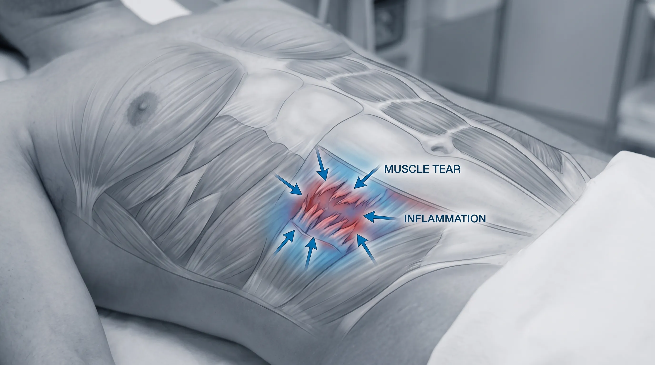
An abdominal muscle tear occurs when the tensile strength of the core musculature is exceeded, resulting in partial or complete disruption of muscle fibers and their surrounding connective tissue. Unlike a mild cramp or temporary tightness, an actual tear involves structural damage that triggers an inflammatory cascade, leading to localized swelling, bruising, and significant pain upon contraction. The condition is clinically referred to as a rectus abdominis strain or oblique strain, depending on the specific anatomical region affected.
Free calculatorLab Values Reference ChartComplete reference chart for medical lab valuesOpen the calculatorAnatomy of the Core Wall
To fully understand the injury, it is essential to recognize the architecture of the anterior and lateral abdominal wall. The rectus abdominis, commonly known as the six-pack muscle, runs vertically along the midline and is primarily responsible for trunk flexion. Flanking this are the external and internal obliques, which facilitate rotation and lateral flexion, while the deepest layer, the transversus abdominis, acts as a natural corset, stabilizing the spine and increasing intra-abdominal pressure. These muscle groups are anchored by fascial layers and interconnected by the linea alba. When force is applied rapidly or repetitively beyond physiological limits, the weakest fibers rupture first, typically at the musculotendinous junction where muscle transitions to tendon.
Degrees of Severity
Medical professionals classify these injuries into three distinct grades, which dictate treatment protocols and recovery expectations. Grade I involves microscopic fiber disruption with mild pain and minimal functional loss. Grade II represents a partial tear affecting a larger portion of the muscle bundle, accompanied by noticeable swelling, tenderness, and reduced strength. Grade III indicates a complete rupture or avulsion, where the muscle detaches entirely from its attachment point, often requiring surgical repair and an extended rehabilitation timeline. Accurately identifying the grade early on prevents premature loading, which can convert a manageable strain into a chronic, debilitating condition.
Recognizing the Signs and Symptoms
The clinical presentation of an abdominal muscle tear varies based on severity, mechanism of injury, and individual pain tolerance. However, several hallmark indicators consistently appear in patients presenting with this condition. Immediate, sharp pain during or shortly after a specific movement is the most common initial complaint. This pain typically intensifies with core engagement, coughing, sneezing, laughing, or changing positions from lying to sitting.
Immediate vs Delayed Pain
In many cases, the initial sensation is a sudden tearing or popping feeling followed by a burning ache that spreads across the affected quadrant. Delayed symptoms may emerge 24 to 48 hours post-injury as inflammation peaks. Patients frequently report stiffness, localized swelling, and ecchymosis (bruising) that can migrate downward due to gravity. Palpation reveals distinct tender spots along the muscle fibers, and attempting to contract the abdomen against resistance elicits sharp discomfort. Unlike visceral pain, which tends to be diffuse and accompanied by systemic symptoms, muscular injury pain remains localized and worsens with mechanical stress. Understanding this distinction helps individuals avoid unnecessary alarm while still prompting appropriate care.
When to Seek Emergency Care
While most tears are musculoskeletal in origin, certain red flags warrant immediate medical evaluation. If you experience severe, unrelenting pain that does not subside with rest, accompanied by nausea, vomiting, fever, or abdominal distension, these may indicate gastrointestinal distress, appendicitis, or an internal organ issue. Additionally, the appearance of a palpable bulge that cannot be gently pushed back in could suggest a hernia rather than an isolated muscle tear. Neurological symptoms like numbness, tingling, or radiating pain down the legs should also be evaluated promptly. Trusting clinical assessment over self-diagnosis ensures that life-threatening or structurally significant conditions are not overlooked.
Primary Causes and Risk Factors
The biomechanics of an abdominal muscle tear often revolve around the mismatch between muscular demand and tissue capacity. When eccentric loading overwhelms contractile ability, microtrauma accumulates until a macroscopic failure occurs. Identifying the underlying triggers is crucial for both recovery and long-term injury prevention.
Sports and High-Intensity Training
Athletes in rotational and explosive sports are disproportionately affected. Golfers, tennis players, baseball pitchers, and combat athletes generate immense torque through the core, placing tremendous shear stress on the obliques. Weightlifters performing compound movements like squats, deadlifts, or overhead presses often compensate for poor bracing technique by over-relying on superficial abdominal muscles, leading to strain. CrossFit enthusiasts and functional fitness athletes are also at heightened risk due to the combination of high-volume repetitions and maximal effort. Sudden spikes in training load without adequate conditioning or progressive overload are the primary culprits in athletic populations.
Everyday Movements and Posture
You do not need to be a competitive athlete to sustain an abdominal muscle tear. Everyday activities such as lifting heavy boxes improperly, slipping on a wet surface, or experiencing a severe, prolonged bout of coughing from respiratory infections can generate enough force to rupture weakened fibers. Chronic poor posture, prolonged sitting, and sedentary lifestyles contribute to muscle atrophy and reduced fascial resilience, making the abdominal wall more susceptible to acute failure. Previous injuries that healed without proper rehabilitation leave behind scar tissue, which is less elastic and more prone to re-injury under load. Addressing biomechanical inefficiencies and strengthening the deep stabilizing muscles are foundational steps in mitigating risk across all demographics.
Diagnosis and Medical Evaluation
Accurate diagnosis is the cornerstone of effective management. Because abdominal pain can stem from numerous sources, a systematic clinical approach is necessary to differentiate muscular injury from visceral, neurological, or structural pathologies.
Physical Examination
A qualified healthcare provider or sports medicine physician will begin with a thorough history, focusing on the mechanism of injury, pain characteristics, and functional limitations. The physical exam typically involves palpation to identify tender regions, swelling, or fascial defects. A key diagnostic maneuver is Carnett sign, where the patient tenses the abdominal muscles while the examiner presses on the painful area. If pain increases during tension, it strongly suggests a musculoskeletal origin rather than an intra-abdominal problem. Range of motion testing, resisted flexion, and lateral bending further isolate the affected muscle group and grade the functional impairment.
Imaging and Differential Diagnosis
For Grade I injuries, imaging is rarely necessary, as clinical presentation suffices for diagnosis. However, persistent pain, suspected Grade III tears, or ambiguous symptoms often warrant diagnostic imaging. Ultrasound provides a real-time, dynamic assessment of muscle architecture, allowing clinicians to visualize fiber continuity, hematoma formation, and fluid accumulation. Magnetic resonance imaging (MRI) offers superior soft tissue contrast, pinpointing the exact location and extent of the tear while ruling out concomitant pathology. Differential diagnosis is critical, as conditions like sports hernia (athletic pubalgia), rib cartilage inflammation, gallbladder disease, or even myocardial ischemia can mimic muscular pain. Collaborating with specialists ensures that no underlying condition is missed, paving the way for targeted intervention. For authoritative clinical guidelines on muscle injury assessment, visit Mayo Clinic Muscle Strains and Cleveland Clinic Abdominal Wall Strain.
Treatment and Recovery Strategies
Rehabilitation from an abdominal muscle tear follows a phased approach designed to respect tissue healing timelines while progressively restoring strength, flexibility, and neuromuscular control. Rushing through stages is the most common cause of chronic pain and recurrent injury.
The Acute Phase (RICE Protocol)
During the first 48 to 72 hours, the primary goal is inflammation control and pain management. The RICE method remains the gold standard: Rest, Ice, Compression, and Elevation. Avoid activities that provoke pain, and consider a light abdominal binder or supportive tape to limit excessive movement during the initial inflammatory stage. Apply ice packs for 15 to 20 minutes every 2 to 3 hours, ensuring a cloth barrier between the cold source and skin. Over-the-counter non-steroidal anti-inflammatory drugs (NSAIDs) can reduce pain and swelling, though prolonged use may interfere with long-term tissue remodeling. Focus on diaphragmatic breathing and gentle pelvic tilts to maintain baseline mobility without straining the healing fibers.
Progressive Physical Therapy
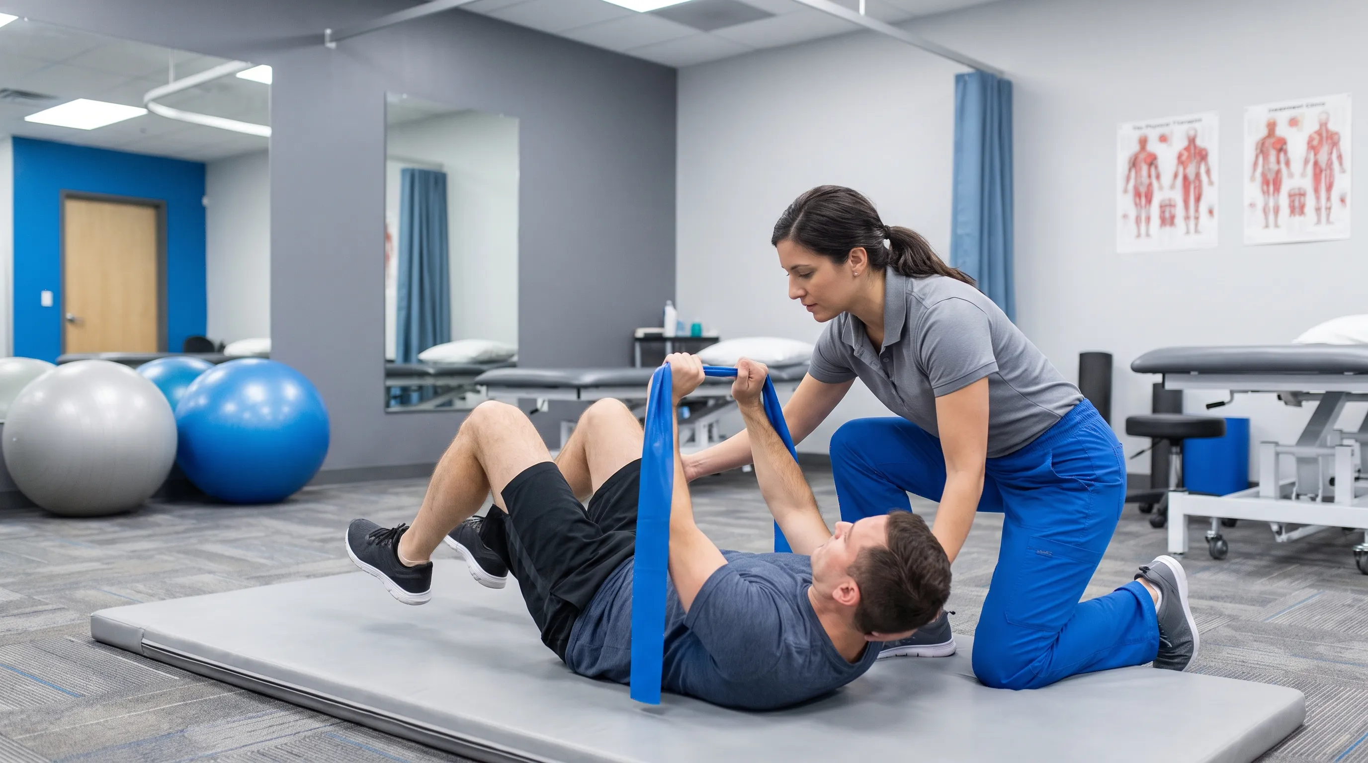
Once acute symptoms subside, typically around day 5 to 7, structured rehabilitation begins under the guidance of a licensed physical therapist. Phase one focuses on pain-free isometric contractions to reactivate neural pathways and prevent muscular atrophy. Phase two introduces controlled dynamic movements like modified planks, bird-dog variations, and supine heel slides. Phase three emphasizes eccentric loading, rotational control, and sport-specific drills to rebuild tissue tolerance. Consistency and proper form outweigh intensity; pushing through sharp pain disrupts collagen alignment and prolongs recovery. Integrating breath work and core bracing techniques ensures that intra-abdominal pressure is managed safely during functional movements. For expert-led rehabilitation demonstrations, refer to this foundational core recovery guide:
Surgical Intervention
While conservative management successfully treats the vast majority of cases, surgery becomes necessary in specific scenarios. Complete rectus sheath hematomas, tendon avulsions from the pubic symphysis, or chronic tears that fail to respond to 3 to 6 months of structured physical therapy may require operative repair. Surgical techniques involve debriding necrotic tissue, reattaching torn tendons using suture anchors, and reinforcing fascial defects. Post-operative recovery demands strict adherence to rehabilitation protocols, with full return to contact sports typically taking 4 to 6 months. Consulting a sports orthopedic surgeon or abdominal wall specialist ensures appropriate intervention when conservative measures fall short. Clinical literature on surgical outcomes can be explored through AAOS Acute Muscle Strains and NIH PMC Research.
| Grade | Tissue Damage | Symptoms | Recovery Timeline | Treatment Focus |
|---|---|---|---|---|
| Grade I | Microscopic fiber disruption | Mild localized pain, minimal swelling, normal range of motion | 2 to 4 weeks | Pain management, gentle mobility, isometric activation |
| Grade II | Partial fiber tear | Moderate to severe pain, visible bruising, strength deficits | 4 to 8 weeks | Progressive loading, eccentric training, physical therapy |
| Grade III | Complete rupture or avulsion | Sudden tearing sensation, severe functional loss, palpable defect | 3 to 6 months | Surgical evaluation, prolonged rehab, gradual return to sport |
Prevention and Long-Term Core Health
Recovering from an abdominal muscle tear is only half the equation; preventing recurrence requires a proactive approach to core conditioning, movement mechanics, and lifestyle optimization. The abdominal wall thrives on balanced strength, adequate recovery, and proper biomechanical alignment.
Proper Warm-Up Routines
Skipping dynamic preparation before intense activity dramatically increases injury risk. A structured warm-up elevates core temperature, increases blood flow to the musculature, and primes neuromuscular pathways for coordinated movement. Begin with 5 to 10 minutes of light cardiovascular activity followed by dynamic stretches like cat-cow, torso rotations, and hip circles. Progress to activation drills such as dead bugs, hollow body holds, and lateral band walks. Never perform explosive movements or heavy loading on cold, stiff tissue. Integrating a consistent warm-up routine reduces microtrauma accumulation and enhances tissue elasticity.
Nutritional Support for Muscle Healing
Optimal tissue repair relies heavily on nutritional adequacy. Protein provides the amino acids necessary for collagen synthesis and myofibrillar regeneration. Aim for 1.2 to 1.6 grams of high-quality protein per kilogram of body weight daily, distributed across multiple meals to sustain an anabolic environment. Micronutrients like vitamin C, zinc, and copper are essential cofactors in cross-linking new collagen fibers. Staying adequately hydrated maintains fascial pliability and supports cellular nutrient transport. Consider incorporating omega-3 fatty acids from fish or algae to naturally modulate inflammation, and ensure adequate caloric intake to avoid catabolic states during recovery periods. Long-term adherence to balanced nutrition not only accelerates healing but fortifies the abdominal wall against future stress.
Additionally, addressing lifestyle factors like sleep quality and stress management plays a subtle but profound role in muscular recovery. Chronic elevation of cortisol impairs tissue repair and increases pain sensitivity. Prioritizing 7 to 9 hours of uninterrupted sleep allows growth hormone secretion to peak, facilitating overnight muscle regeneration. Incorporating mindfulness practices or structured relaxation techniques reduces systemic tension, allowing the abdominal musculature to remain supple and responsive. For comprehensive guidance on integrating recovery-focused habits, explore evidence-based protocols and recommendations that align with modern sports medicine standards. The journey to lasting core resilience is cumulative, built through consistent, intelligent training and disciplined self-care. By respecting your body's signals and investing in foundational strength, you transform a temporary setback into a catalyst for lifelong physical optimization.
Frequently Asked Questions
How long does an abdominal muscle tear take to heal?
Healing time is directly correlated with injury severity. Mild strains (Grade I) typically resolve within 2 to 4 weeks with rest and light mobility work. Moderate partial tears (Grade II) require 4 to 8 weeks of progressive rehabilitation. Severe or complete ruptures (Grade III) may take 3 to 6 months and occasionally necessitate surgical repair. Patience and adherence to phased recovery protocols significantly reduce the risk of chronic pain or recurrent injury.
Can I exercise with an abdominal muscle tear?
In the acute inflammatory phase, strenuous exercise should be strictly avoided to prevent further fiber disruption. However, once pain subsides and a healthcare professional clears you, gentle isometric contractions and pain-free range-of-motion movements are encouraged. A structured physical therapy program will gradually reintroduce dynamic loading, rotational stability, and functional training. Returning to full workouts too quickly can transform a minor strain into a long-term debilitating condition.
What is the difference between an abdominal muscle tear and a hernia?
An abdominal muscle tear involves damage to the contractile muscle fibers or tendon attachments, resulting in localized pain, swelling, and bruising without structural protrusion. A hernia, on the other hand, occurs when internal tissue or an organ pushes through a weakened area in the abdominal fascia or muscle layer, often creating a visible bulge that may change size with coughing or straining. While both cause abdominal discomfort, they require distinctly different diagnostic approaches and treatment pathways.
When should I see a doctor for abdominal pain?
Immediate medical evaluation is warranted if you experience sudden, severe pain accompanied by dizziness, nausea, fever, or vomiting. Seek care if the pain radiates to your chest, back, or groin, or if you notice a firm, immovable mass in your abdomen. Additionally, if pain persists beyond a week despite rest and ice, or if it significantly impairs daily function, a clinical assessment is necessary to rule out serious conditions and establish an accurate recovery plan.
Does an abdominal muscle tear require surgery?
The majority of abdominal muscle tears heal successfully through conservative management, including rest, physical therapy, and gradual loading. Surgical intervention is generally reserved for Grade III complete ruptures, tendon avulsions from bone, or cases where conservative rehabilitation fails to restore function after 3 to 6 months. A sports medicine physician or orthopedic surgeon can determine surgical necessity through physical examination and advanced imaging.
Conclusion
An abdominal muscle tear is a challenging but highly manageable injury when approached with clinical accuracy and disciplined rehabilitation. By recognizing the warning signs, respecting tissue healing timelines, and committing to a structured physical therapy regimen, most individuals achieve complete functional recovery. Prioritizing proper warm-up protocols, balanced nutrition, and progressive core strengthening transforms a temporary setback into an opportunity for long-term musculoskeletal resilience. Always consult a qualified healthcare provider for personalized assessment and treatment planning, especially if symptoms persist or worsen. With patience, professional guidance, and consistent effort, you can safely return to full activity and maintain a robust, injury-resistant core for years to come.
How we work
Researched and drafted with AI assistance, then reviewed by a human editor against the cited sources.
This article is general health information, not medical advice. Consult a qualified healthcare professional about your own situation.