Thin Uterine Lining: Causes, Symptoms, Fertility Impact & Proven Treatments
Key points
- Vitamin E (400-800 IU daily): Antioxidant properties protect endometrial cells from oxidative stress while improving vascular density.
- L-Arginine (3-6 grams daily): Precursor to nitric oxide, enhancing blood flow and tissue oxygenation.
- Coenzyme Q10 (200-300 mg daily): Supports mitochondrial energy production in rapidly dividing endometrial cells.
- Omega-3 Fish Oil (1-2 grams daily): Reduces systemic inflammation and supports hormonal synthesis.
- Myo-Inositol: Improves insulin sensitivity and ovarian responsiveness in patients with PCOS-related thinning.
Have you been struggling to understand your menstrual cycle, navigating unexplained infertility, or recovering from a gynecological procedure only to hear the same concerning diagnosis? Learning that the lining of the uterus is thin can feel overwhelming, especially when you are actively trying to conceive or seeking optimal hormonal balance. The endometrium plays a pivotal role in reproductive health, menstrual regularity, and overall pelvic wellness, yet it remains one of the most misunderstood aspects of women’s physiology. Fortunately, modern reproductive medicine, combined with lifestyle interventions, offers a clear pathway forward. By understanding the underlying mechanisms, recognizing subtle symptoms, and implementing targeted treatments, you can actively support your body’s natural regenerative processes. This comprehensive guide explores the science behind endometrial thinning, evidence-based medical interventions, nutritional strategies, and fertility-focused approaches. Whether you are just beginning your wellness journey or seeking advanced clinical options, the information provided here is grounded in peer-reviewed research and designed to empower your next steps toward optimal uterine health.
Understanding the Endometrium and Its Crucial Role
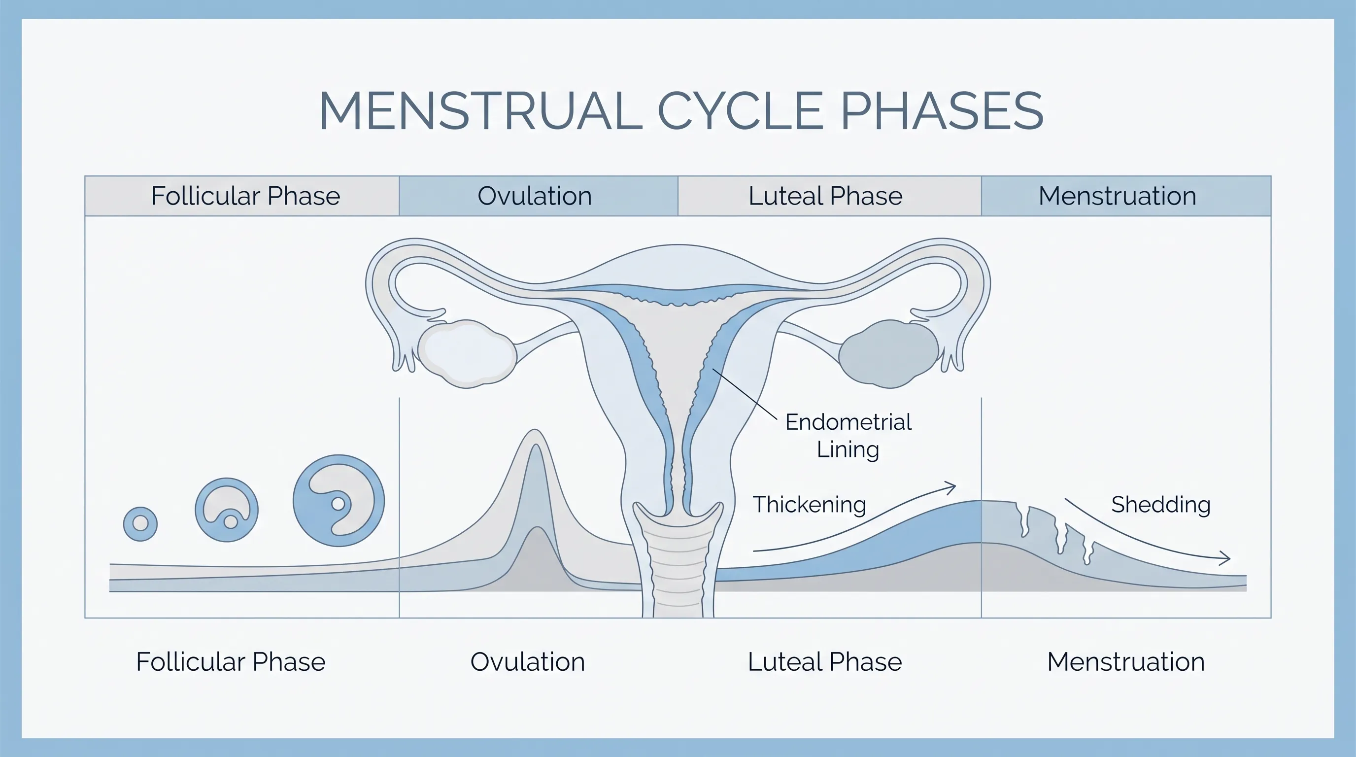
The uterine lining, clinically referred to as the endometrium, is a highly dynamic mucous membrane that undergoes cyclical transformation throughout your reproductive years. It is composed of two distinct layers: the stratum basalis, a deep permanent layer that serves as the foundation for regeneration, and the stratum functionalis, a superficial layer that thickens, sheds, and rebuilds during each menstrual cycle. When a woman learns that the lining of the uterus is thin, it specifically refers to inadequate development of the functionalis layer, which compromises the environment required for embryo implantation and healthy menstrual flow.
Free calculatorOvulation CalculatorCalculate ovulation periodOpen the calculatorWhat Is Endometrial Thickness?
Endometrial thickness is measured in millimeters using transvaginal ultrasound and serves as a primary clinical indicator of uterine receptivity. A robust endometrium typically exhibits a trilaminar (three-line) appearance on imaging, reflecting healthy estrogen-driven glandular proliferation, stromal expansion, and optimal vascular development. Thickness is not a static measurement; it fluctuates dramatically depending on your cycle phase. During menstruation, the lining naturally measures only 1 to 4 mm. As estrogen rises during the follicular phase, the tissue proliferates rapidly. By mid-cycle, thickness should ideally range between 7 mm and 12 mm. Values consistently below 7 mm during the peri-ovulatory window are clinically significant and warrant further investigation.
How the Uterine Lining Changes Throughout Your Cycle
Your endometrium operates under precise endocrine regulation. The hypothalamus releases gonadotropin-releasing hormone (GnRH), stimulating the pituitary gland to secrete follicle-stimulating hormone (FSH) and luteinizing hormone (LH). FSH prompts ovarian follicles to mature and produce estradiol. Estradiol binds to estrogen receptors in the endometrial stroma, triggering cellular mitosis and angiogenesis. Following ovulation, the corpus luteum secretes progesterone, which converts the proliferative lining into a secretory state. This transition increases glycogen production, stabilizes vascular structures, and prepares the tissue for potential embryo implantation. When the lining of the uterus is thin, this delicate hormonal cascade is often disrupted, leading to poor receptivity and altered cycle physiology.
Recognizing the Signs: When the Lining of the Uterus Is Thin
Identifying endometrial thinning early can significantly improve treatment outcomes. While some women experience no overt symptoms, others notice distinct physiological changes that signal underlying tissue compromise. Understanding these clinical markers helps bridge the gap between subtle discomfort and timely medical intervention.
Common Symptoms to Watch For
The most prominent indicator is hypomenorrhea, or unusually light menstrual bleeding. Women with a thin lining often report flow lasting fewer than three days, minimal pad or tampon usage, and pale pink or brownish discharge instead of rich red blood. Some experience secondary amenorrhea, where menstruation stops entirely due to inadequate tissue buildup. Others report pelvic heaviness, mild cramping without bleeding, or difficulty achieving orgasm due to reduced pelvic congestion. When tracking fertility, women may notice a lack of fertile-quality cervical mucus, which correlates with low systemic estrogen. These signs frequently prompt diagnostic evaluation when the lining of the uterus is thin and begins to impact daily wellness or reproductive planning.
When to Seek Medical Evaluation
If your menstrual flow consistently falls below normal parameters, or if you experience unexplained infertility despite regular ovulation, consult a gynecologist or reproductive endocrinologist. Additional red flags include painful intercourse, recurrent early miscarriages, or a history of pelvic infections or uterine surgeries. Diagnostic imaging should be scheduled during the mid-follicular phase to capture baseline proliferation accurately. Early intervention prevents progressive tissue fibrosis, optimizes hormonal balance, and preserves reproductive potential.
Primary Causes and Contributing Factors
Endometrial thinning rarely occurs in isolation. It typically stems from a combination of hormonal, structural, pharmacological, and lifestyle influences. Identifying the root cause is essential for selecting effective therapies.
Hormonal Imbalances and Low Estrogen
Estrogen is the primary architect of endometrial growth. Hypoestrogenism can result from perimenopause, premature ovarian insufficiency (POI), hypothalamic dysfunction, or extreme athletic training. Conditions like hyperprolactinemia or uncontrolled thyroid disorders also suppress estrogen production, directly limiting tissue proliferation. When estrogen receptors are downregulated due to chronic stress or metabolic dysfunction, the uterine lining becomes unresponsive to circulating hormones, leaving the lining of the uterus is thin despite normal blood tests.
Iatrogenic Factors: D&C and Previous Surgeries
Dilation and curettage (D&C) procedures, particularly when performed multiple times or aggressively, can damage the basalis layer. This often leads to Asherman syndrome, characterized by intrauterine adhesions and scar tissue that physically block endometrial regeneration. Myomectomy, hysteroscopic resections, and cesarean deliveries can also compromise vascular integrity. Surgical trauma triggers localized fibrosis, reducing the available tissue surface and impairing natural thickening mechanisms.
Medications, Age, and Lifestyle Influences
Certain fertility medications, particularly clomiphene citrate, exert anti-estrogenic effects on the endometrium while stimulating ovulation. Long-term contraceptive use, corticosteroids, and certain antidepressants may alter hormone metabolism. Advanced maternal age naturally reduces ovarian reserve and estradiol output. Lifestyle factors like chronic smoking, excessive alcohol consumption, extreme caloric restriction, and high-intensity endurance training further suppress reproductive hormones. These cumulative stressors create a physiological environment where the lining of the uterus is thin and less capable of supporting implantation.
Infections and Chronic Inflammation
Pelvic inflammatory disease (PID), endometritis, and untreated sexually transmitted infections can cause chronic inflammatory changes. Persistent cytokine release and immune cell infiltration disrupt normal glandular development and promote fibrotic remodeling. Tuberculosis of the reproductive tract, though rare in certain regions, causes severe endometrial destruction. Addressing underlying infections is a prerequisite for successful tissue restoration.
Diagnostic Procedures and How Doctors Assess Thickness
Accurate diagnosis requires precise imaging, timing, and sometimes invasive evaluation. Reproductive specialists utilize a stepwise approach to differentiate physiological thinning from pathological compromise.
Transvaginal Ultrasound and Sonohysterography
Transvaginal ultrasonography (TVUS) remains the gold standard for measuring endometrial thickness. The probe provides high-resolution images of the uterine cavity, allowing clinicians to assess the trilaminar pattern and measure the anterior-to-posterior distance. When initial scans reveal irregularities, saline infusion sonography (SIS) may be performed. By introducing sterile fluid into the uterine cavity, sonohysterography distends the tissue, highlighting polyps, submucosal fibroids, or adhesions that standard ultrasound might miss.
Hysteroscopy and Endometrial Biopsy
Hysteroscopy involves inserting a narrow, illuminated camera through the cervix to directly visualize the uterine interior. This procedure is both diagnostic and therapeutic, enabling surgeons to remove adhesions, clear polyps, or obtain targeted tissue samples. An endometrial biopsy evaluates cellular architecture, ruling out hyperplasia or malignancy while assessing glandular-stromal ratio. These interventions provide definitive data when the lining of the uterus is thin and standard imaging proves inconclusive. For further clinical guidance, consult ACOG Guidelines on Endometrial Evaluation and review peer-reviewed research via PubMed.
Evidence-Based Treatment Options and Therapies
Modern gynecology offers a spectrum of interventions tailored to the underlying etiology of endometrial thinning. Treatment selection depends on fertility goals, symptom severity, and diagnostic findings.
Hormonal Supplements and Estrogen Therapy
Estrogen replacement remains the cornerstone of treatment. Delivery methods include oral micronized estradiol, transdermal patches, vaginal tablets, and intracavitary estrogen creams. Vaginal administration achieves high local tissue concentrations while minimizing systemic side effects. Clinicians typically prescribe continuous estrogen during the follicular phase, followed by progesterone to induce secretory transformation. Dosage adjustments are guided by serial ultrasounds and serum hormone panels. For patients unresponsive to standard therapy, compounded estrogen gels or extended-release formulations may be explored under specialist supervision.
Advanced Medical Interventions
When conventional hormones fail, adjuvant therapies enhance vascularization and tissue growth. Low-dose aspirin improves endometrial perfusion by inhibiting platelet aggregation and promoting nitric oxide production. Vaginal sildenafil and pentoxifylline dilate pelvic microvasculature, supporting nutrient delivery to the endometrium. Platelet-rich plasma (PRP) intrauterine infusions and granulocyte colony-stimulating factor (G-CSF) represent cutting-edge regenerative approaches. PRP contains concentrated growth factors that stimulate angiogenesis and cellular proliferation. Clinical trials show promising results in refractory cases, though these therapies remain investigational in many regions. Always verify protocols through Mayo Clinic Fertility Guidelines.
Natural and Lifestyle Approaches to Support Uterine Health
Complementary strategies significantly enhance medical outcomes. Acupuncture increases pelvic blood flow by modulating sympathetic nervous activity. Transcutaneous electrical nerve stimulation (TENS) applied to the sacral region promotes vasodilation. Mind-body practices like guided meditation and breathwork lower cortisol, indirectly supporting estrogen balance. Consistent sleep hygiene regulates circadian hormone secretion, while moderate exercise prevents both sedentary stagnation and overtraining-induced suppression.
Fertility Implications and Reproductive Outcomes
The relationship between endometrial thickness and pregnancy success is complex but well-documented. Understanding the nuances helps patients set realistic expectations and pursue targeted fertility optimization.
Implantation Challenges and Early Pregnancy Risks
A receptive lining must reach a critical threshold to support embryo attachment. Measurements below 6 mm correlate with significantly lower implantation rates, increased chemical pregnancy risk, and higher early miscarriage probability. The endometrium secretes leukemia inhibitory factor (LIF), integrins, and cytokines essential for trophoblast invasion. Thin tissue often exhibits altered molecular signaling, creating a suboptimal microenvironment. However, thickness alone does not guarantee failure. Lining quality, vascular pattern, and immune tolerance play equally vital roles.
Assisted Reproductive Technology (ART) Considerations
In vitro fertilization (IVF) programs carefully monitor endometrial development before embryo transfer. Frozen embryo transfer (FET) cycles allow extended estrogen preparation, enabling gradual tissue buildup. Artificial cycle protocols use exogenous hormones for precise control, while natural or modified natural cycles rely on endogenous ovulation. When the lining of the uterus is thin during ART, clinicians may delay transfer, switch to FET, or incorporate adjuvant therapies. Some clinics employ endometrial scratching to provoke a localized healing response, though evidence remains mixed. Comprehensive counseling ensures patients understand success probabilities and alternative pathways like gestational surrogacy or adoption if medical interventions reach their limits. Review Cleveland Clinic Reproductive Medicine Protocols for additional insights.
Practical Strategies to Improve Uterine Lining Naturally
While medical interventions provide targeted correction, daily lifestyle choices profoundly influence endometrial regeneration. Integrating evidence-based nutritional and behavioral habits creates a sustainable foundation for uterine wellness.
Dietary Adjustments and Nutritional Supplements
Anti-inflammatory nutrition forms the cornerstone of endometrial support. Prioritize omega-3-rich foods like wild salmon, flaxseeds, and walnuts to modulate prostaglandin production and enhance cellular membrane fluidity. Nitric oxide-boosting vegetables such as beets, spinach, and arugula promote vasodilation and improve pelvic perfusion. Complex carbohydrates stabilize insulin, preventing hyperinsulinemia that disrupts ovarian function.
Supplementation should be individualized but commonly includes:
- Vitamin E (400-800 IU daily): Antioxidant properties protect endometrial cells from oxidative stress while improving vascular density.
- L-Arginine (3-6 grams daily): Precursor to nitric oxide, enhancing blood flow and tissue oxygenation.
- Coenzyme Q10 (200-300 mg daily): Supports mitochondrial energy production in rapidly dividing endometrial cells.
- Omega-3 Fish Oil (1-2 grams daily): Reduces systemic inflammation and supports hormonal synthesis.
- Myo-Inositol: Improves insulin sensitivity and ovarian responsiveness in patients with PCOS-related thinning.
Always consult a healthcare provider before initiating supplements, particularly if taking fertility medications or anticoagulants.
Exercise, Stress Management, and Blood Flow Optimization
Physical activity must be carefully balanced. Moderate aerobic exercise (30-40 minutes, 4-5 times weekly) improves systemic circulation and insulin sensitivity without suppressing reproductive hormones. Pelvic floor yoga, gentle stretching, and restorative practices specifically target uterine vascular networks. Conversely, excessive high-intensity interval training (HIIT) or marathon-level endurance work elevates cortisol and reduces gonadotropin release, potentially worsening thinning.
Stress management directly impacts the hypothalamic-pituitary-adrenal (HPA) axis. Daily meditation, journaling, progressive muscle relaxation, and therapy reduce chronic sympathetic dominance. Adequate hydration (2.5-3 liters daily) maintains cellular turgor and nutrient transport. Eliminating endocrine-disrupting chemicals found in plastics, non-organic pesticides, and certain cosmetics further supports hormonal equilibrium. When the lining of the uterus is thin, consistency in these daily practices often yields measurable improvements within two to three menstrual cycles.
| Clinical Parameter | Normal Endometrial Profile | Thin Endometrial Profile | Recommended Intervention Focus |
|---|---|---|---|
| Ovulation Thickness | 7-12 mm, trilaminar | <7 mm, homogeneous or irregular | Estrogen optimization, vascular support |
| Menstrual Flow | 30-80 mL, 3-7 days | <30 mL, 1-2 days, spotting | Rule out adhesions, assess hormone metabolism |
| Ultrasound Appearance | Clear three-line pattern | Thin, single-layer, or scarred | Sonohysterography, hysteroscopy |
| Vascularity | Rich subendometrial perfusion | Sparse or absent Doppler signal | Aspirin, L-arginine, sildenafil |
| Fertility Impact | High implantation potential | Reduced receptivity, higher chemical pregnancy risk | FET cycles, PRP, lifestyle modification |
Frequently Asked Questions
What is considered a normal uterine lining thickness during ovulation?
During the mid-luteal phase or ovulation window, a receptive uterine lining typically measures between 7 mm and 12 mm. Thickness below 7 mm at this stage is clinically classified as thin and may impact implantation success.
Can stress really affect the thickness of my uterine lining?
Yes. Chronic psychological stress elevates cortisol levels, which can disrupt the hypothalamic-pituitary-ovarian axis. This hormonal disruption reduces circulating estrogen, ultimately impairing endometrial proliferation and resulting in a thinner lining.
Does a thin uterine lining always mean infertility?
Not necessarily. While a thin lining presents challenges, many women still conceive naturally or through assisted reproduction. The lining's vascular pattern, cellular receptivity, and absence of scarring often matter as much as millimeter measurements alone.
What are the most effective natural supplements for improving endometrial thickness?
Clinical research supports the use of L-arginine, vitamin E, omega-3 fatty acids, and Coenzyme Q10. These compounds improve pelvic microcirculation, reduce oxidative stress, and support healthy cellular proliferation within the endometrium.
How long does it take to thicken a thin uterine lining with estrogen therapy?
Most patients see measurable improvements within one to two menstrual cycles of consistent estrogen supplementation. Physicians typically monitor progress via serial ultrasounds and adjust dosage, delivery method, or adjunct therapies accordingly.
Conclusion
Discovering that the lining of the uterus is thin can feel discouraging, but it is rarely a permanent roadblock. Modern reproductive medicine, combined with targeted nutritional strategies and mindful lifestyle adjustments, provides multiple pathways to restore endometrial health. Whether your goal is achieving regular menstrual cycles, optimizing fertility, or simply supporting long-term pelvic wellness, understanding the underlying mechanisms empowers informed decision-making. Prioritize comprehensive diagnostic evaluation, collaborate with qualified specialists, and integrate evidence-based habits into your daily routine. Your uterus is remarkably resilient, and with consistent care, it can regenerate the healthy tissue needed to thrive. Take the first step today by tracking your cycle, discussing your imaging results with a trusted provider, and implementing gentle, sustainable practices that nurture your reproductive vitality.
For additional authoritative resources on women’s health and hormonal wellness, explore National Institutes of Health Women’s Health Initiative and American Society for Reproductive Medicine Patient Resources.
How we work
Researched and drafted with AI assistance, then reviewed by a human editor against the cited sources.
This article is general health information, not medical advice. Consult a qualified healthcare professional about your own situation.