Pelvic Kidney: Causes, Symptoms, Diagnosis & Management Guide

The human body is a masterpiece of anatomical precision, yet it occasionally presents with fascinating structural variations that challenge conventional expectations. One such variation is the pelvic kidney, a congenital condition where one or both kidneys fail to ascend to their typical position in the upper abdomen during fetal development. While the term might sound alarming, the vast majority of individuals with this anatomical arrangement never experience symptoms and lead entirely normal, active lives. Understanding how a pelvic kidney develops, how it is detected, and what management strategies are available empowers patients to navigate their health confidently. Learn more about ectopic kidney conditions at the Cleveland Clinic. Whether you recently received an incidental diagnosis, are navigating a pregnancy with this condition, or simply want to deepen your medical knowledge, this comprehensive guide provides evidence-based insights, clinical perspectives, and actionable lifestyle recommendations to support long-term renal wellness.
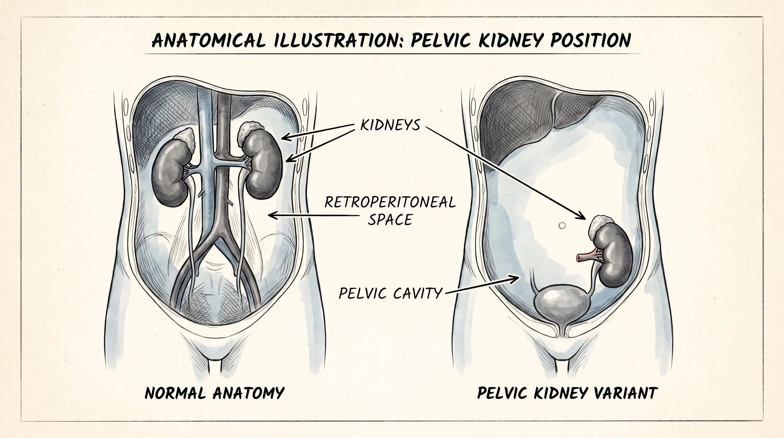
Understanding Pelvic Kidney: What It Is and How It Develops
A pelvic kidney is classified as a type of ectopic kidney, meaning it resides outside its expected anatomical location. In a typical pregnancy, the kidneys originate in the lower pelvic region during the fifth to sixth week of gestation. From this starting point, they gradually migrate upward, a process known as renal ascent, eventually settling beneath the diaphragm on either side of the spine by the eighth to ninth week of development. When this migration is partially or completely interrupted, the kidney remains in or near the pelvic cavity. This anatomical variation occurs in approximately 1 in 400 to 1 in 1,000 live births, making it one of the more common congenital urological findings. Importantly, a pelvic kidney is usually fully functional. It contains the same essential structures as a normally positioned kidney, including nephrons, glomeruli, and a collecting system. The primary difference lies in its location, altered vascular supply, and the trajectory of its ureter.
Free calculatorHigh-Fiber Foods ChartChart of high-fiber foods for digestive healthOpen the calculatorEmbryological Origins of Kidney Migration
To fully appreciate why a pelvic kidney forms, we must examine the intricate choreography of early embryology. Kidney development proceeds through three overlapping stages: the pronephros, mesonephros, and metanephros. The permanent kidneys, or metanephros, arise from the metanephric mesoderm and are initially induced by the ureteric bud, a tubular outgrowth from the mesonephric duct. As the embryo grows, the ureteric bud repeatedly branches while the metanephric mesenchyme differentiates into nephrons. Simultaneously, the developing kidneys undergo a remarkable cranial migration. This ascent is not a passive drift but an active process driven by differential growth of the embryo's caudal regions and the formation of new, higher vascular connections from the abdominal aorta. As the kidneys move upward, the original pelvic blood vessels typically regress. When a pelvic kidney occurs, this vascular remodeling fails to complete, resulting in abnormal arterial and venous supply that originates from the lower aorta, iliac arteries, or pelvic vessels. The ureter, which normally enters the bladder at a relatively steep angle to facilitate unidirectional urine flow, instead follows a longer, more horizontal course from the pelvis to the bladder. This altered anatomical geometry is a critical factor in understanding both the benign nature of most cases and the occasional predisposition to urinary complications. Explore kidney development and urologic conditions through the NIH.
Why the Kidney Gets "Stuck" in the Pelvis
The precise etiology of interrupted renal ascent remains multifactorial, with genetics, embryological timing, and mechanical constraints all playing potential roles. Current medical literature suggests that disruptions in the signaling pathways involving the GDNF-RET receptor system can impair ureteric bud formation and subsequent kidney migration. Environmental factors, such as maternal nutritional deficiencies, certain teratogenic exposures, or localized ischemic events during early gestation, may also contribute. Additionally, mechanical barriers like abnormal pelvic vessel positioning or a narrow embryonic space can physically impede upward movement. In rare instances, the kidneys ascend normally but later descend into the pelvis due to trauma, surgical complications, or severe musculoskeletal abnormalities, though this acquired form is exceedingly uncommon. Most pelvic kidneys are unilateral, meaning only one kidney is affected, while bilateral pelvic kidneys occur in fewer than 10 percent of ectopic cases. Importantly, this condition rarely occurs in isolation; it is sometimes associated with other congenital anomalies, particularly those involving the genitourinary tract, such as vesicoureteral reflux, ureteropelvic junction obstruction, or genital malformations. Routine prenatal screening and postnatal imaging help identify these concurrent findings early, allowing for comprehensive, individualized care planning.
Prevalence and Demographic Factors
Epidemiological data consistently show that pelvic kidneys occur with relatively equal frequency in males and females, though some studies note a slight male predominance in clinical presentations due to differences in pelvic anatomy and healthcare-seeking behaviors. The condition spans all ethnic groups and geographic regions, confirming its classification as a fundamental developmental variation rather than a population-specific trait. Interestingly, while a pelvic kidney is congenital, the age of diagnosis varies widely. Approximately 60 to 70 percent of cases are identified incidentally in adulthood during imaging for unrelated conditions such as back pain, gastrointestinal complaints, or routine health screenings. Prenatal diagnosis has increased dramatically over the past two decades due to standardized obstetric ultrasound protocols, as outlined by Mayo Clinic, which typically include fetal renal visualization at the 20-week anatomy scan. Pediatric cases are frequently caught during evaluations for recurrent fevers, urinary tract infections, or abdominal masses. Regardless of when it is discovered, the clinical significance remains largely tied to the presence or absence of functional impairment rather than the anatomical anomaly itself.
Signs, Symptoms, and Clinical Presentation
One of the most defining characteristics of a pelvic kidney is its frequent clinical silence. The majority of individuals never experience symptoms directly attributable to their renal position. However, when symptoms do arise, they typically stem from secondary anatomical consequences rather than the kidney's location itself. Understanding the spectrum of clinical presentation helps differentiate benign variants from pathological conditions requiring intervention.
The Asymptomatic Majority
In most cases, a pelvic kidney functions perfectly well, filtering blood, regulating electrolytes, maintaining blood pressure, and producing urine without any detectable issues. The body adapts remarkably well to this variation, compensating for altered blood supply and drainage pathways. Many patients live their entire lives without knowing they have a pelvic kidney until it appears on a radiograph, ultrasound, or MRI performed for entirely unrelated reasons. Medical professionals often emphasize that an asymptomatic pelvic kidney requires no treatment, only periodic monitoring to ensure stable renal function through standard blood tests (like serum creatinine and blood urea nitrogen) and urine analysis. This asymptomatic nature is crucial for patient education; receiving a diagnosis can understandably cause anxiety, but comprehensive medical counseling consistently reinforces that the condition does not inherently threaten longevity, vitality, or daily functioning.
Pain and Discomfort Patterns
When symptoms do manifest, they most commonly present as vague, intermittent pain in the lower abdomen, groin, or deep pelvic region. Unlike flank pain associated with upper abdominal kidneys, pelvic kidney discomfort is often localized to the suprapubic area or radiates toward the inner thighs. The pain mechanism usually involves capsular stretching from mild hydronephrosis, ureteral spasm, or local inflammatory responses. Some individuals report a sensation of pelvic fullness or pressure, particularly after consuming large volumes of fluid or during periods of intense physical exertion. In rare instances, the altered vascular anatomy can lead to intermittent vascular compression, causing dull, aching discomfort that fluctuates with posture or activity levels. Differentiating this pain from gynecological conditions (in women), prostatitis (in men), or musculoskeletal strain requires careful clinical evaluation and targeted imaging.
Urinary and Gastrointestinal Overlaps
The altered trajectory of the ureter from a pelvic kidney can create mild urinary drainage inefficiencies. Patients may notice slightly delayed urine flow into the bladder, increased urinary frequency, or occasional difficulty fully emptying the bladder. These symptoms often overlap with common urological and gastrointestinal conditions, leading to potential diagnostic confusion. Constipation, a frequent complaint in pelvic kidney patients, can sometimes exert external pressure on the ureter or bladder neck, temporarily worsening urinary symptoms. Conversely, urinary stasis in the distal ureter can occasionally trigger referred bowel discomfort. Recognizing these interconnected symptom patterns is essential for clinicians and patients alike. Maintaining a symptom diary, tracking fluid intake, noting pain triggers, and communicating these observations to a healthcare provider significantly streamline the diagnostic process and prevent unnecessary testing or interventions.
Diagnostic Pathways and Imaging Techniques
Accurate diagnosis of a pelvic kidney relies heavily on modern imaging modalities, each offering unique advantages depending on the clinical scenario and patient demographics. The diagnostic pathway typically begins with non-invasive screening and progresses to specialized studies only when anatomical or functional clarification is necessary.
Prenatal Detection and Fetal Ultrasound
Prenatal ultrasound is the cornerstone of early identification. During the standard second-trimester anatomy scan, sonographers systematically evaluate fetal renal size, position, and echogenicity. A pelvic kidney appears as a reniform structure located near the fetal bladder or within the iliac fossa, lacking the typical suprarenal positioning. When detected prenatally, specialists often schedule follow-up scans in the third trimester to monitor renal growth, amniotic fluid volume (a proxy for fetal urine production), and potential urinary tract dilation. While prenatal detection does not alter immediate management—since fetal kidneys are not directly treatable in utero—it allows neonatal teams to prepare for postnatal evaluation. Parents receive comprehensive genetic and pediatric urology consultations to establish baseline monitoring protocols and address any questions about long-term outcomes. Current guidelines strongly advise against invasive fetal interventions for isolated pelvic kidneys, emphasizing instead a wait-and-monitor approach after delivery.
Advanced Imaging for Adults and Children
In postnatal settings, ultrasound remains the first-line imaging tool due to its accessibility, safety, and lack of ionizing radiation. A skilled sonographer can visualize the kidney's position, measure parenchymal thickness, and assess for hydronephrosis or calculi. When ultrasound findings are inconclusive or when surgical planning is required, computed tomography (CT) urography provides exceptional anatomical detail. Contrast-enhanced CT maps the exact vascular anatomy, ureteral course, and relationship to surrounding pelvic organs. Magnetic resonance imaging (MRI), particularly MR urography, is increasingly favored in pediatric and reproductive-age populations because it avoids radiation exposure while delivering high-resolution soft tissue differentiation. For functional assessment, nuclear renal scans like DMSA (dimercaptosuccinic acid) or MAG3 evaluate split renal function and drainage efficiency, determining whether each kidney contributes proportionally to overall filtration. These advanced imaging techniques are rarely needed simultaneously; rather, they are deployed strategically based on clinical suspicion and symptom severity.
Differential Diagnosis and Rule-Outs
Because a pelvic kidney occupies space typically reserved for other structures, radiologists and clinicians must carefully rule out mimicking pathologies. Ovarian cysts, uterine fibroids, pelvic lymphadenopathy, appendiceal abscesses, and retroperitoneal tumors can all present as mass-like pelvic findings on initial screening. Additionally, a horseshoe kidney with an abnormally low isthmus or an ectopic bladder tumor can complicate imaging interpretation. To avoid misdiagnosis, multidisciplinary teams utilize cross-sectional imaging combined with clinical correlation. The following table highlights key distinguishing features that separate a pelvic kidney from other pelvic masses:
| Feature | Pelvic Kidney | Ovarian Cyst | Pelvic Lymphadenopathy | Retroperitoneal Sarcoma |
|---|---|---|---|---|
| Primary Location | Anterior/lateral to bladder or iliac vessels | Adnexal/ovarian region | Along iliac chains or periaortic space | Retroperitoneal, often posterior |
| Vascular Supply | Ectopic aortic/iliac branches | Ovarian artery (from abdominal aorta) | Mesenteric/iliac tributaries | Multiple irregular feeding vessels |
| Functional Activity | Filters blood, produces urine, shows cortical-medullary differentiation | Hormonal/fluid-filled, no filtration | Immune tissue response, no urine output | Metabolically active tissue, no renal function |
| Imaging Hallmark | Reniform shape, collecting system visible on contrast phases | Thin-walled, anechoic/complex fluid | Rounded/lobulated soft tissue nodes | Heterogeneous mass, infiltrative margins |
| Clinical Correlation | Usually asymptomatic or mild urinary symptoms | Menstrual irregularities, pelvic pain | Infection, malignancy, systemic inflammation | Progressive pain, weight loss, bowel/bladder compression |
Understanding these differentials ensures that incidental findings are accurately classified, preventing unnecessary biopsies, surgeries, or prolonged anxiety for patients.
Complications and Associated Health Risks
While most cases remain benign, the altered anatomy of a pelvic kidney can occasionally predispose individuals to specific urological and systemic complications. Recognizing these risks enables proactive management and minimizes long-term renal strain.
Increased Risk of Infections
Urinary tract infections (UTIs) represent the most common complication associated with a pelvic kidney. The elongated, less direct ureteral pathway, combined with a more horizontal drainage angle, can create mild urinary stasis. When urine pools or flows sluggishly, bacterial colonization becomes easier to establish. Learn more about UTI prevention and treatment from the CDC. Women are particularly susceptible due to anatomical proximity between the urethra and vaginal/perineal flora. Recurrent UTIs, if left untreated, can ascend to the kidney, causing pyelonephritis, which may temporarily impair renal function or lead to scarring over time. Preventive strategies focus heavily on hydration, proper perineal hygiene, prompt antibiotic treatment of early infections, and, in severe cases, low-dose prophylactic antibiotics prescribed by a urologist. Regular urine cultures help monitor bacterial patterns and guide targeted therapy.
Obstruction, Hydronephrosis, and Stones
The altered vascular and ureteral anatomy occasionally creates mechanical kinks or compression points, particularly at the ureteropelvic junction (UPJ) or where the ureter crosses major pelvic vessels. This can lead to UPJ obstruction, characterized by delayed drainage and subsequent hydronephrosis (swelling of the kidney due to urine backup). Chronic, high-grade hydronephrosis may progressively damage nephrons, though most cases are mild and stable. Additionally, urinary stasis increases the concentration of stone-forming minerals like calcium, oxalate, and uric acid, elevating the risk of nephrolithiasis. Kidney stones in a pelvic kidney can present atypically, causing pelvic or groin pain rather than classic flank pain. Management typically involves conservative hydration and pain control, but larger or obstructive stones may require extracorporeal shock wave lithotripsy (ESWL), ureteroscopy, or percutaneous nephrolithotomy, all adapted to accommodate the kidney's lower position.
Impact on Pregnancy and Reproductive Health
Pregnancy introduces unique physiological demands that interact with pelvic kidney anatomy. As the uterus expands, it can compress the pelvic kidney or its ureter, leading to gestational hydronephrosis. This is often physiological and resolves postpartum, but in some cases, it triggers recurrent UTIs, hematuria, or discomfort. Hormonal changes during pregnancy, particularly progesterone-induced ureteral smooth muscle relaxation, further slow urine transit. Mayo Clinic guidelines emphasize close multidisciplinary coordination during pregnancy with renal anomalies. Obstetricians coordinate closely with maternal-fetal medicine specialists and urologists to monitor renal function through serial ultrasounds and urine cultures. Vaginal delivery is generally safe, though cesarean sections may be recommended if severe obstruction or recurrent infections threaten maternal or fetal well-being. Postpartum, the urinary tract typically normalizes as uterine involution relieves pelvic pressure. Women with a pelvic kidney are encouraged to maintain open communication with their healthcare team throughout pregnancy, ensuring timely interventions and peace of mind.
Management Strategies and Treatment Options
The overarching principle in managing a pelvic kidney is "less is more." Intervention is reserved strictly for symptomatic complications or documented functional decline. Conservative approaches dominate the treatment landscape, with surgical and medical therapies deployed only when necessary.
Conservative Monitoring and Lifestyle Adjustments
For asymptomatic individuals, the primary management strategy involves periodic surveillance. This typically includes annual blood pressure checks, serum creatinine monitoring, and urinalysis to detect early signs of infection, proteinuria, or hematuria. Lifestyle modifications play a crucial role in supporting renal health: following NIDDK recommendations for maintaining a balanced, low-sodium diet rich in vegetables and lean proteins, consuming 2 to 3 liters of water daily (adjusted for body weight and climate), avoiding excessive caffeine and alcohol, and practicing stress-reduction techniques that support healthy blood pressure. Weight management is equally important, as obesity increases the risk of hypertension, diabetes, and kidney disease. Incorporating these habits creates a resilient physiological foundation that minimizes stress on the ectopic kidney and promotes overall systemic wellness.
Medical Interventions for Complications
When complications arise, targeted medical therapies provide effective relief. Antibiotic courses are prescribed for confirmed UTIs, with selection guided by urine culture sensitivity reports. Alpha-blockers or anticholinergics may be used to ease ureteral spasms or improve bladder emptying in patients experiencing urinary frequency or hesitancy. Pain management focuses on non-narcotic analgesics like NSAIDs or acetaminophen, reserved for acute flares to avoid chronic medication dependency. In cases of recurrent stones, thiazide diuretics, citrate supplements, or allopurinol may be introduced to alter urinary chemistry and prevent crystallization. Medical management is highly individualized, requiring ongoing collaboration between patients, primary care physicians, and nephrologists or urologists.
Surgical Correction and Nephropexy
Surgery is a last resort, indicated only for severe, refractory complications. Ureteropelvic junction obstruction that causes progressive hydronephrosis may be corrected with minimally invasive pyeloplasty, often performed laparoscopically or with robotic assistance. The procedure reconstructs the UPJ to restore smooth urine flow while preserving renal parenchyma. In rare instances where the kidney causes chronic pain due to mobility or vascular compression, nephropexy (surgical fixation of the kidney to surrounding tissues) can stabilize its position. Surgical success rates exceed 90 percent in experienced hands, with rapid recovery and minimal long-term restrictions. However, the decision to operate requires careful risk-benefit analysis, weighing potential complications like bleeding, infection, or urinary leakage against the severity of existing symptoms. Most patients never require surgical intervention, underscoring the importance of conservative, evidence-based management as the first and often only line of defense.
Living with a Pelvic Kidney: Practical Guidance
Navigating life with a congenital anatomical variation requires practical awareness without allowing it to dictate daily choices. By adopting preventive habits and recognizing warning signs, individuals can maintain optimal health while fully engaging in work, recreation, and personal milestones.
Daily Habits and Preventive Measures
Consistent daily routines form the foundation of long-term renal wellness. Establish a hydration schedule that spreads water intake throughout the day rather than consuming large volumes at once, which can overwhelm drainage pathways. Limit dietary sodium to under 2,300 milligrams daily to support healthy blood pressure and reduce kidney filtration strain. Incorporate fiber-rich foods to prevent constipation, which can exert indirect pressure on the pelvic kidney and ureters. Practice mindful urination habits: avoid holding urine for extended periods, empty the bladder completely, and maintain proper hygiene to prevent ascending infections. Regular sleep, stress management, and routine medical checkups complete a holistic wellness framework that protects both the pelvic kidney and overall systemic health.
Exercise, Travel, and Trauma Precautions
Physical activity is strongly encouraged, as cardiovascular fitness directly supports renal perfusion and blood pressure regulation. However, high-impact contact sports or activities with significant blunt abdominal trauma risk (like rugby, martial arts, or extreme cycling) warrant caution. Wearing protective padding, avoiding direct blows to the lower abdomen/pelvis, and consulting a sports medicine physician can mitigate injury risk. During travel, particularly long flights or road trips, staying hydrated, walking periodically to promote circulation, and using restroom breaks to prevent prolonged bladder distension are simple yet effective strategies. Packing a basic medical summary that notes the pelvic kidney condition, along with current medications and specialist contacts, ensures prompt, accurate care if unexpected medical attention is needed while away from home.
When to Seek Immediate Medical Attention
While most symptoms are mild and manageable, certain red flags demand urgent evaluation. These include high fever with chills, severe or rapidly worsening pelvic/abdominal pain, visible blood in the urine, sudden inability to urinate, persistent nausea/vomiting, or sudden swelling in the lower extremities or face. These symptoms may indicate acute pyelonephritis, complete ureteral obstruction, kidney stones causing backup, or early signs of systemic complications. Contacting a healthcare provider immediately, visiting an urgent care clinic, or proceeding to the emergency department with a list of current medications ensures timely intervention. Delaying care during acute episodes can temporarily or permanently damage renal function, so proactive communication and rapid response are essential components of responsible self-advocacy.
Frequently Asked Questions
Can you live a normal life with a pelvic kidney?
Absolutely. The overwhelming majority of individuals with a pelvic kidney experience no symptoms, maintain normal renal function, and engage in all typical life activities, including careers, athletics, and family planning. Routine monitoring and healthy lifestyle choices are generally sufficient for lifelong wellness.
Is a pelvic kidney considered a birth defect?
It is classified as a congenital anatomical variant rather than a traditional birth defect. The kidneys develop normally but simply halt their natural migration during fetal development. It does not impair cognitive development, physical capabilities, or overall life expectancy.
How is a pelvic kidney diagnosed in adults?
Adults typically receive the diagnosis incidentally during imaging studies like abdominal ultrasounds, CT scans, or MRIs performed for unrelated complaints. Radiologists identify the kidney's lower position, and urologists may order functional scans to confirm normal filtration and drainage.
Does a pelvic kidney cause complications during pregnancy?
Most women carry full-term pregnancies without issues. Occasionally, the growing uterus compresses the ureter, leading to mild hydronephrosis or increased urinary frequency. Close prenatal monitoring, hydration, and prompt infection treatment ensure safe maternal and fetal outcomes.
When is surgery required for a pelvic kidney?
Surgery is reserved for severe, persistent complications like chronic urinary obstruction, recurrent debilitating infections, large obstructive stones, or intractable pain. Procedures like pyeloplasty or nephropexy are highly successful when conservative measures fail, but most patients never require operative intervention.
Conclusion
A pelvic kidney represents a remarkable example of human anatomical diversity. Rather than a disease or a dangerous abnormality, it is a common developmental variation that typically functions seamlessly alongside the body's other systems. Through modern prenatal screening, advanced imaging, and patient-centered urological care, individuals receive accurate diagnoses, clear guidance, and reassurance that long-term health is entirely achievable. By understanding the embryological origins, recognizing when symptoms warrant attention, embracing conservative monitoring, and adopting proactive lifestyle habits, patients can navigate life with confidence. Regular checkups, consistent hydration, mindful physical activity, and open communication with healthcare providers form a sustainable framework for protecting renal function. If you or a loved one has been diagnosed with a pelvic kidney, remember that this condition rarely dictates your daily life or future prospects. With evidence-based management and a focus on overall wellness, you can thrive just as successfully as anyone else. Always consult qualified medical professionals for personalized advice, but take comfort in knowing that millions of people live fully, vibrantly, and healthily with this very same anatomical variation.
How we work
Researched and drafted with AI assistance, then reviewed by a human editor against the cited sources.
This article is general health information, not medical advice. Consult a qualified healthcare professional about your own situation.