Lump on Ankle: Comprehensive Guide to Causes, Diagnosis & Treatment

Discovering an unexpected lump on ankle can be an unsettling experience, prompting a wave of questions about its origin, potential risks, and necessary interventions. The human ankle is a complex anatomical junction where bones, tendons, ligaments, blood vessels, and nerves converge to support weight-bearing mobility and maintain balance. When a localized swelling or mass appears in this region, it often reflects an underlying physiological response to mechanical stress, trauma, inflammation, or cellular proliferation. While the immediate instinct may be to worry about serious pathology, clinical evidence consistently shows that the vast majority of these formations are benign and highly manageable with appropriate medical guidance. Understanding the precise nature of a lump on ankle is essential for distinguishing between harmless soft-tissue accumulations and conditions requiring prompt intervention. This comprehensive guide explores the multifaceted causes, diagnostic pathways, evidence-based treatments, and self-care strategies associated with ankle masses, empowering you with actionable knowledge to navigate your healthcare decisions confidently. By examining current clinical research and orthopedic best practices, we will demystify the evaluation process and provide clear, practical steps for monitoring, managing, and preventing recurrence. Whether you have recently noticed a small nodule after an intense workout, observed gradual swelling following minor trauma, or simply seek reassurance about a long-standing bump, this resource offers medically sound insights grounded in anatomical science and patient-centered care.
Understanding the Causes of an Ankle Lump
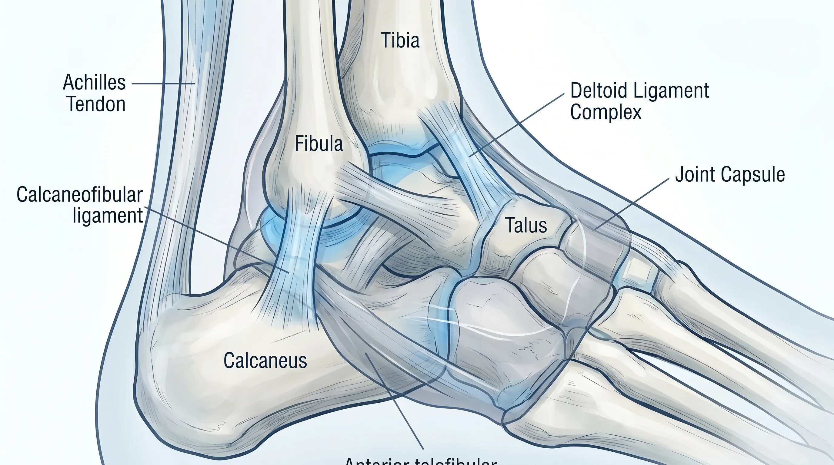
The etiology of ankle masses spans a wide spectrum, ranging from benign cystic formations to inflammatory nodules and degenerative changes. Accurate identification begins with understanding how different tissues within and around the ankle joint respond to various internal and external stimuli. Each category of mass presents distinct clinical characteristics, growth patterns, and symptomatic profiles that guide further evaluation and management strategies.
Free calculatorBMI CalculatorCalculate Body Mass IndexOpen the calculatorBenign Soft Tissue Masses
Ganglion cysts represent one of the most common benign lesions encountered in clinical practice, frequently manifesting as a firm, smooth, and occasionally fluctuant mass near the ankle joint capsule or tendon sheaths according to Mayo Clinic. These fluid-filled sacs contain viscous synovial fluid that herniates through the joint lining or tendon covering, often triggered by repetitive microtrauma, joint instability, or localized irritation. While many ganglion cysts remain asymptomatic and resolve spontaneously, others may cause discomfort during specific movements or compress adjacent neurovascular structures, leading to tingling or numbness in the surrounding foot. Another prevalent soft-tissue formation is the lipoma, a benign proliferation of mature adipocytes that typically presents as a soft, mobile, and painless subcutaneous nodule. Lipomas grow slowly over years and rarely undergo malignant transformation, though their location near weight-bearing areas can occasionally lead to mechanical irritation from footwear or pressure during ambulation.
Tendon and Ligament Pathologies
Chronic overuse, acute sprains, or degenerative tendon diseases can precipitate localized thickening or nodule formation along the tendons traversing the ankle region. Tendinopathies involving the Achilles, peroneal, or posterior tibial tendons often develop nodular swellings due to collagen disorganization, neovascularization, and localized edema as detailed by Cleveland Clinic. These tendon nodules may feel firm, exhibit tenderness to palpation, and cause pain during resisted movement or weight-bearing activities. Similarly, post-traumatic scar tissue formation or fibrous bands following severe ligamentous injuries can create palpable irregularities that persist long after the acute healing phase has concluded. Understanding the biomechanical stressors that contribute to these tendon and ligament alterations is crucial for implementing targeted rehabilitation and preventing further structural compromise when evaluating a lump on ankle.
Bone and Joint Abnormalities
Osseous prominences, commonly referred to as bone spurs or osteophytes, frequently develop around the ankle joint as a compensatory response to chronic joint degeneration, osteoarthritis, or prior fractures. These bony outgrowths may feel hard, fixed, and immovable upon examination, often correlating with reduced range of motion and crepitus during joint articulation. In younger populations, osteochondromas—benign cartilage-capped bone tumors—can occasionally arise near the distal tibia or fibula, presenting as painless, slow-growing masses that may alter local biomechanics. Vascular anomalies such as hemangiomas or arteriovenous malformations can also manifest as palpable swellings with characteristic warmth, compressibility, or pulsatility, requiring specialized vascular imaging for accurate diagnosis.
Inflammatory and Systemic Conditions
Rheumatoid arthritis and other autoimmune connective tissue disorders frequently produce rheumatoid nodules around joint margins, characterized by firm, subcutaneous masses containing granulomatous inflammation and fibrinoid necrosis. These nodules often indicate active systemic disease and may coincide with morning stiffness, polyarticular swelling, and elevated inflammatory markers, per the National Institutes of Health. Gouty tophi represent another inflammatory etiology, resulting from monosodium urate crystal deposition in periarticular tissues following years of uncontrolled hyperuricemia. According to the CDC, these chalky, yellowish masses typically develop around the Achilles insertion or lateral malleolus, causing significant discomfort during footwear application and potentially eroding underlying bone if left untreated. Infectious processes, including septic bursitis or deep soft-tissue abscesses, can also present as rapidly enlarging, erythematous, and exquisitely tender masses requiring urgent antimicrobial therapy and surgical drainage.
When to Seek Immediate Medical Attention
Recognizing clinical red flags is paramount when evaluating any new or changing mass, as certain symptoms warrant expedited medical assessment. Rapid enlargement over days to weeks, particularly without a clear traumatic antecedent, raises suspicion for aggressive pathology and necessitates prompt imaging. Persistent pain that disrupts sleep, worsens at rest, or remains unresponsive to conservative analgesia may indicate nerve compression, vascular compromise, or underlying neoplastic activity. Skin changes overlying the mass, such as ulceration, spontaneous drainage, hyperpigmentation, or temperature alterations, should prompt immediate dermatological and orthopedic evaluation. Systemic manifestations, including unexplained weight loss, night sweats, persistent fever, or profound fatigue, suggest potential systemic involvement requiring comprehensive diagnostic workup. Additionally, neurological deficits such as progressive numbness, tingling, motor weakness, or altered gait patterns indicate neurovascular impingement that demands urgent intervention to prevent irreversible functional impairment.
Diagnostic Process and Imaging Techniques
A systematic diagnostic approach ensures accurate characterization of the mass, guiding appropriate therapeutic interventions while minimizing unnecessary procedures. Clinical evaluation integrates patient history, physical examination findings, and targeted diagnostic studies to establish a definitive diagnosis and rule out concerning pathology.
Physical Examination and Medical History
The initial clinical assessment focuses on documenting the onset, duration, and evolution of the mass alongside associated symptoms and relevant medical history. Palpation techniques evaluate consistency (soft, firm, or hard), mobility (fixed versus mobile), margins (well-defined versus infiltrative), and tenderness. Transillumination testing can differentiate cystic from solid lesions, as fluid-filled structures typically allow light penetration. Assessment of adjacent joint stability, tendon integrity, and neurovascular status provides crucial context for determining structural involvement. Reviewing prior injuries, occupational demands, athletic activities, systemic illnesses, and medication use helps identify predisposing factors and guides further diagnostic testing.
Ultrasound, MRI, and Radiography
Ultrasonography serves as an excellent first-line imaging modality due to its accessibility, cost-effectiveness, and superior visualization of superficial soft-tissue structures. Dynamic scanning capabilities allow real-time assessment of tendon movement, fluid dynamics, and vascularity using color Doppler imaging. Magnetic resonance imaging (MRI) provides unparalleled soft-tissue contrast, enabling detailed evaluation of mass composition, internal architecture, tendon involvement, bone marrow edema, and neurovascular relationships. MRI is particularly valuable for preoperative planning and characterizing indeterminate lesions. Conventional radiography remains essential for evaluating underlying bony architecture, detecting osteophytes, fracture malunion, calcifications within the mass, or aggressive bone destruction suggestive of malignant processes.
Laboratory Testing and Biopsy Procedures
When clinical suspicion points toward inflammatory, infectious, or neoplastic etiologies, targeted laboratory studies including complete blood counts, erythrocyte sedimentation rate, C-reactive protein, rheumatoid factor, anti-CCP antibodies, and serum uric acid levels provide critical diagnostic insights. Core needle or excisional biopsy may be indicated for lesions demonstrating atypical features, rapid growth, or imaging characteristics suggestive of malignancy. Histopathological analysis confirms tissue origin, cellular differentiation, and proliferative activity, ensuring precise diagnostic classification and appropriate therapeutic planning.
Evidence-Based Treatment Options
Management strategies are tailored to the specific diagnosis, symptom severity, functional impact, and patient preferences, emphasizing a stepwise approach that prioritizes conservative interventions before considering invasive procedures.
Conservative Management and Observation
Asymptomatic or minimally symptomatic masses often require only periodic monitoring and activity modification to prevent mechanical irritation. Clinicians frequently recommend watchful waiting for ganglion cysts and lipomas that do not impair function or cause significant discomfort. Education regarding natural disease progression, expected outcomes, and warning signs ensures patients remain informed and prepared for potential changes. Modifying exercise routines, avoiding repetitive stress, and utilizing supportive devices can effectively mitigate symptom exacerbation while maintaining overall joint health. For individuals managing a lump on ankle conservatively, regular self-assessment and documentation of changes prove invaluable for tracking disease trajectory.
Aspiration and Intra-Articular Injections
For symptomatic cystic lesions, ultrasound-guided aspiration offers immediate volume reduction and symptom relief, though recurrence rates remain notably high without adjunctive therapies. Combining aspiration with intralesional corticosteroid injections can decrease local inflammation and reduce recurrence probability by modifying the cyst wall environment. Sclerotherapy using agents like hypertonic saline or sodium tetradecyl sulfate represents an emerging alternative for refractory cases, promoting cyst wall fibrosis and fluid reabsorption. These minimally invasive procedures provide valuable therapeutic options for patients seeking symptom control without surgical intervention.
Surgical Excision and Rehabilitation
Surgical intervention becomes necessary when conservative measures fail, neurological compromise exists, malignancy is suspected, or the mass causes significant functional impairment. Open or arthroscopic excision aims for complete removal with clear margins to minimize recurrence risk while preserving critical anatomical structures. Intraoperative nerve monitoring and meticulous hemostasis enhance safety and optimize functional outcomes. Postoperative rehabilitation emphasizes protected weight-bearing, progressive range-of-motion exercises, edema management, and gradual strengthening protocols. Physical therapy integration restores proprioception, improves dynamic stability, and facilitates safe return to athletic or occupational activities, ensuring comprehensive recovery and long-term joint preservation.
Home Care and Self-Management Strategies
Implementing evidence-based self-care practices empowers patients to actively participate in their recovery while minimizing symptom progression and preventing recurrence. Consistent adherence to structured home management protocols significantly enhances treatment efficacy and overall functional outcomes.
The Modified RICE Protocol and Activity Adjustment
The foundational RICE protocol—Rest, Ice, Compression, and Elevation—remains highly relevant for managing acute inflammation and mechanical irritation surrounding ankle masses per Mayo Clinic first-aid guidelines. Modified implementation involves brief, frequent cold therapy sessions to reduce localized swelling without causing tissue ischemia, combined with lightweight compression garments that support venous return without restricting arterial perfusion. Strategic activity modification requires temporarily avoiding high-impact exercises, uneven terrain, or footwear that exerts direct pressure on the affected area. Low-impact alternatives such as swimming, stationary cycling, or elliptical training maintain cardiovascular fitness while minimizing mechanical stress on the ankle joint during the healing phase.
Footwear Optimization and Orthotic Support
Selecting appropriate footwear plays a pivotal role in managing ankle masses and preventing mechanical aggravation. Shoes with wide toe boxes, cushioned midsoles, flexible uppers, and adequate arch support distribute plantar pressures evenly and reduce friction over vulnerable structures. Custom or over-the-counter orthotic inserts can correct biomechanical imbalances, improve hindfoot alignment, and decrease abnormal tendon loading that contributes to nodule formation. Avoiding excessively tight lacing, rigid heel counters, or minimalist footwear styles ensures optimal joint mechanics and minimizes localized irritation around existing masses.
Targeted Physical Therapy and Mobility Drills
Structured exercise programs focusing on proprioceptive training, eccentric strengthening, and controlled mobility restoration significantly improve ankle joint resilience and functional capacity. Isometric strengthening of peroneal and tibial muscles enhances dynamic joint stabilization, reducing abnormal tendon strain during ambulation. Range-of-motion exercises including ankle alphabet writing, controlled dorsiflexion/plantarflexion progressions, and resistance band eversion/inversion movements restore tissue extensibility and prevent joint stiffness. Progressive balance training using unstable surfaces or single-leg stance exercises rebuilds neuromuscular coordination, ensuring safe navigation of daily activities and reducing future injury risk. For comprehensive visual guidance on proper technique, refer to the educational demonstration below.

Prevention and Long-Term Ankle Health
Proactive strategies focused on biomechanical optimization, injury prevention, and lifestyle modification significantly reduce the likelihood of developing new ankle masses while preserving long-term joint function. Implementing consistent preventive measures ensures sustained musculoskeletal health across all age groups and activity levels.
Regular stretching and mobility routines targeting the calf musculature, Achilles tendon, and peroneal complex maintain optimal tissue elasticity and prevent excessive tension that predisposes to tendinopathy and cyst formation. Gradual progression when introducing new exercise programs, modifying training volume, or transitioning to different athletic surfaces minimizes cumulative microtrauma and allows adaptive tissue remodeling. Maintaining a healthy body weight reduces excessive joint loading, decreasing mechanical stress on articular cartilage, tendons, and supporting ligaments. Incorporating anti-inflammatory dietary patterns rich in omega-3 fatty acids, antioxidants, and adequate hydration supports tissue repair processes and modulates systemic inflammatory responses. Routine foot and ankle self-examinations enable early detection of new swellings, skin changes, or functional alterations before they progress to symptomatic stages. Prompt consultation with healthcare providers ensures timely evaluation, accurate diagnosis, and appropriate management, preventing complications and optimizing recovery trajectories. Commitment to consistent biomechanical care, intelligent activity modification, and proactive medical follow-up establishes a foundation for lifelong ankle health and uninterrupted mobility.
Comparative Analysis of Ankle Masses
Understanding the clinical distinctions between benign and concerning formations assists patients and clinicians in determining appropriate monitoring frequency and intervention urgency. The following comparison outlines key differentiating characteristics that inform diagnostic decision-making and treatment prioritization.
| Feature | Benign Masses (e.g., Ganglion Cyst, Lipoma) | Concerning Pathologies (e.g., Malignancy, Infection) |
|---|---|---|
| Growth Pattern | Slow, stable over months to years | Rapid progression over weeks to days |
| Mobility | Mobile or slightly movable under skin | Fixed, adherent to underlying structures |
| Consistency | Soft, fluctuant, or rubbery | Firm, hard, or irregular |
| Pain Level | Often painless or mild mechanical discomfort | Persistent, rest pain, or severe tenderness |
| Skin Appearance | Normal overlying skin | Redness, warmth, ulceration, or drainage |
| Systemic Symptoms | None present | Fever, weight loss, night sweats, fatigue |
| Response to Rest | Improves or remains stable | Continues to enlarge or worsen despite rest |
Frequently Asked Questions
Can a lump on ankle disappear without medical treatment?
Many benign soft-tissue formations, particularly ganglion cysts and small lipomas, frequently resolve spontaneously without intervention. Clinical observations indicate that approximately thirty to fifty percent of ganglion cysts disappear within one to two years through natural fluid reabsorption and wall remodeling. Watchful waiting combined with activity modification often suffices for asymptomatic lesions. However, persistent or progressively enlarging masses require professional evaluation to rule out underlying pathology that necessitates targeted intervention.
What imaging test provides the most accurate diagnosis for an ankle mass?
Magnetic resonance imaging remains the gold standard for comprehensive soft-tissue characterization, offering exceptional contrast resolution that differentiates cystic, solid, fatty, and vascular components. Ultrasound serves as an excellent initial modality for dynamic evaluation of superficial masses and tendon relationships. Conventional radiography effectively identifies underlying osseous abnormalities, calcifications, or joint degeneration. Healthcare providers typically select imaging sequences based on clinical presentation, lesion depth, and suspected tissue origin to ensure precise diagnostic accuracy.
Is it safe to attempt home drainage or puncture of an ankle cyst?
Self-directed drainage attempts are strongly discouraged due to significant risks of infection, neurovascular injury, tissue scarring, and cyst recurrence. Non-sterile techniques introduce pathogenic bacteria into deep soft tissues, potentially triggering severe cellulitis or septic arthritis. Professional aspiration utilizes ultrasound guidance, sterile technique, and appropriate needle selection to maximize safety and therapeutic efficacy. If you suspect a cystic formation, consult a qualified healthcare provider who can perform evidence-based interventions under controlled clinical conditions.
How long does surgical recovery take for ankle mass removal?
Recovery timelines vary based on lesion size, anatomical location, surgical technique, and individual healing capacity. Superficial mass excisions typically allow protected weight-bearing within one to two weeks, with suture removal occurring around ten to fourteen days postoperatively. Comprehensive soft-tissue healing requires four to six weeks, during which physical therapy emphasizes gradual range-of-motion restoration and progressive strengthening. Full return to high-impact activities or competitive sports generally occurs between eight to twelve weeks, contingent upon functional assessments and clinician clearance.
When should I consult a specialist versus my primary care provider?
Initial evaluation by a primary care clinician is appropriate for most newly discovered masses, as they can perform initial assessments, order appropriate imaging, and implement conservative management. Referral to an orthopedic surgeon, podiatrist, or rheumatologist becomes necessary when lesions demonstrate rapid growth, cause significant functional impairment, involve neurovascular structures, or require surgical intervention. Complex cases with suspected malignancy, severe inflammatory arthritis, or intricate anatomical involvement warrant multidisciplinary specialist consultation to ensure comprehensive diagnostic accuracy and optimal treatment outcomes.
Conclusion
Discovering a lump on your ankle often triggers understandable concern, but understanding the underlying causes and evidence-based management pathways provides clarity and reassurance. Most ankle masses stem from benign processes that respond effectively to conservative care, targeted interventions, or minimally invasive procedures when necessary. By recognizing warning signs, seeking timely professional evaluation, and adhering to structured self-management strategies, individuals can confidently navigate their diagnostic journey while preserving long-term joint function and mobility. Implementing proactive prevention measures, optimizing footwear mechanics, and engaging in consistent rehabilitation exercises establishes a sustainable foundation for ongoing ankle health. Whether your situation requires simple observation or specialized intervention, partnering with qualified healthcare professionals ensures personalized, safe, and effective care tailored to your unique anatomical and lifestyle needs.
How we work
Researched and drafted with AI assistance, then reviewed by a human editor against the cited sources.
This article is general health information, not medical advice. Consult a qualified healthcare professional about your own situation.