Knee Hyperlaxity: Causes, Diagnosis, Stabilization Exercises & Long-Term Joint Health
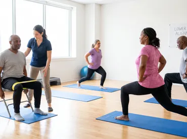
Do you ever feel like your knees just bend backward or shift slightly during everyday movements like walking down stairs or standing up from a chair? For some individuals, the joints exhibit an extraordinary range of motion that goes far beyond typical flexibility. This phenomenon, clinically recognized as knee hyperlaxity, is not merely a sign of being limber. While it can offer temporary advantages in certain athletic or artistic disciplines like gymnastics, dance, and martial arts, it frequently predisposes individuals to chronic instability, accelerated cartilage wear, recurrent ligamentous strain, and complex musculoskeletal pain syndromes. Understanding the underlying biomechanics, learning how to identify subtle warning signs, and implementing a targeted, evidence-based management strategy are absolutely essential for preserving long-term joint health and maintaining an active, pain-free lifestyle.
If you suspect that your joints possess excessive mobility, you are not navigating this alone. Millions of people worldwide navigate daily life with hypermobile connective tissue, often completely unaware that their natural flexibility requires specific care, deliberate strengthening, and protective biomechanical adjustments. This comprehensive guide explores the science behind excessive joint mobility, provides actionable rehabilitation protocols backed by clinical research, and equips you with practical strategies to stabilize your knees, reduce injury risk, and optimize overall musculoskeletal function for decades to come.
What Is Knee Hyperlaxity?
Knee hyperlaxity refers to an abnormal increase in the physiological range of motion around the knee joint, caused primarily by excessive elasticity or structural laxity in the surrounding ligaments, tendons, and joint capsule. Unlike muscular flexibility, which is highly trainable and generally beneficial, ligamentous laxity is largely governed by the composition and arrangement of collagen fibers within connective tissues. Ligaments act as passive stabilizers, functioning like high-tensile cables that guide bone alignment and prevent joints from moving beyond their safe anatomical boundaries. When these structures are inherently more pliable, the knee relies disproportionately on active muscular stabilization and neuromuscular coordination to maintain proper tracking during movement.
Free calculatorBMI CalculatorCalculate Body Mass IndexOpen the calculatorThe condition exists along a broad spectrum. On one end, generalized joint hypermobility (GJH) may be entirely asymptomatic, allowing individuals to lead completely normal, unrestricted lives. On the other end, hypermobility spectrum disorders (HSD) and hereditary connective tissue conditions such as Ehlers-Danlos syndrome (hypermobility type) can present with widespread systemic manifestations, including chronic pain, fatigue, joint subluxations, and gastrointestinal or cardiovascular involvement. Recognizing where you fall on this spectrum is the critical first step toward personalized intervention. For authoritative clinical definitions and diagnostic criteria, you can review the comprehensive guidelines published by the American Academy of Orthopaedic Surgeons AAOS Clinical Guidelines and peer-reviewed literature on connective tissue mechanics available through the National Institutes of Health NIH Connective Tissue Research.
The Biomechanics of Hypermobile Knees
To fully grasp why knee hyperlaxity matters, it is essential to understand joint biomechanics. A healthy knee functions as a modified hinge joint with six degrees of freedom: flexion/extension, varus/valgus angulation, internal/external rotation, and anterior/posterior/posterolateral translation. In a typical joint, passive restraints (ligaments, menisci, capsule) share the load with active stabilizers (quadriceps, hamstrings, gastrocnemius, gluteals). When passive restraints are compromised by excessive elasticity, the active stabilizers must compensate continuously. This creates a neuromuscular overload scenario.
Muscles designed primarily for movement (phasic muscles) are forced to act as postural stabilizers, leading to early fatigue, altered firing patterns, and compensatory movement strategies. For example, individuals with knee hyperlaxity often exhibit quadriceps dominance, over-reliance on the iliotibial band, or excessive tibial internal rotation. Over time, these compensations can lead to patellofemoral pain syndrome, iliotibial band friction, premature meniscal degeneration, and secondary osteoarthritis. The proprioceptive deficit is equally critical. Ligaments house mechanoreceptors that send spatial feedback to the cerebellum. Excessively lax ligaments provide delayed or inaccurate signals, resulting in poor joint position sense and delayed reflexive muscle contractions during sudden directional changes or uneven terrain navigation.
Genetic vs. Acquired Joint Laxity
The etiology of knee hyperlaxity generally falls into two primary categories: constitutional (genetic/hereditary) and acquired (environmental/hormonal/traumatic). Constitutional hyperlaxity is predominantly inherited through autosomal dominant patterns, though incomplete penetrance and variable expressivity mean that family members may exhibit vastly different degrees of laxity and symptom severity. Mutations in genes regulating collagen synthesis, particularly Type I, III, and V collagen, directly impact the tensile strength and cross-linking of connective tissues.
Acquired hyperlaxity, conversely, develops over time due to external or systemic factors. Hormonal fluctuations play a massive role; relaxin and estrogen levels significantly increase during pregnancy, temporarily reducing ligamentous stiffness to prepare the pelvis for childbirth. This systemic relaxation often affects all synovial joints, including the knees, leaving postpartum individuals temporarily vulnerable to instability and injury. Additionally, repetitive microtrauma, prior joint dislocations, prolonged immobilization followed by aggressive rehabilitation, and certain pharmacological agents (like fluoroquinolone antibiotics) can degrade ligament integrity. Distinguishing between these origins is crucial, as genetic hyperlaxity typically requires lifelong neuromuscular conditioning, while acquired forms may partially resolve with targeted tissue remodeling and time.
Recognizing the Signs and Symptoms
Knee hyperlaxity does not always announce itself with dramatic pain. In fact, many individuals remain asymptomatic for years, only noticing subtle quirks in their movement patterns or experiencing discomfort after prolonged activity. However, as the cumulative stress on joint structures increases, the clinical picture becomes increasingly pronounced. Recognizing the early warning signs allows for proactive intervention before irreversible structural changes occur.
Common Clinical Presentations
The most recognizable physical marker of knee hyperlaxity is hyperextension, often visually described as the knee bending backward or exhibiting a noticeable recurvatum posture during standing. Beyond this obvious sign, individuals frequently report a vague sense of the knee feeling loose, untrustworthy, or prone to buckling. Daily activities that involve weight transfer, such as descending stairs, pivoting while walking, or standing up from a seated position, may trigger sudden sensations of joint shifting or near-subluxation.
Pain presentation is typically mechanical and activity-dependent. Rather than inflammatory joint pain, individuals describe aching along the joint line, sharp discomfort behind the patella, or deep fatigue within the quadriceps and calf muscles due to constant compensatory contraction. Swelling may occur intermittently following unaccustomed exertion, often described as a heavy or stiff sensation rather than acute effusion. Fatigue is another frequently overlooked symptom; the neuromuscular system expends significantly more energy maintaining stability, leading to disproportionate muscle exhaustion compared to peers. Many patients also develop secondary conditions, including chronic lower back pain from altered pelvic tilt, foot pronation issues from compensatory arch collapse, and widespread myofascial trigger points.
When Hyperlaxity Becomes Pathological
Not all hypermobility is pathological. The medical community differentiates between asymptomatic hypermobility (a benign anatomical variant) and symptomatic hypermobility (a functional impairment requiring intervention). The transition from benign to pathological typically occurs when the body's compensatory mechanisms begin to fail, leading to structural breakdown. Key indicators of pathological progression include recurrent joint subluxations or partial dislocations, frequent soft tissue injuries (sprains, strains, tendinopathies), progressive cartilage thinning detectable via imaging, chronic pain unresponsive to standard anti-inflammatory protocols, and significant functional limitation affecting activities of daily living.
Another critical warning sign is the development of widespread central sensitization, where chronic joint strain alters pain processing in the nervous system, leading to disproportionate pain responses to non-threatening stimuli. If you experience locking, giving way, persistent effusion, night pain, or a noticeable decrease in joint stability despite adequate rest and basic strengthening, clinical evaluation is strongly warranted. The Hypermobility Association and related patient advocacy groups provide excellent resources for distinguishing normal flexibility from clinically significant laxity The Ehlers-Danlos Society Resources.
Diagnostic Process and Medical Evaluation
A thorough diagnostic approach to knee hyperlaxity combines clinical observation, standardized scoring systems, functional movement analysis, and selective imaging to rule out confounding pathologies. The process is largely clinical, meaning it relies heavily on the examiner's expertise and the patient's symptom narrative rather than advanced technological scans.
Physical Examination Techniques
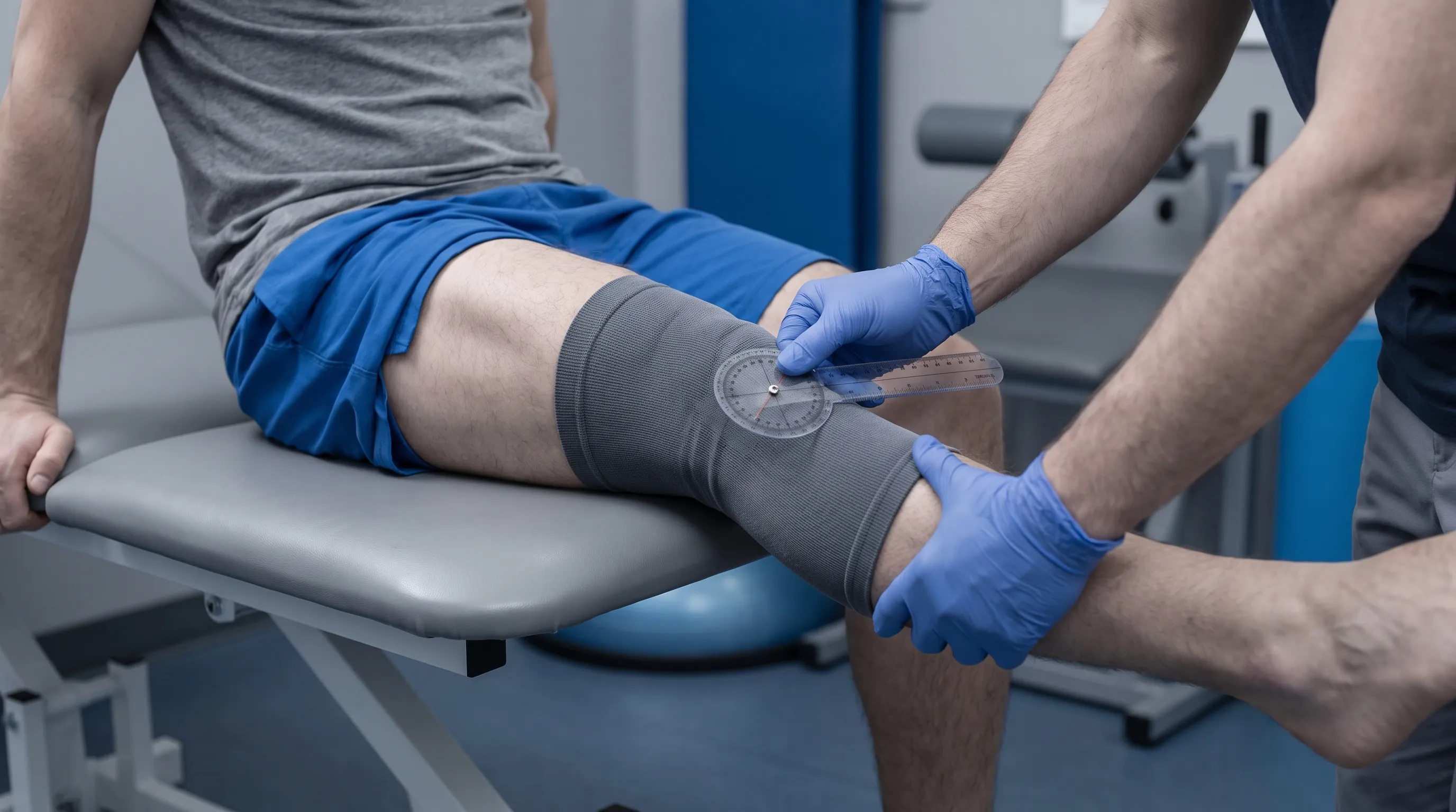
The cornerstone of hypermobility assessment is the Beighton Score, a nine-point screening tool widely utilized in rheumatology and orthopedics to quantify generalized joint laxity. The test evaluates bilateral flexibility across specific joints: passive apposition of the little fingers beyond 90 degrees, passive hyperextension of the thumbs to touch the forearms, bilateral elbow hyperextension greater than 10 degrees, bilateral knee hyperextension greater than 10 degrees, and the ability to bend forward with knees straight and palms flat on the floor. A score of 4/9 or higher in adults typically indicates generalized hypermobility, while pediatric thresholds are slightly higher due to natural childhood flexibility.
Beyond the Beighton assessment, clinicians perform targeted knee-specific provocation tests. The anterior and posterior drawer tests evaluate cruciate ligament integrity, though in hyperlax patients, these may yield false-positive laxity findings due to inherent tissue elasticity rather than traumatic rupture. The valgus and varus stress tests assess collateral ligament stability. A crucial component is observing functional movement patterns. Clinicians look for dynamic valgus collapse (knee caving inward), excessive tibial rotation, quadriceps avoidance gait, and abnormal foot pronation during single-leg stance and squatting maneuvers. The Lower Limb Assessment Score (LLAS) and similar functional scales often supplement clinical exams to quantify instability severity.
Imaging and Differential Diagnosis
Radiographs and magnetic resonance imaging (MRI) are generally normal in isolated knee hyperlaxity. Their primary utility lies in differential diagnosis and ruling out structural mimics. Plain X-rays can identify osseous alignment anomalies, early degenerative joint changes, or patellar tracking deviations. MRI provides exquisite detail of soft tissue, allowing clinicians to differentiate between true ligamentous laxity and partial tears, meniscal degeneration, or chondral lesions. Ultrasound may occasionally be employed for dynamic assessment of tendon gliding and real-time ligament behavior under stress.
Differential diagnosis is critical. Conditions such as generalized joint hypermobility syndrome, hypermobile Ehlers-Danlos syndrome, Marfan syndrome, rheumatoid arthritis, and acquired ligamentous insufficiency from trauma share overlapping symptoms. Blood tests may be ordered if systemic connective tissue disorders are suspected, looking for inflammatory markers or specific genetic panels. A multidisciplinary approach involving rheumatologists, orthopedic specialists, and licensed physical therapists often yields the most accurate diagnosis and tailored management plan.
| Assessment Tool | What It Measures | Clinical Significance |
|---|---|---|
| Beighton Score | Generalized joint hypermobility (9 points) | Scores ≥4 in adults indicate systemic laxity; predicts joint injury risk |
| Valgus/Varus Stress Test | Collateral ligament stability (MCL/LCL) | Identifies directional instability and excessive medial/lateral gapping |
| Anterior/Posterior Drawer | Cruciate ligament tension (ACL/PCL) | Differentiates traumatic laxity from constitutional hyperlaxity |
| Dynamic Gait Observation | Neuromuscular control during movement | Reveals compensatory patterns like dynamic valgus, hyperextension, or pronation |
| Lower Limb Assessment Score (LLAS) | Functional instability and joint-specific laxity | Guides rehabilitation intensity and predicts need for orthotic intervention |
Managing Knee Hyperlaxity: Conservative Approaches
The vast majority of individuals with symptomatic knee hyperlaxity achieve excellent functional outcomes through conservative, non-surgical management. The treatment philosophy centers on transforming the knee from a passive, ligament-dependent joint into an active, muscle-supported structure. This requires a paradigm shift from stretching and passive therapy toward targeted strengthening, proprioceptive re-education, and intelligent activity modification.
Targeted Strengthening and Stabilization Exercises
Muscular reinforcement is the single most effective intervention for knee hyperlaxity. The goal is not merely to increase bulk, but to enhance neuromuscular recruitment patterns, improve rate of force development, and build endurance within the stabilizing musculature. Exercises should prioritize closed-chain movements (feet fixed) over open-chain movements (feet free), as closed-chain activities promote co-contraction of agonist and antagonist muscles, significantly enhancing joint compression and stability.
Begin with foundational movements that emphasize proper alignment and control over range of motion. Wall sits are excellent for building quadriceps endurance without shearing forces. Focus on maintaining a slight micro-bend in the knees rather than locking into extension. Glute bridges activate the posterior chain, addressing the common gluteal inhibition seen in hypermobile individuals. Progress to single-leg glute bridges to introduce proprioceptive challenge. Clamshells and side-lying leg raises strengthen the gluteus medius, which is critical for preventing dynamic knee valgus. When incorporating squats, utilize box squats or controlled mini-squats to prevent descending beyond 90 degrees initially, gradually building depth as stability improves.
Eccentric training deserves special emphasis. Eccentric contractions (muscle lengthening under load) stimulate collagen synthesis and improve tendon stiffness, directly addressing the tissue laxity component. Nordic hamstring progressions, eccentric calf raises, and slow-tempo step-downs are highly effective. Always prioritize form over load. If form deteriorates or joint alignment shifts, reduce resistance or regress the movement. A certified physical therapist can provide individualized progression models based on your specific biomechanical profile and tissue tolerance. For detailed exercise protocols and clinical recommendations, consult the American Physical Therapy Association's joint stability guidelines APTA Rehabilitation Resources.
Proprioceptive Training and Balance Work
Proprioception is the body's internal GPS system. In knee hyperlaxity, this system requires frequent recalibration. Balance training should progress through structured phases: stable surface to unstable surface, double-leg to single-leg, eyes open to eyes closed, and static to dynamic. Begin with simple tandem stance (heel-to-toe) and single-leg standing on firm ground, aiming for 30-60 seconds per side with perfect posture. Progress to foam pads, BOSU balls, or wobble boards, focusing on maintaining knee alignment over the second toe.
Incorporate reactive neuromuscular training. Light perturbations, such as gentle taps to the shoulder or thigh while balancing, force the neuromuscular system to react quickly and re-stabilize the joint. Agility drills like controlled lateral shuffles, figure-eight walking, and step-over exercises improve multi-directional stability. Plyometric training should be approached cautiously and only introduced after months of foundational strength and balance work. Low-level hops with emphasis on soft, silent landings teach the muscles to absorb impact efficiently, reducing ligamentous strain. Consistency is paramount; proprioceptive gains diminish rapidly without regular maintenance. Even ten minutes of daily balance practice yields substantial long-term benefits.
Bracing and Supportive Footwear
External support serves as a valuable adjunct to internal strengthening, particularly during the initial rehabilitation phases or high-demand activities. However, the type and duration of bracing significantly impact long-term outcomes. Rigid hinged braces should be reserved for acute instability episodes or post-surgical protection, as continuous use promotes quadriceps atrophy and further proprioceptive degradation. Neoprene sleeves, kinesiology taping, and compression garments are generally preferred for daily management. They provide mild proprioceptive feedback, compress soft tissue to reduce mechanoreceptor noise, and offer psychological confidence without restricting movement.
Kinesiology tape applied by a trained professional can facilitate specific muscle firing patterns, gently guide patellar tracking, and provide sensory input that reduces perceived instability. Footwear selection is equally critical. Hypermobile individuals frequently exhibit pes planus (flat feet) or excessive foot pronation, which internally rotates the tibia and compromises knee alignment. Shoes with moderate arch support, firm heel counters, and structured midsoles provide a stable foundation for the entire kinetic chain. Custom or over-the-counter orthotics may be necessary for severe biomechanical deviations. The goal is always to use external supports as training wheels, gradually weaning off as internal muscular control improves.
Advanced Interventions and Medical Treatments
While conservative management resolves symptoms for most individuals, a subset of patients with severe, refractory knee hyperlaxity may require advanced therapeutic interventions. These are typically considered when comprehensive physical therapy fails to restore functional stability, when recurrent subluxations cause progressive joint damage, or when pain significantly impairs quality of life despite optimal rehabilitation.
When Physical Therapy Is Not Enough
Persistent symptoms despite months of dedicated strengthening, proprioceptive training, and lifestyle modification warrant advanced evaluation. In these cases, clinicians may explore regenerative medicine options. Prolotherapy involves injecting an irritant solution (commonly dextrose) into lax ligaments to stimulate localized inflammation and subsequent tissue remodeling. The inflammatory cascade recruits fibroblasts, which lay down new collagen, effectively tightening and strengthening the ligamentous structure over several treatment sessions. Platelet-rich plasma (PRP) therapy utilizes concentrated growth factors from the patient's own blood to accelerate connective tissue repair and modulate chronic inflammation. While promising, evidence for regenerative injections in hypermobility remains evolving, and these treatments should only be administered by board-certified physicians experienced in managing connective tissue disorders. Comprehensive reviews of these modalities are available through the Journal of Orthopaedic Surgery and Research Prolotherapy and PRP Clinical Evidence.
Surgical Considerations and Post-Op Rehabilitation
Surgical intervention for isolated knee hyperlaxity is exceptionally rare and generally contraindicated. Operating on inherently lax tissue often yields disappointing results, as the repaired or reconstructed structures remain prone to stretching under the same biomechanical forces. However, surgery becomes necessary when hyperlaxity leads to specific, localized structural failures. For instance, severe, recurrent patellar instability requiring medial patellofemoral ligament (MPFL) reconstruction, advanced meniscal tears causing mechanical locking, or end-stage osteoarthritis necessitating partial or total knee arthroplasty may be indicated.
Post-operative rehabilitation in hypermobile patients requires specialized protocols. Surgeons and therapists must balance tissue protection with early mobilization. Weight-bearing restrictions are often more conservative, and bracing periods extended. The emphasis shifts aggressively to restoring proprioception and preventing post-surgical muscular atrophy. Patients must be educated that surgery corrects acute structural damage but does not cure underlying hyperlaxity. Lifelong neuromuscular maintenance remains essential to protect surgical repairs and prevent contralateral or adjacent joint breakdown.
Lifestyle Modifications and Long-Term Joint Protection
Managing knee hyperlaxity extends far beyond the clinic or gym. It requires a holistic lifestyle approach that integrates ergonomic awareness, activity pacing, nutritional optimization, and psychological resilience. Long-term joint protection hinges on daily habits that minimize cumulative microtrauma and maximize tissue resilience.
Activity Modification and Ergonomics
Not all activities are created equal when joints are inherently mobile. High-impact sports, repetitive jumping, and aggressive end-range stretching should generally be minimized or modified. Instead, favor low-impact, controlled resistance activities like swimming, cycling, Pilates, and controlled strength training. If you participate in sports requiring agility or contact, invest in quality protective gear, prioritize proper warm-up routines emphasizing neuromuscular activation, and schedule adequate recovery periods. Pacing is crucial; learn to recognize early fatigue signals and rest before joint instability occurs.
Ergonomic adjustments in daily life significantly reduce joint strain. Avoid prolonged standing on hard surfaces; use anti-fatigue mats and alternate weight shifts. When sitting, ensure feet are flat, knees are slightly below hips, and chairs provide adequate lumbar support to maintain pelvic neutrality. Lift objects using proper biomechanics: hinge at the hips, engage the core, keep loads close to the body, and never twist while bearing weight. Modify exercise routines to avoid exercises that promote extreme ranges of motion, such as deep lunges with poor alignment or hyperextended yoga poses. Instead, focus on mid-range stability and controlled tempo.

Nutrition for Connective Tissue Health
Diet plays a surprisingly significant role in ligamentous integrity and recovery. Connective tissue relies on specific micronutrients to synthesize and cross-link collagen effectively. Vitamin C is absolutely essential as a cofactor for collagen hydroxylation; deficiency directly impairs tissue strength and delays healing. Incorporate citrus fruits, bell peppers, broccoli, kiwis, and strawberries daily. High-quality protein provides the amino acids glycine, proline, and hydroxyproline necessary for collagen structure. Bone broth, collagen peptides, lean poultry, fish, and legumes are excellent sources.
Omega-3 fatty acids exert potent anti-inflammatory effects, reducing chronic synovial irritation and supporting cell membrane fluidity. Fatty fish, walnuts, flaxseeds, and chia seeds should be dietary staples. Hydration is equally critical; interstitial fluid provides the medium through which nutrients reach avascular tissues like ligaments and menisci. Dehydration increases tissue stiffness and impairs shock absorption. Additionally, consider targeted supplementation under professional guidance. Glucosamine and chondroitin may support cartilage health, while magnesium aids muscle relaxation and neuromuscular signaling. Always prioritize whole foods, as synergistic nutrient interactions outperform isolated supplements. For evidence-based nutritional guidance, refer to the National Institutes of Health dietary supplements database NIH Dietary Supplements for Joint Health.
Frequently Asked Questions
Can knee hyperlaxity be cured completely?
While the underlying genetic or connective tissue structure causing knee hyperlaxity cannot be permanently altered or cured, the condition can be effectively managed. Through targeted neuromuscular re-education, progressive strengthening, and proprioceptive training, individuals can achieve near-complete joint stability, eliminate symptoms, and maintain full functional capacity throughout their lives.
Should I stretch if I have hypermobile knees?
Traditional static stretching is generally not recommended for individuals with pronounced knee hyperlaxity, as it can further compromise already lax ligamentous structures. Instead, the focus should shift to dynamic mobility, eccentric strengthening, and controlled range-of-motion exercises that build muscular support without overextending the joint capsule.
How is knee hyperlaxity diagnosed by a healthcare professional?
Diagnosis typically involves a comprehensive physical examination utilizing the Beighton scoring system, clinical stress tests like the valgus/varus and anterior/posterior drawer tests, and a thorough review of medical history. Imaging studies such as X-rays or MRIs are usually reserved for ruling out structural damage, ligament tears, or degenerative changes rather than diagnosing hyperlaxity itself.
Does bracing worsen knee laxity over time?
Prolonged, continuous use of rigid braces can potentially lead to muscular deconditioning and reduced proprioceptive feedback. However, when used strategically as a temporary support during high-risk activities, rehabilitation phases, or acute instability episodes, bracing serves as a valuable tool. It should always be paired with active muscle strengthening to prevent dependency.
What exercises are safest for strengthening hypermobile knees?
Low-impact, closed-chain exercises are safest and most effective. These include controlled wall sits, mini-squats, step-ups, glute bridges, clamshells, and single-leg balance progressions. The emphasis should always be on controlled eccentric contractions, proper joint alignment, and avoiding end-range hyperextension to safely build dynamic stability.
Conclusion
Navigating life with knee hyperlaxity requires a fundamental shift in how we approach joint health. Rather than viewing excessive mobility as a flaw to be eliminated, it should be recognized as a distinct biomechanical reality that demands specialized care, intelligent training, and consistent protection. The journey from instability to confidence is not about forcing the body into rigid conformity, but rather empowering it with robust muscular support, refined neuromuscular control, and sustainable lifestyle habits. By prioritizing closed-chain strengthening, dedicating time to proprioceptive refinement, making informed choices about external supports, and fueling connective tissues with targeted nutrition, individuals with knee hyperlaxity can absolutely thrive. The knee is remarkably adaptable, and with evidence-based, proactive management, you can preserve joint integrity, minimize injury risk, and maintain an active, vibrant lifestyle well into the future. Start small, stay consistent, and partner with qualified healthcare professionals to build a personalized strategy that honors your body's unique architecture while safeguarding it for the long haul.
How we work
Researched and drafted with AI assistance, then reviewed by a human editor against the cited sources.
This article is general health information, not medical advice. Consult a qualified healthcare professional about your own situation.