Complete Guide to Recovery from Frenectomy: Healing, Care, and Exercises
Navigating the healing process after an oral procedure requires patience, informed care, and a clear understanding of tissue biology. Recovery from frenectomy is a highly structured physiological journey that varies significantly based on age, procedure type, and post-operative compliance. Whether you are an adult addressing long-standing speech impediments, a parent supporting an infant through breastfeeding optimization, or an adolescent correcting orthodontic complications, the foundational principles of oral wound management remain consistent. The human mouth is a uniquely dynamic environment, constantly bathed in saliva, subjected to muscular movement, and exposed to microbial flora. This complex ecosystem accelerates healing but also introduces specific challenges that demand proactive management. By implementing evidence-based wound care protocols, adhering to targeted mobility exercises, and monitoring tissue progression closely, patients can minimize discomfort, prevent complications, and achieve optimal functional outcomes. This comprehensive guide provides actionable strategies, clinical insights, and step-by-step protocols to ensure your recovery progresses smoothly and safely.
Understanding Frenectomy Recovery: What to Expect
The frenectomy procedure involves the precise removal or modification of restrictive connective tissue bands located in the mouth. When the upper lip, lower lip, or tongue is tethered too closely to the gums or floor of the mouth, functional limitations emerge. These can range from infant feeding difficulties and speech articulation errors to gingival recession and orthodontic relapse. Surgical intervention, whether performed with traditional scalpels, electrosurgery, or advanced laser technology, creates a controlled wound that the body must subsequently repair, as detailed in clinical guidelines from the Mayo Clinic. Understanding the biological timeline of this healing process is crucial for setting realistic expectations and reducing post-operative anxiety.
Free calculatorWater Intake CalculatorCalculate daily water needsOpen the calculatorThe physiological response to soft tissue surgery follows a predictable cascade of events. Immediately following tissue incision or ablation, vascular constriction occurs to limit blood loss, followed rapidly by platelet aggregation and fibrin mesh formation. This biological scaffolding protects the exposed nerve endings and initiates the inflammatory phase, which brings oxygenated blood, white blood cells, and growth factors to the surgical site. During recovery from frenectomy, this initial phase typically manifests as mild swelling, localized tenderness, and the development of a protective fibrin layer. Recognizing these as signs of active cellular repair rather than complications is essential for maintaining compliance with post-operative protocols.
The Immediate Post-Procedure Phase (0-24 hours)
The first twenty-four hours represent the most critical window for establishing a stable healing foundation. Anesthesia gradually dissipates during this period, which can trigger heightened sensitivity and low-grade discomfort. Patients should prioritize rest, avoid speaking excessively, and maintain an elevated head position to minimize edema. Applying a clean, damp gauze pad with gentle pressure for ten to fifteen minutes post-procedure helps secure the initial clot. Ice therapy applied externally in fifteen-minute intervals significantly reduces localized swelling by inducing vasoconstriction. During this window, the oral cavity undergoes rapid hemostasis, and the wound edges begin their initial approximation. Hydration should be maintained through gentle sipping, while solid foods remain strictly avoided to prevent mechanical disruption of the fragile clot matrix, aligning with standard post-operative oral surgery recommendations.
The First Week: Healing and Tissue Remodeling
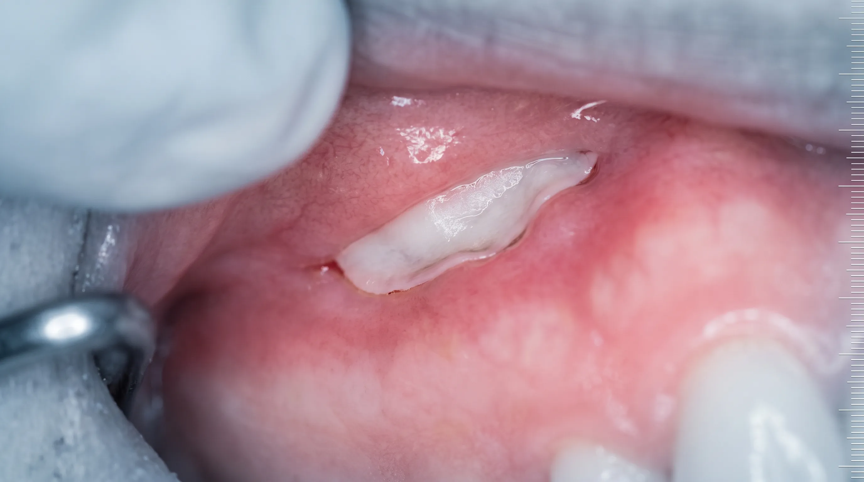
Days two through seven mark the transition from the inflammatory phase into early tissue proliferation. Fibroblasts migrate into the wound bed, synthesizing collagen and establishing the groundwork for new connective tissue formation. During this stage, patients often notice the surgical site developing a white, yellowish, or grayish coating. This fibrinous membrane is entirely normal and serves as a natural biological dressing. It should never be scraped, rubbed, or rinsed aggressively. Swelling typically peaks around day three before gradually subsiding. Saline rinses introduced after the first twenty-four hours help maintain a clean microbial environment without disturbing the delicate regenerative tissue. Mobility exercises commence during this period, usually beginning on day one or two, to prevent the wound edges from migrating together and reforming restrictive tissue.
Long-Term Tissue Adaptation (Weeks 2-4+)
As the healing process matures, collagen fibers undergo reorganization and maturation, aligning along lines of mechanical stress. This remodeling phase is heavily influenced by active stretching and myofunctional retraining. Recovery from frenectomy extends beyond mere wound closure; true success requires neuromuscular adaptation to the newly available range of motion. Speech patterns, swallowing mechanics, and lip seal dynamics require conscious retraining to establish healthy functional habits. By week four, the surgical site typically exhibits mature, pink epithelial tissue with minimal residual tenderness. Full structural strength and complete nerve regeneration, however, can take six to eight weeks, particularly in adults with dense connective tissue matrices. Consistent follow-up appointments ensure that myofunctional therapy progresses appropriately and that no subclinical reattachment occurs.
Essential Wound Care Guidelines for Optimal Healing
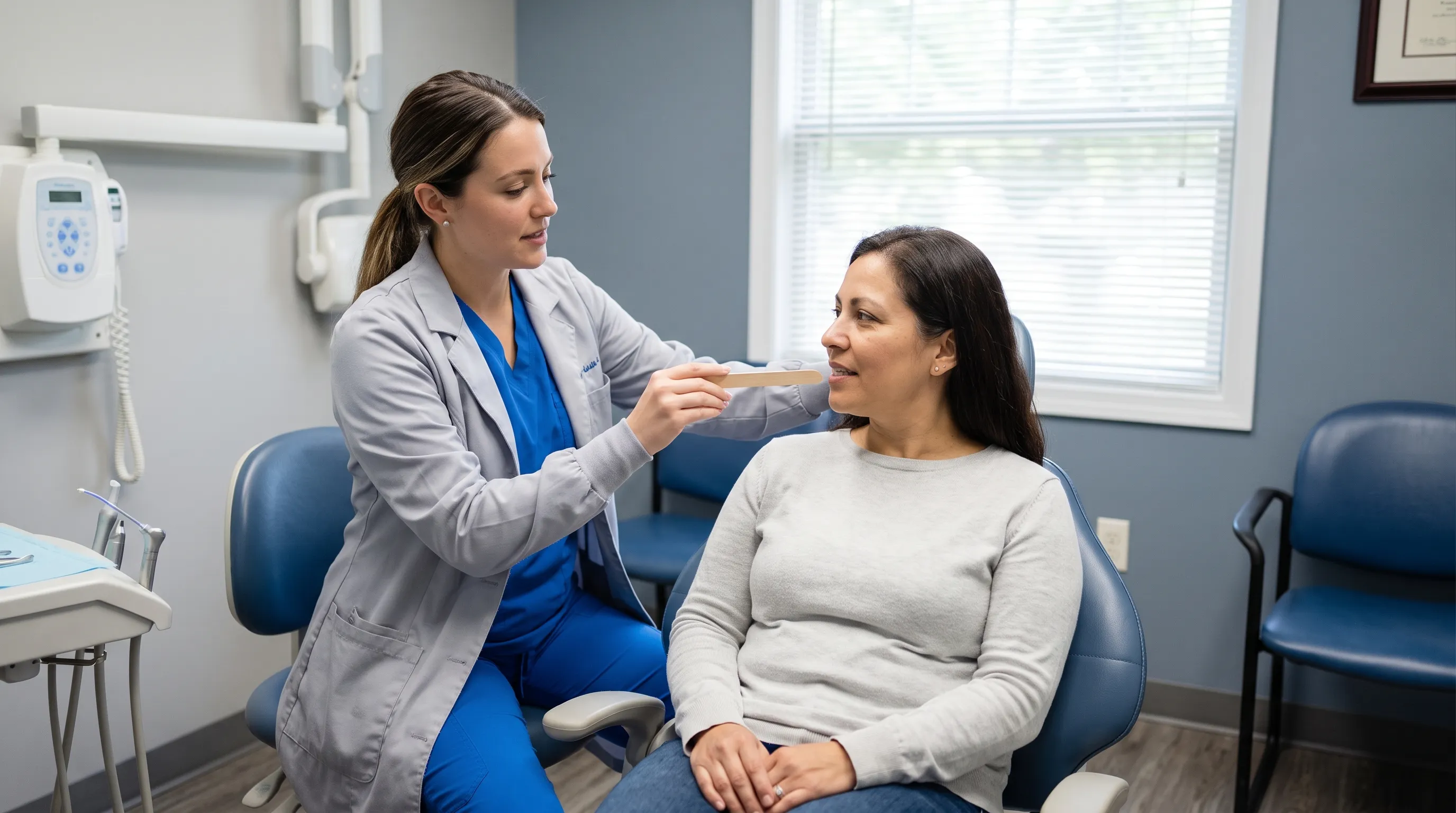
Maintaining a sterile yet moist healing environment is the cornerstone of successful soft tissue recovery in the oral cavity. Unlike skin wounds, intraoral surgical sites cannot be bandaged or kept completely dry, necessitating a specialized hygiene protocol. Effective wound management prevents bacterial colonization, reduces inflammation, and accelerates epithelial migration across the surgical bed, as supported by CDC infection control standards. Implementing a consistent, evidence-based care routine significantly decreases the risk of complications and optimizes the trajectory of recovery from frenectomy.
Proper oral hygiene begins with understanding what to avoid as much as what to actively perform. Commercial mouthwashes containing alcohol or harsh antiseptics disrupt the delicate balance of healing tissues and delay re-epithelialization. Instead, gentle cleansing methods support the body's innate regenerative capabilities while maintaining microbial control. The goal is to remove food debris and reduce biofilm accumulation without applying mechanical force to the healing wound.
Managing Bleeding and Swelling
Minor oozing during the first twenty-four hours is entirely expected and should not cause alarm. To manage persistent bleeding, place a folded piece of moistened gauze or a damp black tea bag directly over the surgical site and apply firm, steady pressure for twenty to thirty minutes. The tannic acid in black tea promotes vasoconstriction and enhances clot stability. Swelling is a natural inflammatory response that can be effectively controlled using cold therapy during the first forty-eight hours. Wrap ice packs in a clean cloth and apply to the external facial region adjacent to the surgical site in fifteen-minute cycles. Avoid heat therapy initially, as vasodilation increases blood flow to the area and exacerbates edema. Sleeping with the head elevated on two pillows further utilizes gravity to reduce fluid accumulation in the oral tissues.
Oral Hygiene Protocols
Gentle cleansing begins twenty-four hours post-procedure. Warm saline rinses, prepared by dissolving half a teaspoon of non-iodized salt in eight ounces of distilled or filtered water, should be swished gently three to four times daily. The key is allowing the solution to passively flow over the surgical area rather than vigorously gargling. Continue routine tooth brushing but avoid the immediate vicinity of the incision site for the first week, gradually reintroducing careful cleaning as tenderness subsides. If chlorhexidine gluconate rinse has been prescribed, use it exactly as directed, typically twice daily, for no longer than seven to ten days to prevent oral candidiasis or staining. Never use electric toothbrushes or water flossers directed at the healing frenum area until complete epithelialization is confirmed by your dental professional.
Nutrition and Hydration Tips
Systemic hydration plays a direct role in mucosal cell turnover and tissue pliability. Aim for at least sixty-four to ninety-six ounces of water daily, consuming liquids at cool or room temperature to minimize thermal irritation. Dehydration thickens saliva, increasing bacterial adhesion to the healing wound and delaying recovery from frenectomy. Incorporating electrolyte-rich fluids during the first few days helps maintain cellular function, especially if caloric intake is temporarily reduced. Avoid carbonated beverages, as the bubbles create mechanical agitation that can disturb early clot formation and introduce unnecessary sugar to the oral microbiome. Consistent hydration supports lymphatic drainage, which accelerates the removal of inflammatory byproducts from the surgical site. Proper nutritional support aligns with WHO dietary guidelines for post-procedural recovery.
The Critical Role of Stretching Exercises
Perhaps the most misunderstood aspect of post-operative care is the mandatory implementation of mobility exercises. Without consistent mechanical intervention, the body's natural wound contraction process can easily pull the separated tissue edges back together, resulting in partial or complete reattachment. The healing process generates myofibroblasts, specialized cells that contract the wound matrix to close the gap. While beneficial for skin lacerations, this contraction is detrimental to frenectomy recovery, as it directly opposes the functional goal of increased tissue mobility. Stretching protocols counteract this biological tendency, physically guiding tissue remodeling along the desired vector of motion.
Research consistently demonstrates that patients who adhere to structured exercise regimens experience significantly lower reattachment rates and faster functional recovery. The exercises are not optional; they are an integral component of the surgical procedure itself. Skipping or inconsistently performing mobility routines undermines the clinical intervention and often necessitates secondary surgical revision. Understanding the biomechanics behind these movements transforms compliance from a tedious obligation into a purposeful therapeutic practice.
Why Stretching Prevents Reattachment
During the proliferation phase of wound healing, fibroblasts lay down disorganized collagen fibers across the defect. If left undisturbed, these fibers mature into dense, restrictive scar tissue. Active stretching applies controlled tensile stress to the developing matrix, aligning collagen fibers parallel to the direction of movement rather than across the wound bed. This alignment preserves tissue flexibility and maintains the surgical gap. Additionally, stretching disrupts early fibrin adhesions before they mature into permanent attachments. The frequency and intensity of these exercises must be calibrated to balance mechanical stimulation with adequate recovery time for cellular repair.
Step-by-Step Tongue Tie Release Exercises
For lingual frenectomy patients, tongue mobility exercises should begin within the first twenty-four to forty-eight hours, depending on provider clearance. Clean your hands thoroughly before each session. Gently lift the tongue upward toward the palate using a clean finger or tongue depressor, applying steady pressure until a mild stretching sensation is felt. Hold for two seconds, release, and repeat ten times. Next, perform lateral stretches by extending the tongue toward each corner of the mouth, holding each position for two seconds. Execute five repetitions per side. The final movement involves sweeping the tongue across the upper teeth and behind the lower teeth in a circular motion to promote neuromuscular coordination. Perform these routines four to six times daily for the first two weeks. Always ensure the surgical site is not bleeding during or immediately after exercises. If bleeding occurs, apply light pressure with clean gauze and pause exercises for one to two hours before resuming.
Lip Tie Post-Op Mobility Routines
Labial frenectomy recovery requires a slightly different approach focused on upper lip elevation and gumline stretching. Using a clean index finger, gently lift the upper lip upward and outward, exposing the surgical site and the inner mucosal surface. Apply gentle, sustained tension toward the nose for two seconds, then release. Repeat this movement ten to fifteen times per session, performing four sessions daily. Avoid pulling downward toward the teeth, as this places stress on the gingival margin rather than the healing frenum. For infants, caregivers perform these stretches by placing a clean finger under the lip and gently rolling it upward during diaper changes or before feeding sessions. Consistency is paramount; lip tissue has a high propensity for rapid reattachment if mobility routines are interrupted.
When to Adjust Exercise Frequency
Monitoring tissue response to stretching prevents overexertion while maintaining therapeutic effectiveness. Mild pink discoloration or slight tenderness immediately following a routine is normal. However, sharp pain, active bleeding, or increased swelling indicates excessive force or frequency. In such cases, reduce exercise duration to five-second holds and decrease frequency to three times daily for two to three days until inflammation subsides. Conversely, if tissue mobility plateaus after the second week, slightly increase stretch intensity and add lateral or rotational movements to challenge different fiber orientations. Always communicate significant changes in tissue appearance or range of motion to your provider or myofunctional therapist for professional protocol modification.
Pain Management and Comfort Strategies
Post-operative discomfort is a natural consequence of tissue trauma, but modern evidence-based approaches allow patients to manage symptoms effectively without compromising healing. Pain perception varies widely among individuals due to genetic factors, nerve density, and psychological stress levels. Implementing a multi-modal pain management strategy addresses different physiological pathways of discomfort, providing comprehensive relief during recovery from frenectomy. Balancing pharmacological interventions with non-pharmacological comfort measures ensures optimal symptom control while minimizing systemic side effects.
Effective pain management begins with proactive scheduling rather than reactive treatment. Maintaining therapeutic levels of analgesics in the bloodstream prevents pain from escalating to a threshold where higher doses become necessary. Combining medication with localized cooling, gentle tissue relaxation, and stress reduction techniques creates a synergistic effect that accelerates comfort and improves sleep quality, which directly supports tissue regeneration.
Safe Over-the-Counter Medication Use
Non-steroidal anti-inflammatory drugs (NSAIDs) such as ibuprofen are typically first-line recommendations for frenectomy pain management due to their dual action as analgesics and anti-inflammatory agents. When approved by a physician, adults can follow standard dosing schedules, ensuring consumption with food to protect gastric mucosa, per Mayo Clinic medication guidelines. Acetaminophen serves as an excellent alternative for patients with NSAID contraindications, such as gastrointestinal ulcers, asthma, or renal impairment. Alternating ibuprofen and acetaminophen every three to four hours can provide continuous relief without exceeding maximum daily limits. Always consult healthcare providers regarding medication interactions, particularly if taking anticoagulants, antidepressants, or chronic prescription therapies. Avoid aspirin in pediatric and young adult populations due to the risk of Reye syndrome.
Natural Soothing Methods
Beyond pharmaceutical options, several evidence-supported natural interventions significantly reduce post-operative discomfort. Honey possesses documented antimicrobial and wound-healing properties. For patients over one year of age, applying a small drop of medical-grade manuka honey directly to the surgical site after exercises creates a protective barrier while reducing inflammation, as supported by NCBI clinical studies. Cold compresses applied externally reduce nerve conduction velocity in peripheral tissues, providing immediate numbing relief. Herbal saline rinses infused with chamomile or calendula can be used after the initial forty-eight hours to leverage mild anti-inflammatory phytochemicals without disrupting the healing microbiome. Mindfulness breathing techniques and progressive muscle relaxation reduce cortisol levels, which directly correlates with decreased pain perception and accelerated tissue repair.
Sleep Positioning and Comfort
Quality sleep is non-negotiable for cellular regeneration, yet post-operative discomfort frequently disrupts restorative cycles. Elevating the head and shoulders using a wedge pillow or two standard pillows prevents nocturnal fluid accumulation in the oral cavity, significantly reducing morning swelling and pressure-related pain. Sleep on the back to avoid accidental facial pressure against the surgical site, particularly important for labial procedures. Maintaining a consistent room temperature prevents mouth breathing, which dries mucosal tissues and exacerbates irritation. Using a bedside humidifier keeps the oral environment hydrated overnight. If nocturnal teeth clenching or bruxism occurs, request a custom-fitted night guard or soft dental splint from your provider to protect the healing tissues from mechanical stress during sleep.
Dietary Modifications During Recovery from Frenectomy
Nutritional intake directly influences the rate and quality of tissue regeneration following soft tissue surgery. The body requires specific macronutrients and micronutrients to synthesize collagen, maintain immune function, and fuel cellular mitosis in the healing wound bed. Modifying dietary textures and temperatures protects the surgical site while delivering essential building blocks for recovery from frenectomy. Thoughtful meal planning during the first two to three weeks minimizes mechanical trauma while maximizing nutritional support for accelerated healing.
The oral cavity's sensory network is highly sensitive post-operatively, making temperature and texture critical factors in dietary selection. Extreme heat increases vasodilation and bleeding risk, while rough or sharp foods physically disrupt the fibrin matrix. Strategic nutritional choices bridge the gap between healing limitations and metabolic demands, ensuring the body receives continuous support without compromising surgical integrity.
The First 48 Hours: Soft and Cool Foods
Immediately following the procedure, focus on calorie-dense, nutrient-rich liquids and purees that require minimal mastication. Cool or room-temperature foods provide passive analgesic effects while reducing local inflammation. Excellent options include Greek yogurt, blended fruit smoothies without seeds, lukewarm bone broth, mashed avocado, and pureed sweet potatoes. These foods supply complete proteins, healthy fats, and essential vitamins while gliding easily over the surgical site. Avoid straws completely, as the negative intraoral pressure can dislodge early blood clots and trigger dry socket-like symptoms or delayed healing. Consume liquids directly from a cup or use a spoon to ensure gentle ingestion.
Gradual Reintroduction of Solid Foods
Days three through seven mark the transition to slightly more substantial textures as initial tenderness decreases. Introduce soft-scrambled eggs, well-cooked pasta, steamed fish, oatmeal, and ripe bananas. Chew deliberately on the posterior molars, avoiding direct pressure on the anterior surgical area. Continue prioritizing hydration alongside solid meals, aiming for twenty to thirty grams of protein daily to support fibroblast activity and collagen synthesis. Vitamin C and zinc play critical roles in epithelialization and immune defense, so incorporate blended citrus fruits (diluted to reduce acidity), cooked spinach, and fortified cereals. Monitor tissue response to each new food texture, advancing only when no irritation or bleeding occurs.
Foods to Avoid to Prevent Irritation
Certain foods pose significant risks to the healing surgical site and should be eliminated for at least two to three weeks. Highly acidic items like tomatoes, citrus juices, and carbonated sodas sting exposed nerve endings and alter the pH balance of the oral microbiome, potentially slowing epithelial migration. Spicy foods containing capsaicin trigger neurogenic inflammation and increase vascular permeability, worsening swelling. Crunchy or abrasive foods such as chips, nuts, seeds, and hard candies create micro-trauma to the fibrin layer and can physically embed in the healing wound bed. Extremely hot beverages and soups cause thermal injury to newly regenerating cells, delaying tissue maturation. Alcohol and tobacco use severely impair angiogenesis and fibroblast proliferation, dramatically increasing complication rates and extending recovery timelines. Strict adherence to these dietary restrictions during the critical healing window prevents unnecessary setbacks and ensures optimal functional restoration.
Warning Signs and When to Contact Your Provider
While complications are relatively uncommon when post-operative protocols are followed, recognizing early warning signs empowers patients to seek timely medical intervention before minor issues escalate into significant setbacks. Distinguishing between normal physiological healing responses and pathological deviations requires careful observation and a baseline understanding of expected tissue changes. Proactive communication with your dental or surgical provider ensures complications are addressed promptly, preserving the long-term success of recovery from frenectomy.
The oral environment presents unique monitoring challenges due to its concealed location and constant moisture. Patients must rely on visual inspection, sensory feedback, and functional assessment to gauge healing progress. Keeping a daily symptom journal noting pain levels, swelling progression, mobility changes, and dietary tolerance provides valuable data for professional evaluation and helps identify trends that warrant clinical intervention.
Differentiating Normal Healing from Complications
Normal healing follows a predictable trajectory characterized by gradual improvement. Initial swelling peaks on day three, followed by steady reduction. The white or yellowish fibrin layer remains intact without spreading or producing foul odor. Mild discomfort responds predictably to analgesics and decreases incrementally each week. Functional mobility steadily improves with consistent stretching. In contrast, complications typically present with sudden deterioration rather than gradual stagnation. Escalating pain after day four, foul-tasting discharge, fever exceeding 100.4°F, or visible pus indicate infection, warranting evaluation per clinical guidelines on oral infections. Excessive bleeding that does not resolve with pressure after thirty minutes requires immediate evaluation. Persistent numbness beyond twelve hours suggests potential nerve trauma and warrants prompt imaging or neurological assessment. Understanding these distinctions prevents unnecessary emergency visits while ensuring legitimate complications receive urgent care.
Signs of Infection or Excessive Reattachment
Oral infections following frenectomy are rare due to the mouth's robust vascularity but can occur in immunocompromised patients or those neglecting hygiene protocols. Warning signs include spreading redness extending beyond the surgical margins, throbbing pain unrelieved by medication, localized warmth, and swollen lymph nodes beneath the jaw. Reattachment presents differently, characterized by progressive loss of tongue or lip mobility despite consistent exercise compliance. Tissue may appear pale or fibrous rather than pink and elastic. A visible tethering sensation when attempting full extension, coupled with discomfort that localizes to the surgical bed rather than spreading, suggests collagen bridging. Both scenarios require professional intervention; infections demand targeted antimicrobial therapy, while reattachment may require early manual separation by a specialist before complete maturation occurs.
Emergency vs. Routine Follow-Ups
Not every post-operative concern necessitates an emergency visit. Schedule routine follow-ups within the first week for wound inspection and exercise verification, and again at weeks three and four for functional assessment. Contact your provider promptly but not emergently for moderate swelling persisting beyond day five, minor bleeding with exercise, or persistent discomfort interfering with sleep. Seek emergency or same-day urgent care for uncontrollable hemorrhage, difficulty breathing or swallowing, facial asymmetry from rapidly expanding swelling, high fever with chills, or signs of an allergic reaction such as hives or facial rash. Emergency departments can manage acute hemorrhage and airway compromise, while your oral surgeon or dentist should manage wound-specific complications. Maintaining direct contact information and understanding escalation criteria ensures appropriate care pathways are utilized efficiently.
Recovery Timelines by Age and Frenectomy Type
Healing trajectories vary significantly based on patient age, tissue density, and the specific anatomical location of the frenectomy. Infants possess highly vascularized, rapidly regenerating mucosal tissue that typically heals within two to three weeks, though neuromuscular feeding adaptation may take longer. Children and adolescents experience intermediate healing rates, with surface closure occurring within ten to fourteen days and functional stabilization around four weeks. Adults encounter denser collagen networks and slower cellular turnover, often requiring four to six weeks for complete tissue maturation and myofunctional retraining. Understanding these physiological differences helps patients and caregivers set appropriate milestones and avoid unnecessary concern during extended adult recovery phases.
The table below outlines comparative healing benchmarks across different patient demographics and procedural types, serving as a reference for tracking progress during recovery from frenectomy.
| Patient Demographic | Procedure Type | Initial Clot Stabilization | Surface Epithelialization | Functional Mobility Return | Full Tissue Remodeling |
|---|---|---|---|---|---|
| Infants (0-12 months) | Laser Tongue Tie | 6-12 hours | 5-7 days | 7-10 days | 14-21 days |
| Children (2-12 years) | Electrosurgery Lip Tie | 12-18 hours | 7-10 days | 14 days | 28-35 days |
| Adolescents (13-17 years) | Scalpel Tongue Tie | 18-24 hours | 10-12 days | 21 days | 35-42 days |
| Adults (18+ years) | Laser/Laser-Assisted Lip | 24-48 hours | 10-14 days | 21-28 days | 42-56 days |
Infant and Newborn Considerations
Neonatal tissue exhibits remarkable regenerative capacity, but post-operative care relies entirely on caregiver diligence. Breastfeeding or bottle-feeding typically resumes within one to two hours post-procedure, though latch mechanics may initially worsen due to localized tenderness before rapidly improving. Stretching exercises are performed by caregivers using clean fingers, focusing on gentle lip and tongue elevation during diaper changes. White tissue appearance peaks around day three and resolves by day seven. Weight gain monitoring and lactation consultant support ensure nutritional intake remains adequate while the infant adapts to newly available oral range of motion.
Pediatric and Adolescent Recovery
Young patients possess strong healing capacity but require behavioral compliance strategies for stretching protocols. Incorporating games, reward charts, or digital tracking apps transforms repetitive exercises into engaging routines. Speech therapy often complements surgical intervention, addressing compensatory articulation patterns developed over years. Swelling management follows standard ice protocols, with NSAIDs dosed by weight. Return to school typically occurs within twenty-four to forty-eight hours, though physical education classes should be modified for one week to prevent facial trauma during healing.
Adult Frenectomy Healing Process
Adult recovery demands heightened attention to chronic health factors that influence tissue repair. Conditions like diabetes, autoimmune disorders, or long-term tobacco use significantly prolong the inflammatory phase and increase complication risks. Myofunctional therapy is strongly recommended for adults to retrain decades of compensatory swallowing, breathing, and postural habits. Laser-assisted techniques often provide superior hemostasis and reduced initial pain, but underlying tissue density still requires the full four-to-six-week remodeling period. Adults should prioritize protein intake, stress management, and sleep optimization to counteract age-related declines in cellular regeneration speed.
Frequently Asked Questions
How long does it take to fully recover from a frenectomy?
Complete recovery from frenectomy typically spans four to six weeks for adults, while infants and young children often show significant functional improvement within two weeks. The initial surface wound usually closes within seven to ten days, but deeper tissue remodeling and myofunctional adaptation continue for a month or longer. Consistent stretching, proper wound care, and following your provider's guidelines are essential for optimal healing.
Is it normal for the surgical site to turn white or yellow?
Yes, the appearance of a white, gray, or yellowish film over the healing tissue is completely normal and represents a fibrinous exudate, not pus. This protective layer forms as part of the natural secondary intention healing process in the moist environment of the oral cavity. It usually sloughs off naturally as new epithelial tissue regenerates beneath it. Never scrape or aggressively clean this tissue.
What should I do if the frenulum appears to be reattaching?
If you notice the tissue pulling back together or mobility decreasing, increase your prescribed stretching frequency and intensity immediately. Reattachment occurs when wound edges are left immobile for extended periods, allowing collagen fibers to bridge the gap. Consult your provider or a myofunctional therapist to adjust your exercise protocol. Early intervention typically prevents the need for revision surgery.
Can I return to normal activities immediately after the procedure?
Light daily activities can usually resume within twenty-four hours, but vigorous exercise, heavy lifting, and contact sports should be avoided for at least four to seven days to prevent increased bleeding and delayed healing. Adults should follow their surgeon's specific clearance guidelines based on procedure complexity. Listening to your body and prioritizing rest during the initial inflammatory phase supports faster tissue regeneration.
Are stretching exercises painful during frenectomy recovery?
Mild discomfort or stretching tension is expected, but sharp or severe pain indicates excessive force. Exercises should produce a tolerable pulling sensation and should never cause active bleeding. If pain is significant, take prescribed or over-the-counter analgesics thirty minutes before your routine, apply cold therapy beforehand, and ensure your technique matches your provider's instructions precisely.
Conclusion
Recovery from frenectomy is a structured, biologically driven process that demands active participation from patients and caregivers alike. Success hinges on a delicate balance between protecting the healing tissue and applying targeted mechanical stimulation to prevent restrictive reattachment. By adhering to evidence-based wound care protocols, maintaining strict dietary modifications, implementing consistent stretching routines, and recognizing early warning signs, individuals can navigate the post-operative period with confidence and minimal discomfort. The initial investment in disciplined care yields profound long-term benefits, including improved speech articulation, enhanced feeding mechanics, reduced dental complications, and optimized airway function. As the body progresses through inflammation, proliferation, and tissue remodeling phases, professional guidance remains invaluable. Regular follow-up appointments, collaboration with myofunctional therapists, and open communication with surgical providers ensure that any deviations from the expected healing trajectory are promptly addressed. Ultimately, understanding the physiological principles behind soft tissue repair transforms the recovery journey from a passive waiting period into an active, empowered healing experience. With patience, consistency, and informed self-care, patients achieve durable functional restoration and significantly improved oral health outcomes.
How we work
Researched and drafted with AI assistance, then reviewed by a human editor against the cited sources.
This article is general health information, not medical advice. Consult a qualified healthcare professional about your own situation.