Understanding Tubular Breast: Diagnosis, Correction, and Long-Term Wellness

Understanding the physical and emotional dimensions of breast development is essential for anyone navigating body image concerns or medical anomalies. Among the various congenital variations that can affect breast morphology, the tubular breast condition stands out as one of the most frequently misunderstood (Cleveland Clinic). Often described as a developmental anomaly of the breast tissue and supporting fascial structures, this condition affects individuals of all ethnic backgrounds and can manifest with varying degrees of severity. While it does not pose a direct threat to overall physical health or increase malignancy risk, the anatomical constrictions it creates can significantly impact self-perception, intimate relationships, and even physiological functions like lactation. The journey toward understanding, diagnosing, and managing this variation requires a compassionate, evidence-based approach that honors both clinical realities and personal wellness goals. This comprehensive guide will explore the underlying embryology, diagnostic frameworks, surgical innovations, psychological support systems, and practical recovery strategies that empower individuals to make informed decisions about their bodies and their health.
Understanding the Anatomy and Embryology of Breast Development
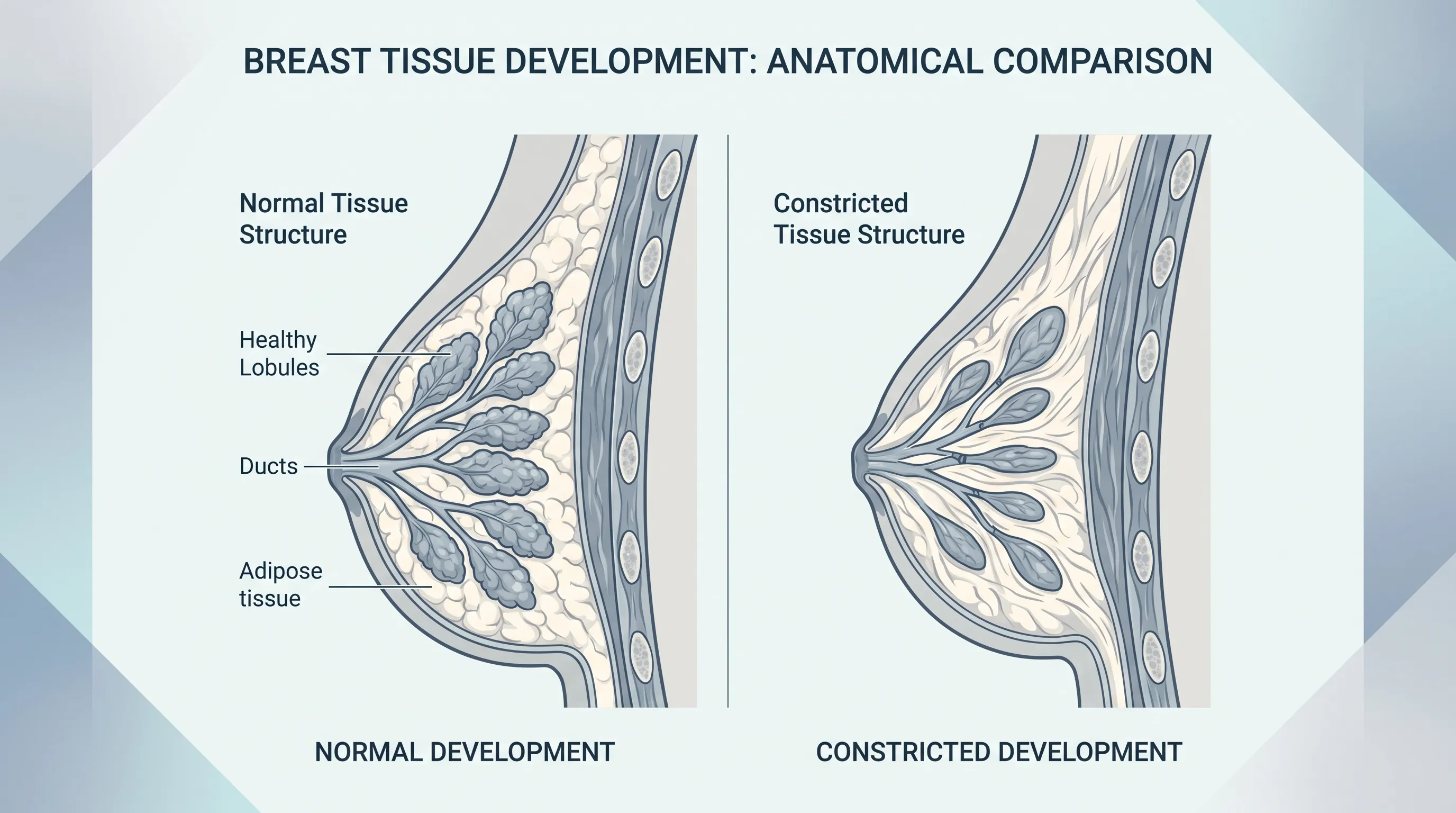
The human breast is a complex organ system composed of glandular tissue, adipose fat, connective stroma, vascular networks, and specialized skin envelopes. During fetal development and puberty, breast tissue undergoes a highly orchestrated series of morphological changes driven by hormonal signaling and genetic expression. In most individuals, the breast bud expands symmetrically, the mammary ridge differentiates into ductal-lobular units, and the inframammary fold settles at the natural chest wall boundary. However, when the fascial envelope surrounding the developing glandular tissue fails to expand proportionally, a constricted phenotype emerges. This developmental divergence results in a breast shape characterized by a narrow base, vertically elongated tissue distribution, and an abnormally high inframammary crease. The underlying pathophysiology involves abnormal thickening of the superficial and deep fascial layers, which restricts outward tissue expansion and forces glandular development to herniate primarily into the areolar complex.
Free calculatorOvulation CalculatorCalculate ovulation periodOpen the calculatorHow Breast Development Normally Occurs
Normal mammary development begins in utero with the formation of the mammary ridge, or milk line, extending from the axilla to the groin. Under the influence of maternal hormones, bilateral breast buds form during weeks six to eight of gestation. At birth, these rudimentary structures contain only primary lactiferous ducts embedded within a small stromal matrix. The most transformative phase occurs during puberty, when estrogen stimulates ductal branching while progesterone and prolactin prepare the tissue for lobuloalveolar development (National Institutes of Health (NIH)). Adipose deposition increases breast volume, while Cooper’s ligaments provide structural support. The inframammary fold typically matures between ages fifteen and eighteen, establishing the foundational contour of the mature breast. When this coordinated expansion proceeds without interruption, the resulting morphology aligns with typical anatomical standards.
The Pathophysiology Behind Constricted Tissue
In cases of a tubular breast, the embryological divergence occurs primarily at the level of the superficial fascial system. Research indicates that localized overexpression of collagen-producing fibroblasts creates a dense, restrictive ring around the developing glandular base. This fascial constriction prevents horizontal expansion, forcing developing tissue to grow vertically toward the path of least resistance. The result is a characteristic conical projection with a disproportionately large areola, reduced lower pole fullness, and a high-set inframammary crease. The degree of tissue constriction directly correlates with the clinical grading of the condition. Importantly, this anomaly does not involve malignant transformation or systemic illness; rather, it represents a localized architectural variation in connective tissue remodeling. Current embryological models suggest a combination of genetic predisposition and localized hormonal receptor distribution differences may contribute to fascial rigidity during critical developmental windows.
Recognizing Physical Characteristics and Clinical Signs
Identifying a tubular breast requires careful observation of both external morphology and internal tissue distribution. While presentation varies widely among individuals, several hallmark features consistently appear across clinical evaluations. These physical characteristics are not merely aesthetic; they reflect underlying structural limitations that can influence comfort, functionality, and long-term tissue behavior. Understanding these signs helps individuals recognize when professional evaluation is warranted and provides a clear foundation for discussing treatment objectives with healthcare providers.
Common Morphological Features
The most recognizable indicator of this condition is a noticeably narrow breast base diameter relative to overall chest width. The tissue often appears elongated vertically, projecting outward in a conical or cylindrical shape rather than maintaining a hemispherical contour. The lower pole frequently exhibits volume deficiency, creating a visual imbalance that makes the upper breast appear disproportionately full. Another defining characteristic is the elevated inframammary fold, which may sit several centimeters higher than typical anatomical positioning. The areolar complex is often enlarged, sometimes demonstrating periareolar herniation where glandular tissue protrudes into the nipple region, creating a stretched or puffy appearance. Bilateral involvement is common, though asymmetry in severity and morphology frequently occurs. In some individuals, the chest wall itself may appear slightly conical, further accentuating the overall shape discrepancy. These features collectively create a distinct silhouette that differs significantly from typical breast architecture.
Functional Implications and Lactation Considerations
Beyond visual presentation, the structural limitations of a constricted breast can impact physiological function. The compressed glandular architecture may reduce the volume of functional milk-producing alveoli, potentially leading to insufficient milk supply during lactation. Additionally, the distorted ductal pathways can impede efficient milk ejection, increasing the risk of engorgement, mastitis, and nipple trauma (Centers for Disease Control and Prevention (CDC)). While many individuals successfully breastfeed with supplemental guidance, others experience partial or complete lactation failure due to the underlying anatomical constraints. Physical discomfort can also arise from tissue tension, particularly during hormonal fluctuations, exercise, or pregnancy when glandular expansion occurs against rigid fascial boundaries. Recognizing these functional realities is crucial for developing holistic care plans that address both structural correction and physiological wellness. Early consultation with lactation specialists and breast health providers can mitigate complications and optimize postpartum experiences.
Classification Systems and Grading Frameworks
To standardize diagnosis, treatment planning, and surgical communication, the medical community relies on established classification systems. These frameworks categorize severity based on fascial restriction extent, areolar herniation presence, lower pole deficiency, and overall tissue displacement. The most widely accepted grading model was developed through extensive clinical observation and retrospective analysis, providing surgeons with predictable outcome benchmarks and helping patients understand their specific anatomical profile.
Type I to IV Categorization
The Von Heimburg and Nygaard classification system remains the gold standard for clinical assessment. Type I presents with mild constrictions, primarily affecting the medial lower quadrant with minimal areolar involvement and only slight inframammary elevation. Type II features more pronounced lower pole deficiency, extending across both medial and lateral quadrants, with moderate areolar enlargement and a clearly elevated fold. Type III demonstrates significant tissue constriction affecting the entire lower pole, substantial periareolar herniation, and a markedly high inframammary crease. Type IV, the most severe presentation, combines extensive fascial restriction with severe areolar herniation, often accompanied by significant chest wall asymmetry and minimal lower pole development. Understanding your specific type directly informs surgical strategy, as mild cases may require simple tissue release and fat grafting, while severe cases demand comprehensive fascial release, implant placement, and staged reconstruction. Accurate classification also helps set realistic expectations regarding correction outcomes and recovery timelines.
Differential Diagnosis and Distinguishing Features
It is crucial to distinguish this developmental variation from other congenital anomalies and acquired conditions. Poland syndrome involves unilateral absence of the pectoralis major muscle alongside ipsilateral breast hypoplasia, creating chest wall asymmetry rather than isolated tissue constriction. Simple breast asymmetry refers to normal variation in size or shape between sides without fascial restriction or conical projection. Hypoplasia involves underdeveloped glandular tissue across all quadrants, lacking the characteristic elongated morphology. Tuberous breast deformity specifically combines narrow base diameter, elevated fold, lower pole deficiency, and areolar herniation. Additionally, acquired changes from trauma, radiation, or aggressive weight fluctuation can mimic certain features but lack the congenital fascial restriction pattern. Proper differential diagnosis requires thorough clinical examination, patient history, and sometimes imaging to rule out underlying musculoskeletal anomalies. Misclassification can lead to inappropriate treatment plans, underscoring the importance of evaluation by a board-certified specialist familiar with congenital breast variations.
| Classification Type | Base Diameter Restriction | Lower Pole Deficiency | Areolar Herniation | Inframammary Fold Position | Recommended Surgical Approach |
|---|---|---|---|---|---|
| Type I | Mild, medial quadrant | Minimal | None or minimal | Slightly elevated | Tissue scoring, fat grafting, minor lift |
| Type II | Moderate, medial and lateral | Noticeable | Moderate | Clearly elevated | Comprehensive release, implant or fat, mastopexy |
| Type III | Severe, entire lower pole | Significant | Pronounced | Markedly high | Multi-stage release, tissue expander, implant, fat transfer |
| Type IV | Extreme, global constriction | Severe | Extensive | Very high | Staged reconstruction, extensive fascial release, custom implant/fat combination |
Evidence-Based Diagnostic and Evaluation Protocols
A thorough diagnostic process ensures accurate classification, rules out comorbid conditions, and establishes a personalized treatment roadmap. Medical evaluation begins with comprehensive history taking, followed by targeted physical examination, and concludes with imaging when clinically indicated. Adhering to standardized protocols minimizes misdiagnosis risk and aligns treatment with patient-specific anatomical realities. Individuals seeking evaluation should prioritize clinics specializing in congenital breast conditions, as expertise directly impacts assessment accuracy and long-term outcomes.
Clinical Assessment and Patient History
The diagnostic journey starts with a detailed medical interview exploring developmental milestones, family history, menstrual patterns, pregnancy intentions, and prior breast procedures. Providers will document symptom onset, progression, physical discomfort, lactation experiences, and psychological impact. During the physical examination, clinicians measure base width, sternal notch-to-nipple distance, nipple-to-inframammary fold distance, and areolar diameter. Tissue mobility, glandular density, skin quality, and fascial pliability are carefully assessed. Providers evaluate breast symmetry, chest wall contour, pectoral muscle function, and axillary lymph node status. Palpation identifies areas of abnormal tension, nodularity, or structural irregularities. The examination often includes posture analysis, shoulder alignment, and spinal curvature assessment, as postural compensation frequently develops to accommodate asymmetry. Standardized photographic documentation in multiple views provides a baseline for tracking changes, planning surgical intervention, and evaluating outcomes. This systematic approach ensures all anatomical and functional dimensions are captured before treatment discussions begin.
Imaging Modalities and Medical Screening
While diagnosis primarily relies on clinical examination, imaging supplements evaluation in specific scenarios. Ultrasound provides detailed visualization of glandular architecture, skin thickness, and underlying fascial constraints without radiation exposure. It is particularly useful for younger patients or those with dense breast tissue. Mammography may be recommended for individuals over thirty to establish baseline screening and rule out incidental findings, though it offers limited value for assessing congenital shape variations (Mayo Clinic). Magnetic resonance imaging (MRI) delivers high-resolution three-dimensional mapping of tissue volume, vascular distribution, and chest wall anatomy, making it invaluable for complex reconstruction planning. MRI can precisely quantify glandular deficiency, measure fascial restriction depth, and simulate postoperative tissue expansion. Additionally, baseline imaging establishes a reference for future breast cancer screening, as structural variations can complicate mammographic interpretation. Routine dermatological examination ensures skin integrity and rules out inflammatory conditions. When combined with clinical findings, these imaging tools create a comprehensive diagnostic profile that guides surgical strategy and long-term health monitoring.
Comprehensive Treatment Pathways and Surgical Correction
Addressing a tubular breast requires a nuanced approach that balances anatomical correction, functional optimization, and psychological well-being. Treatment decisions depend on classification severity, patient age, reproductive plans, aesthetic goals, and overall health status. Modern reconstructive techniques have evolved significantly, moving beyond simple implant placement to comprehensive tissue remodeling strategies that restore natural contour, proportion, and mobility. Patients benefit from multidisciplinary care that integrates surgical precision, physical rehabilitation, and emotional support.
Non-Surgical and Conservative Management Strategies
For individuals with mild constriction, minimal functional impairment, or strong preference for non-operative approaches, conservative management remains viable. Supportive garment therapy utilizes custom-fitted bras or compression sleeves designed to gently stretch fascial boundaries over extended periods. While evidence supporting permanent tissue expansion from external garments remains limited, these devices can improve comfort, reduce skin tension, and enhance daily confidence. Hormonal optimization through targeted nutrition, stress reduction, and balanced endocrine support may help stabilize tissue behavior, particularly during menstrual fluctuations or pregnancy. Lactation preparation programs incorporate manual glandular massage, targeted stretching exercises, and specialized nipple shields to maximize feeding capacity. Psychological counseling and body image therapy play crucial roles in conservative pathways, helping individuals develop healthy self-perception and reduce appearance-related distress. Regular clinical monitoring ensures early detection of skin changes, tissue displacement, or functional decline. While non-surgical methods cannot fundamentally alter congenital fascial architecture, they provide meaningful quality-of-life improvements for those preferring delayed or alternative approaches.
Advanced Surgical Techniques and Tissue Expansion
Surgical correction represents the most definitive intervention for significant anatomical restriction. The foundational principle involves carefully releasing the constrictive fascial ring to allow tissue to expand into a more natural contour. Surgeons employ radial scoring techniques, where precise incisions through the thickened superficial fascia permit horizontal tissue movement without compromising blood supply. This release is typically combined with strategic tissue redistribution to address lower pole deficiency. Tissue expanders may be placed temporarily, gradually inflating over weeks to stretch skin and fascia before permanent reconstruction. Alternatively, some techniques utilize progressive suture suspension and dermal flaps to reposition existing tissue downward. Areolar reduction is frequently performed to address herniation, utilizing circumareolar or purse-string techniques that minimize visible scarring. Mastopexy (breast lift) addresses skin excess and repositioning, while fat grafting enhances contour smoothness and volume distribution. These procedures often require staged approaches, particularly in severe cases, allowing tissues to adapt gradually and reducing complication risks. Advanced surgical planning incorporates three-dimensional imaging simulation, enabling precise volume calculation and symmetry optimization.
Navigating Implant Selection and Fat Grafting
Implant placement remains a cornerstone of moderate to severe correction, providing reliable volume restoration and structural support. Shaped anatomical implants often deliver superior lower pole projection compared to round alternatives, aligning better with natural tissue expansion patterns. Surgeons carefully evaluate implant positioning (subglandular vs. submuscular), shell texture, projection profile, and fill material (saline vs. silicone) based on skin thickness, tissue coverage, and patient anatomy. Autologous fat grafting has emerged as a powerful adjunct, allowing surgeons to transfer processed adipose tissue to refine contours, smooth transitions, and enhance lower pole fullness without synthetic materials. Fat harvesting typically occurs from the abdomen, thighs, or flanks, with centrifugation removing impurities before multi-plane injection. Graft survival rates average sixty to seventy percent, often requiring secondary sessions for optimal volume. Combining implants with fat transfer leverages the structural predictability of prosthetics alongside the natural softness and adaptability of autologous tissue. Patients should understand that implant longevity varies, typically requiring replacement or revision after ten to fifteen years due to capsular contracture, positional shifts, or aesthetic changes. Comprehensive preoperative education ensures realistic expectations and informed consent.

Recovery Protocols, Long-Term Care, and Psychological Wellness
The postoperative phase represents a critical window for tissue adaptation, complication monitoring, and emotional adjustment. Successful outcomes extend beyond surgical precision to encompass structured rehabilitation, diligent self-care, and comprehensive psychological support. Individuals who follow evidence-based recovery guidelines typically experience smoother healing, fewer complications, and higher satisfaction with aesthetic and functional results. Establishing a proactive care framework before surgery significantly improves the overall experience.
Immediate Post-Operative Guidelines and Complication Monitoring
The first seventy-two hours require strict adherence to prescribed protocols to minimize bleeding, swelling, and infection risk. Patients must maintain semi-elevated positioning, utilize cold therapy in twenty-minute intervals, and wear surgical compression garments continuously for four to six weeks. Drain care involves monitoring fluid output volume and color, typically lasting three to seven days. Incision hygiene includes gentle cleansing with approved antiseptics, avoiding submersion until complete epithelialization, and applying silicone-based scar therapy once sutures dissolve. Pain management follows a stepwise approach, transitioning from prescription analgesics to non-steroidal anti-inflammatory medications and acetaminophen. Patients must avoid upper body lifting, strenuous exercise, and sleeping on the stomach for a minimum of six weeks. Warning signs requiring immediate medical attention include sudden asymmetry, severe escalating pain, fever above one hundred one degrees, foul-smelling drainage, or visible implant malposition. Regular follow-up appointments at one, three, six, and twelve weeks ensure proper tissue settling, drain removal, and early complication detection. Gradual return to activity follows phased milestones, with light walking encouraged immediately and resistance training postponed until three months post-surgery.
Mental Health Support and Body Image Resources
The emotional journey accompanying breast correction deserves equal attention to physical recovery. Many individuals experience complex feelings including relief, vulnerability, grief over previous discomfort, and adjustment anxiety during swelling resolution (World Health Organization (WHO)). Body dysmorphia, if present preoperatively, may persist or shift toward new areas of focus without proper psychological preparation. Evidence-based recommendations include engaging in cognitive-behavioral therapy focused on body acceptance, joining moderated support groups with individuals sharing similar experiences, and practicing mindfulness techniques that redirect focus from appearance to function and vitality. Journaling emotional responses, setting realistic recovery expectations with medical teams, and establishing open communication with partners and family members foster resilience. Professional counselors specializing in surgical transitions can help patients navigate identity shifts, intimacy concerns, and social reintegration. Long-term wellness incorporates regular self-examination, annual clinical screenings, and ongoing dialogue with healthcare providers about changing needs, particularly during life transitions such as pregnancy, perimenopause, or significant weight changes. Prioritizing mental health alongside physical recovery ensures sustainable well-being and authentic self-acceptance.
Frequently Asked Questions
Is a tubular breast a health risk or purely cosmetic?
While primarily a developmental variation rather than a disease, this condition can sometimes impact lactation function, cause physical discomfort due to tissue tension, and lead to significant psychological distress. It does not increase breast cancer risk. Medical evaluation focuses on both anatomical correction and functional optimization rather than treating pathology.
At what age is surgical correction typically recommended?
Board-certified surgeons generally recommend waiting until breast development is complete, usually around age eighteen to twenty-one. Operating before full maturation can result in asymmetry, recurrent constriction, or suboptimal scar placement as tissues continue to change during late adolescence. Individual maturity and psychological readiness are equally important considerations.
Can fat grafting completely replace implants in correction procedures?
For mild to moderate cases, autologous fat grafting can successfully improve volume and contour without implants. However, severe constrictions often require a combination of tissue expansion, implant placement, and fat transfer for optimal, lasting results. Fat grafting enhances natural softness but has limited structural capacity for severe volume deficiency.
Does this condition affect breastfeeding capabilities?
The constricted glandular tissue and elevated inframammary fold can reduce milk-producing capacity and alter ductal pathways. While some individuals can partially or fully breastfeed, many experience insufficient supply and benefit significantly from prenatal lactation consulting, supplemental nursing systems, and specialized feeding strategies.
Is surgical correction typically covered by health insurance?
Coverage varies significantly by provider, region, and specific policy language. Most insurers classify correction as elective cosmetic surgery; however, some may offer partial or full coverage if documented physical symptoms, functional impairment, or severe psychological distress are medically substantiated. Pre-authorization appeals with detailed clinical documentation often improve approval odds.
Conclusion
Navigating the complexities of a tubular breast requires informed decision-making, compassionate clinical care, and a holistic approach to physical and emotional well-being. From understanding the underlying embryological mechanisms and standardized classification systems to exploring advanced surgical techniques and comprehensive recovery protocols, individuals now have access to evidence-based pathways that prioritize both anatomical correction and long-term wellness. Modern reconstructive innovations, combined with multidisciplinary support, offer highly personalized solutions that address functional concerns, aesthetic goals, and psychological resilience. By engaging with qualified healthcare providers, utilizing available mental health resources, and adhering to structured postoperative guidelines, individuals can achieve sustainable outcomes that honor their unique bodies and life journeys. Prioritizing thorough education, realistic expectations, and ongoing self-care ensures that every step of this journey supports lasting confidence and optimal health. Whether pursuing surgical intervention, conservative management, or a combination of both, empowered patients who partner with experienced professionals consistently experience the most transformative and enduring results.
How we work
Researched and drafted with AI assistance, then reviewed by a human editor against the cited sources.
This article is general health information, not medical advice. Consult a qualified healthcare professional about your own situation.