Hazy Vision in One Eye: Causes, Diagnosis, and Treatment Guide

Experiencing visual disturbances can be deeply unsettling, especially when symptoms appear unevenly across your field of sight. When you notice hazy vision in one eye while the other remains perfectly clear, it is natural to feel concerned. This specific symptom often prompts a rush to search online, but understanding the underlying mechanisms, potential triggers, and appropriate medical responses requires a structured approach. Unilateral vision loss is not a standalone disease; rather, it serves as a clinical indicator pointing to various physiological processes, ranging from benign ocular surface dryness to serious neurological events. Recognizing the distinction between temporary optical interference and progressive pathological changes empowers patients to seek timely care and avoid unnecessary complications.
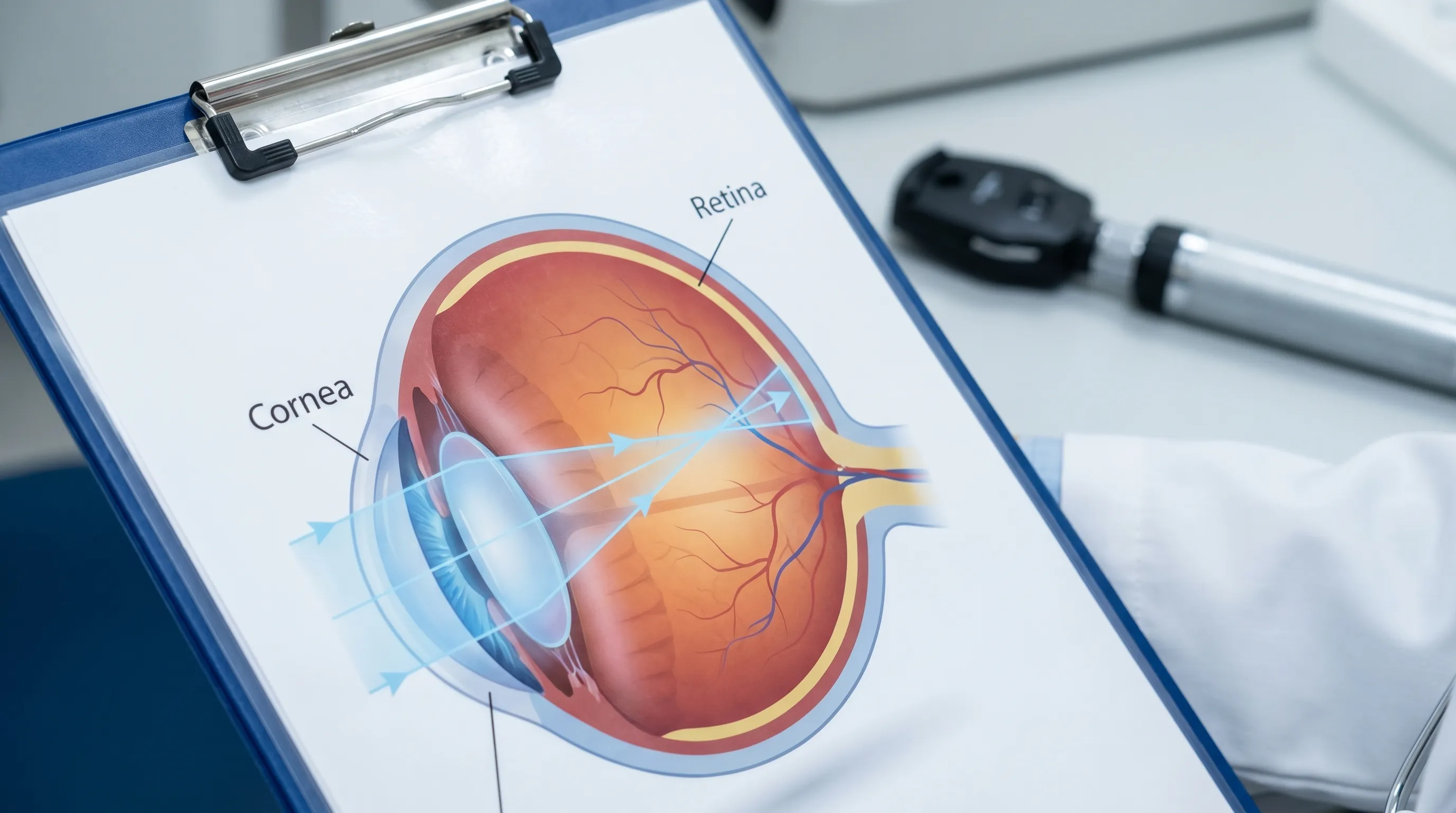
The human visual system relies on precise light refraction through the cornea and lens, followed by accurate image transmission along the optic nerve to the occipital cortex. Any disruption along this intricate pathway can manifest as cloudiness, dimming, or distortion. Because each eye functions independently, a unilateral symptom immediately localizes the issue to that specific anatomical unit. This isolation actually provides clinicians with valuable diagnostic clues, streamlining the investigative process and guiding targeted interventions. Whether the onset is gradual over months or abrupt within minutes, documenting the accompanying signs, duration, and triggering factors becomes essential for accurate diagnosis and effective management.
Understanding Hazy Vision in One Eye: Mechanisms and Manifestations
To comprehend why unilateral haziness occurs, we must first examine how the eye processes visual information. Light enters through the transparent cornea, passes through the aqueous humor, travels through the pupil, and is focused by the crystalline lens onto the retina. The retina then converts this optical signal into electrical impulses that travel via the optic nerve. When any segment of this system becomes compromised, the resulting image quality degrades. If only one pathway is affected, the brain receives mismatched visual data, which it attempts to reconcile, often leading to eye strain, headaches, or a persistent sense of visual fatigue.
The Optical Pathway and Unilateral Distortion
The anterior segment of the eye, comprising the cornea and lens, is primarily responsible for focusing light. Surface irregularities, dehydration, or early lens opacification scatter incoming photons, reducing contrast sensitivity. This scattering effect is frequently described by patients as looking through a frosted window or a mild fog. When the cornea experiences localized edema or develops microscopic irregularities from dry eye syndrome, light refracts unevenly. The brain compensates by squinting or increasing tear production, providing temporary relief. However, chronic interference leads to sustained visual degradation that only resolves once the structural or environmental trigger is addressed.
Posterior segment disturbances originate deeper within the ocular anatomy. The vitreous humor, normally a clear gel, can develop opacities or detach from the retinal surface. These vitreous changes cast shadows on the retina, perceived as floaters or generalized haze. When combined with retinal vascular leakage or ischemic events, the macular region loses its ability to resolve fine details, resulting in a pronounced central blur. Understanding whether the interference is anterior or posterior helps specialists prioritize diagnostic modalities and tailor therapeutic strategies effectively.
Differentiating Temporary Blur from Progressive Decline
Not every episode of unilateral haziness warrants panic, but distinguishing fleeting optical interference from genuine pathological progression is critical. Temporary blurring often correlates with environmental factors, such as prolonged screen exposure, low humidity, contact lens overwear, or allergic reactions. These episodes typically resolve within minutes to hours after removing the irritant or applying artificial tears. Patients who experience fluctuating clarity throughout the day, with symptoms improving after rest or blinking, usually suffer from ocular surface dysfunction or accommodative fatigue.
In contrast, progressive visual decline follows a predictable trajectory. It begins as mild cloudiness during high-contrast activities like night driving and gradually extends to routine reading or facial recognition. Accompanying signs may include color desaturation, increased glare sensitivity, or persistent peripheral shadowing. Tracking these patterns through a symptom journal allows ophthalmologists to map disease evolution accurately. Early documentation of when the symptom started, how it fluctuates, and what improves or worsens it forms the foundation of evidence-based diagnosis and personalized care planning.
Primary Medical Causes of Unilateral Visual Changes
Unilateral haziness stems from diverse etiologies spanning metabolic, degenerative, inflammatory, and vascular domains. Clinicians categorize these causes based on anatomical localization and progression speed. Refractive shifts, ocular surface disorders, and age-related structural changes dominate the benign spectrum. Conversely, retinal vascular pathology, optic nerve inflammation, and intraocular pressure spikes represent sight-threatening conditions requiring urgent intervention. A thorough differential diagnosis considers systemic health markers, medication history, and occupational exposures.
Refractive Shifts and Ocular Surface Disorders
Refractive errors, including myopia, hyperopia, astigmatism, and presbyopia, develop asymmetrically in many adults. When one eye undergoes a sudden focal shift, the brain struggles to merge the disparate images, creating a sense of unilateral blur. This is particularly common in individuals over forty as the crystalline lens loses elasticity. Corneal irregularities from dry eye syndrome, blepharitis, or contact lens-induced keratopathy further distort light transmission. The tear film comprises three essential layers: lipid, aqueous, and mucin. Deficiencies in any layer disrupt smooth light refraction, causing intermittent haziness that fluctuates with blinking frequency or environmental humidity.
Management focuses on restoring optical surface integrity. Preservative-free artificial tears, lipid-based lubricants, and omega-3 supplementation improve tear stability. For refractive asymmetry, updated corrective lenses, orthokeratology, or laser vision correction can restore balanced visual input. Addressing underlying allergies, optimizing indoor climate control, and implementing the 20-20-20 rule during screen use significantly reduce symptomatic episodes.
Cataract Development and Lens Opacification
Cataracts represent the most prevalent cause of gradual unilateral vision loss globally. Protein aggregation within the crystalline lens creates light-scattering regions that progressively obscure the retinal image. While bilateral development is typical, one eye often advances faster due to anatomical variations, prior trauma, prolonged corticosteroid use, or asymmetric ultraviolet exposure. Patients describe a yellowish tint, heightened glare from headlights or sunlight, and diminishing contrast sensitivity. Reading small print becomes challenging, and colors appear washed out.
Early-stage management relies on magnification aids, anti-glare coatings, and lighting adjustments. Once cataracts impair daily functioning or increase fall risk, phacoemulsification surgery becomes the gold standard. The procedure involves ultrasonic fragmentation of the cloudy lens, aspiration of the material, and insertion of a customized intraocular lens. Modern multifocal and toric lenses can simultaneously correct presbyopia and astigmatism, delivering exceptional postoperative visual quality. Regular monitoring ensures optimal timing for surgical intervention.
Macular Degeneration and Retinal Vascular Disease
Age-related macular degeneration (AMD) and diabetic retinopathy target the central retina, directly compromising detailed vision. Dry AMD manifests through drusen accumulation and gradual photoreceptor atrophy, creating a central scotoma with peripheral haze. Wet AMD involves abnormal choroidal neovascularization that leaks fluid and blood beneath the macula, producing rapid distortion and severe haziness. Diabetic microvascular damage leads to capillary leakage, macular edema, and ischemic zones. Hypertensive retinopathy similarly compromises vascular integrity, causing exudates and cotton-wool spots that scatter light.
Treatment protocols differ based on pathology type. Anti-vascular endothelial growth factor (anti-VEGF) intravitreal injections suppress abnormal vessel growth and reduce fluid accumulation. Photodynamic therapy and focal laser coagulation target leaking vessels directly. Nutritional interventions, including AREDS2-formula supplements containing vitamin C, vitamin E, zinc, copper, lutein, and zeaxanthin, slow dry AMD progression. Strict glycemic and blood pressure control remain foundational for diabetic and hypertensive retinopathy management.
Optic Neuropathies and Inflammatory Conditions
The optic nerve transmits visual signals from the retina to the brain. Inflammation, compression, or ischemia along this pathway disrupts signal fidelity, causing unilateral haziness, color vision deficits, and pain with eye movement. Optic neuritis frequently precedes multiple sclerosis diagnoses, presenting with subacute vision loss and relative afferent pupillary defect. Ischemic optic neuropathy arises from compromised posterior ciliary artery blood flow, often triggered by giant cell arteritis or severe hypotension. Compressive lesions from pituitary adenomas or thyroid eye disease exert mechanical pressure on the nerve fibers.
Diagnosis relies on high-resolution MRI, optical coherence tomography (OCT) retinal nerve fiber layer analysis, and comprehensive visual field testing. High-dose corticosteroid therapy accelerates recovery in inflammatory cases, while urgent temporal artery biopsy confirms giant cell arteritis, requiring immediate immunosuppression to prevent bilateral involvement. Surgical decompression addresses space-occupying lesions. Prompt recognition and intervention preserve neural integrity and optimize long-term functional recovery.
When Hazy Vision Signals a Medical Emergency
Certain presentations demand immediate emergency department or ophthalmology clinic evaluation. Delaying care during sight-threatening events reduces the window for effective treatment and increases the likelihood of irreversible damage. Recognizing red-flag symptoms ensures patients navigate urgent care pathways efficiently and avoid catastrophic outcomes. Time-sensitive interventions preserve retinal viability, prevent glaucomatous nerve atrophy, and mitigate stroke-related neurological deficits.
Retinal Detachment and Hemorrhagic Events
Retinal detachment occurs when the neurosensory retina separates from the underlying retinal pigment epithelium, disrupting photoreceptor oxygen and nutrient supply. Patients experience sudden unilateral haziness accompanied by flashing lights, a dramatic increase in floaters, and a descending curtain-like shadow across the visual field. Hemorrhagic events, including vitreous hemorrhage or central retinal vein occlusion, introduce blood into the optical pathway, causing abrupt cloudiness and severe vision loss. Both conditions require same-day specialist evaluation.
Management involves laser photocoagulation, cryotherapy, or surgical vitrectomy to reattach the retina and drain accumulated fluid or blood. Anti-VEGF agents and thrombolytics address vascular occlusions when administered within the therapeutic window. Patients with high myopia, prior ocular trauma, lattice degeneration, or family history of detachment face elevated risks and should undergo regular peripheral retinal examinations. Immediate immobilization and rapid transport optimize surgical outcomes.
Acute Angle-Closure Glaucoma
Acute angle-closure glaucoma arises when the iris blocks aqueous humor drainage through the trabecular meshwork, causing rapid intraocular pressure escalation. Symptoms include profound unilateral haziness, severe ocular pain radiating to the brow or temple, nausea, vomiting, halos around lights, and a visibly fixed, mid-dilated pupil. The optic nerve suffers rapid ischemic damage, and untreated cases progress to permanent blindness within days. This condition constitutes an absolute ophthalmologic emergency.
Initial management focuses on aggressive pressure reduction using systemic osmotic agents, topical beta-blockers, alpha-agonists, and carbonic anhydrase inhibitors. Peripheral iridotomy creates an alternative aqueous outflow pathway, preventing recurrence. Prophylactic laser treatment in the fellow eye mitigates bilateral risk. Patients with shallow anterior chambers, hyperopic refractive error, or Asian ancestry should undergo routine gonioscopy to assess angle anatomy and implement preventive strategies.
Transient Ischemic Attacks and Amaurosis Fugax
Amaurosis fugax describes temporary monocular vision loss caused by embolic material transiently occluding the retinal artery. Patients report a sudden, painless graying or blacking out of vision in one eye, typically resolving within minutes. This symptom frequently serves as a harbinger of carotid artery stenosis or impending cerebrovascular accident. The CDC notes that transient ischemic attacks (TIAs) are critical warning signs requiring urgent neurological and cardiovascular evaluation to prevent permanent damage.
Diagnostic workup includes carotid duplex ultrasonography, echocardiography, lipid profiling, and magnetic resonance angiography. Antiplatelet therapy, statins, and carotid endarterectomy reduce future stroke risk. Lifestyle modifications addressing hypertension, atrial fibrillation, diabetes, and smoking cessation significantly lower recurrence probability. Treating amaurosis fugax extends far beyond ocular health, representing a critical opportunity to prevent life-threatening neurological events.
The Diagnostic Pathway: How Professionals Evaluate Unilateral Blur
Accurate diagnosis requires a systematic, multi-modal approach integrating patient history, clinical examination, and advanced imaging. Ophthalmologists and optometrists utilize standardized protocols to isolate the etiology of hazy vision in one eye, ensuring targeted interventions rather than empirical guesswork. Each diagnostic step builds upon the previous findings, creating a comprehensive clinical picture that guides personalized treatment planning.
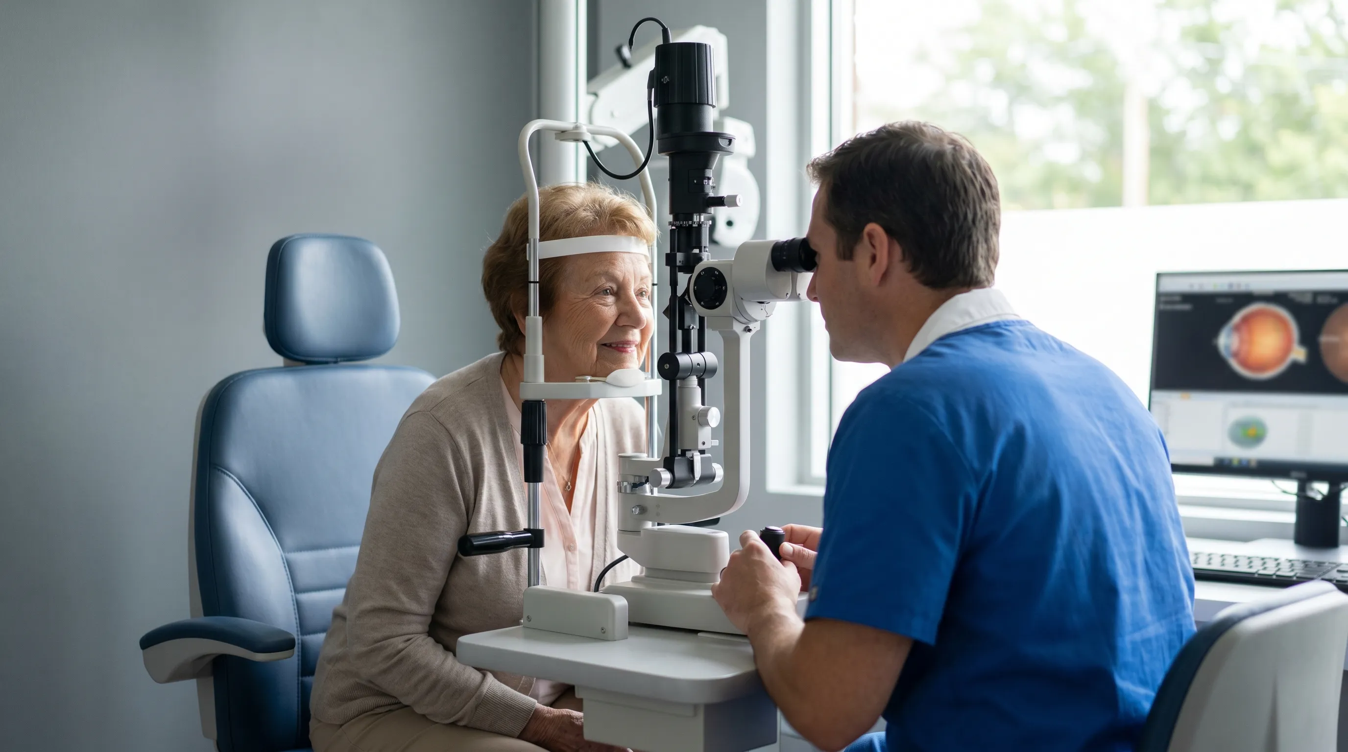
Clinical Assessment and Slit-Lamp Examination
The foundational evaluation begins with a detailed symptom history. Clinicians inquire about onset timing, duration, progression pattern, associated pain, systemic conditions, medication usage, and family ocular history. Visual acuity testing quantifies the degree of impairment, while color vision assessment detects optic nerve compromise. Pupillary examination evaluates symmetry and constriction response, identifying relative afferent defects indicative of retrobulbar pathology.
Slit-lamp biomicroscopy provides magnified, three-dimensional visualization of the anterior segment. It reveals corneal edema, keratic precipitates, cataract morphology, anterior chamber inflammation, and intraocular lens positioning. Tonometry measures intraocular pressure, screening for glaucomatous risk. Dilated fundus examination allows direct observation of the optic disc, macula, and peripheral retina, detecting hemorrhages, exudates, vascular attenuation, or pigmentary changes. These clinical maneuvers form the cornerstone of accurate diagnosis and risk stratification.
Imaging Modalities and Functional Testing
Advanced imaging transforms diagnostic precision by revealing microscopic structural alterations invisible to conventional examination. Optical coherence tomography (OCT) generates high-resolution cross-sectional scans of the retina and optic nerve, quantifying macular thickness, subretinal fluid, and nerve fiber layer integrity. Optical coherence tomography angiography (OCTA) maps retinal and choroidal vasculature without contrast dyes, identifying ischemic zones and neovascular networks.
Fluorescein angiography tracks dye circulation through the retinal vasculature, highlighting leakage, occlusion, or abnormal perfusion. Visual field testing maps functional deficits, differentiating between glaucomatous peripheral loss and macular central scotomas. Ultrasound biomicroscopy evaluates the ciliary body and posterior segment when media opacity prevents direct visualization. Electrodiagnostic testing measures retinal and optic nerve electrical responses. Integrating these modalities ensures comprehensive anatomical and functional assessment, enabling precise diagnosis and optimized therapeutic targeting.
Evidence-Based Treatment Approaches and Management
Treatment selection depends entirely on the confirmed diagnosis, disease stage, and patient-specific factors. Management strategies span conservative monitoring, pharmacological therapy, minimally invasive procedures, and surgical correction. Patient education remains integral to successful outcomes, ensuring adherence to treatment regimens and lifestyle modifications. Multidisciplinary coordination with primary care, endocrinology, cardiology, and neurology optimizes systemic risk factor control alongside targeted ocular therapy.
Pharmacological Interventions and Anti-VEGF Therapies
Medication protocols address inflammation, infection, vascular leakage, and intraocular pressure dysregulation. Topical corticosteroids and nonsteroidal anti-inflammatory drugs resolve anterior segment inflammation and cystoid macular edema. Antimicrobial therapy targets bacterial, viral, or fungal keratitis and endophthalmitis. Glaucoma management employs prostaglandin analogs, beta-blockers, alpha-agonists, carbonic anhydrase inhibitors, and Rho kinase inhibitors, often combined into single-dose formulations to enhance compliance.
Anti-VEGF injections represent a paradigm shift in retinal disease management. Ranibizumab, aflibercept, and faricimab bind vascular endothelial growth factor, halting abnormal vessel proliferation and reducing macular edema. Patients undergo loading doses followed by maintenance injections tailored to disease activity. Extended dosing intervals and treat-and-extend protocols minimize visit frequency while maintaining visual stability. Biosimilar availability improves accessibility and reduces long-term treatment burden. Consistent monitoring ensures timely intervention during disease flare-ups.
Surgical Correction and Laser Procedures
When conservative measures prove insufficient, surgical intervention restores optical clarity and structural integrity. Phacoemulsification with intraocular lens implantation remains the most performed ocular surgery worldwide, delivering rapid visual rehabilitation. Vitrectomy addresses non-clearing hemorrhage, epiretinal membranes, macular holes, and retinal detachment. Gas or silicone oil tamponade maintains retinal apposition during healing.
Laser therapies include selective laser trabeculoplasty for open-angle glaucoma, peripheral iridotomy for angle closure, and panretinal photocoagulation for proliferative diabetic retinopathy. Photodynamic therapy combines light activation with targeted medication to regress choroidal neovascularization. Micro-incisional glaucoma surgery implants stents to enhance aqueous outflow. Each procedure requires precise patient selection, meticulous preoperative optimization, and structured postoperative rehabilitation to maximize success rates and minimize complications.
Supportive Care and Symptom Alleviation
Complementary management strategies enhance comfort, preserve remaining vision, and improve quality of life. Low vision rehabilitation employs magnifiers, telescopic devices, electronic readers, and high-contrast materials to maximize functional independence. Occupational therapy adapts home environments, reducing fall risk and enhancing daily task performance. Psychological counseling addresses the emotional impact of vision loss, fostering resilience and adaptive coping mechanisms.
Dry eye management escalates from artificial tears to punctal plugs, autologous serum drops, and intense pulsed light therapy for meibomian gland dysfunction. Scleral lenses create a fluid reservoir over irregular corneas, stabilizing vision in severe ectasia or post-transplant cases. Regular follow-up ensures timely intervention during disease progression. Patient empowerment through structured education programs improves self-management and reduces emergency department utilization.
Long-Term Eye Health and Preventive Strategies
Proactive eye care extends far beyond symptom resolution. Implementing sustainable lifestyle practices, nutritional optimization, and routine screening mitigates the risk of future visual impairment. Prevention remains the most cost-effective strategy for preserving lifelong ocular function and maintaining independence. Evidence-based guidelines emphasize modifiable risk factor control and early detection of subclinical pathology.
Nutritional Optimization and Antioxidant Support
Diet profoundly influences ocular tissue health and inflammatory regulation. Leafy green vegetables supply lutein and zeaxanthin, concentrating in the macula to filter high-energy blue light and neutralize oxidative stress. Omega-3 fatty acids from fatty fish stabilize the tear film lipid layer, reducing evaporative dry eye symptoms. Vitamin C, vitamin E, and zinc support collagen integrity and photoreceptor function. Zinc deficiency correlates with accelerated drusen formation and retinal degeneration.
Hydration maintains aqueous humor volume and corneal transparency. Limiting refined carbohydrates and saturated fats reduces systemic inflammation and endothelial dysfunction. AREDS2-formula supplementation demonstrates statistically significant reductions in advanced AMD progression among high-risk individuals. However, supplements complement rather than replace whole-food nutrition. Consulting registered dietitians ensures balanced intake aligned with metabolic requirements and medication interactions.
Lifestyle Modifications and Ergonomic Practices
Digital environments impose unprecedented visual demands, accelerating accommodative fatigue and tear film instability. Implementing the 20-20-20 rule—looking twenty feet away for twenty seconds every twenty minutes—reduces ciliary muscle strain. Adjusting screen brightness, enabling blue light filters, and positioning monitors slightly below eye level minimize evaporative exposure and neck tension. Humidifiers counteract dry indoor climates, preserving ocular surface moisture.
Physical activity enhances retinal blood flow and systemic vascular health, reducing glaucoma progression and diabetic complication rates. Wearing polycarbonate safety glasses during sports, home repairs, or laboratory work prevents traumatic ocular injuries. Avoiding tobacco exposure eliminates a major risk factor for cataract formation, optic neuropathy, and macular degeneration. UV-protective sunglasses with wraparound designs shield the lens and retina from photochemical damage during peak sunlight hours.
Regular Screenings and Risk Management
Early detection transforms disease trajectories by enabling intervention before irreversible damage occurs. The National Eye Institute emphasizes that comprehensive dilated eye exams are crucial for identifying silent, sight-threatening conditions early. Adults without risk factors should undergo comprehensive eye examinations every two years until age forty, then annually. Individuals with diabetes, hypertension, autoimmune disorders, or family history of glaucoma require annual or biannual monitoring. Baseline optical coherence tomography and visual field testing establish normative data for future comparison.
Carotid artery ultrasound, echocardiography, and lipid panels identify systemic embolic sources and atherosclerotic burden. Managing blood pressure below 130/80 mmHg and maintaining HbA1c levels under 7% significantly reduces microvascular retinopathy progression. Genetic counseling benefits families with hereditary retinal dystrophies or congenital glaucoma. Integrating routine ocular screenings into primary care wellness visits ensures seamless monitoring and timely specialist referral when anomalies emerge.
| Condition Category | Primary Symptoms | Diagnostic Gold Standard | Primary Treatment Approach |
|---|---|---|---|
| Refractive & Surface Errors | Fluctuating haze, dryness, glare | Slit-lamp, corneal topography | Lubrication, updated prescriptions, ergonomic adjustments |
| Cataracts | Progressive cloudiness, color desaturation, glare sensitivity | Dilated exam, OCT, lens grading | Phacoemulsification with intraocular lens implantation |
| Retinal Vascular Disease | Central distortion, hemorrhage shadows, rapid decline | OCT angiography, fluorescein angiography | Anti-VEGF injections, laser coagulation, glycemic control |
| Optic Neuropathies | Color loss, pain with movement, central scotoma | MRI brain/orbits, visual evoked potentials | High-dose corticosteroids, immunosuppression, surgical decompression |
| Acute Glaucoma | Severe pain, halos, fixed pupil, nausea | Tonometry, gonioscopy, anterior segment OCT | Pressure-lowering drops, oral osmotics, laser iridotomy |
Frequently Asked Questions
Is sudden hazy vision in one eye a medical emergency?
Yes, sudden or rapidly developing hazy vision in one eye requires immediate medical evaluation. It can indicate retinal detachment, acute glaucoma, stroke, or a transient ischemic attack, all of which need prompt intervention to prevent permanent sight loss.
Can dry eyes cause blurriness in just one eye?
Absolutely. An unstable tear film can disrupt light entering the eye, causing temporary haziness or fluctuating vision. If only one tear duct or gland is compromised, the blurriness may remain isolated to that eye until the ocular surface is properly lubricated.
How is optic neuritis diagnosed when vision is hazy?
Diagnosis involves a comprehensive neurological and ophthalmic evaluation, including visual evoked potentials, MRI scans of the brain and optic nerves, and pupil reaction tests. These help identify inflammation and rule out multiple sclerosis or other autoimmune triggers.
What lifestyle changes can prevent progressive vision decline?
Maintaining stable blood pressure and glucose levels, wearing UV-protective sunglasses, adopting a diet rich in lutein and omega-3 fatty acids, quitting smoking, and taking regular screen breaks significantly reduce the risk of degenerative eye conditions.
Will cataract surgery restore clear vision in the affected eye?
Yes, cataract extraction with intraocular lens implantation is highly effective. The cloudy natural lens is removed and replaced with a clear artificial lens, typically restoring sharp vision within days. Postoperative care ensures optimal healing and visual outcomes.
Conclusion
Navigating the experience of hazy vision in one eye requires balancing vigilance with informed action. While occasional optical interference stems from benign environmental or surface-level factors, persistent or progressive symptoms demand professional evaluation to identify underlying pathology. The diagnostic landscape has advanced significantly, offering precise imaging, targeted pharmacotherapy, and minimally invasive procedures that preserve and restore visual function. Patients who engage actively in their care, maintain consistent screening schedules, and implement evidence-based lifestyle modifications dramatically reduce the risk of sight-threatening complications.
Prioritizing eye health extends beyond treating individual symptoms; it encompasses comprehensive systemic wellness and proactive risk management. By recognizing early warning signs, understanding diagnostic pathways, and collaborating closely with qualified eye care professionals, individuals safeguard their visual independence for decades to come. Whether addressing refractive shifts, managing chronic retinal conditions, or responding to acute ocular emergencies, timely intervention remains the cornerstone of successful outcomes. Your vision is irreplaceable, and investing in its preservation yields lifelong dividends in safety, mobility, and overall quality of life.

About the author
David Chen, DO, is a board-certified neurologist specializing in neuro-oncology and stroke recovery. He is the director of the Comprehensive Stroke Center at a New Jersey medical center and has published numerous articles on brain tumor treatment.