Broken Capillaries on Nose: Causes, Treatments & Expert Skincare Guide
You glance into the mirror on a quiet morning and notice a faint, reddish web-like pattern resting just beneath the skin of your nose. It does not wipe away with cleanser, and no matter how diligently you apply makeup, the fine lines persist. What you are seeing are commonly referred to as broken capillaries on the nose, but the clinical reality is far more nuanced than the name suggests. These delicate, superficial vascular networks are a widespread dermatological concern that affects millions of adults worldwide, often carrying a complex mix of cosmetic frustration and underlying physiological triggers. Understanding the precise medical mechanisms behind their formation, recognizing the lifestyle and genetic factors that accelerate their development, and exploring clinically validated treatment pathways are essential steps toward restoring a balanced, even complexion. In this comprehensive guide, we will examine the evidence-based science behind these visible vessels, outline actionable prevention strategies, and provide clear guidance on when professional dermatological intervention is warranted. Whether you are managing early-stage facial redness, navigating a diagnosed inflammatory skin condition, or simply seeking to optimize your daily skincare protocol, you will find thoroughly researched, medically accurate information to help you make informed decisions about your skin health.
What Are Broken Capillaries on the Nose?
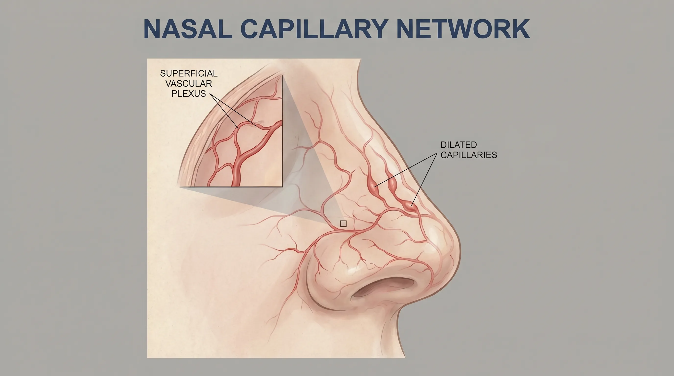
Despite their popular name, broken capillaries on the nose are rarely, if ever, truly "broken" in the literal sense. Instead, they represent a vascular phenomenon where tiny blood vessels near the skin's surface become permanently dilated, losing their ability to constrict back to a normal resting state. Medically classified as telangiectasias, these structures typically measure between 0.5 and 1 millimeter in diameter and appear as fine red, purple, or blueish lines that branch outward in a spiderweb or arborizing pattern. The nasal area is particularly susceptible to their development due to its rich vascular supply, constant environmental exposure, and the thin, fragile nature of the overlying epidermis.
Medical Terminology: Understanding Telangiectasias
The term telangiectasia originates from Greek and Latin roots: "tel-" meaning end or distant, "angi-" referring to a blood vessel, and "ectasia" denoting expansion or dilation. From a physiological perspective, these lesions represent functional arterioles, venules, or capillaries that have undergone structural remodeling. The elastic fibers and smooth muscle cells surrounding the vessel walls degrade or become compromised over time, often due to chronic inflammatory mediators, ultraviolet radiation, or repeated hemodynamic stress. When these supportive structures fail, the vessel remains in a chronically engorged state, allowing hemoglobin and deoxygenated blood to become visually prominent through the translucent upper dermis.
It is crucial to distinguish telangiectasias from other vascular markings such as petechiae, purpura, or hemangiomas. Petechiae and purpura result from actual capillary rupture and extravasation of red blood cells into the surrounding tissue, appearing as flat, non-blanching dots that do not respond to pressure. In contrast, broken capillaries on the nose are intact vessels that retain blood flow, making them responsive to targeted vascular interventions.
How to Identify Them: The Diascopy Test and Visual Cues
Dermatologists frequently utilize the diascopy test to differentiate telangiectasias from hemorrhagic lesions. By applying gentle, firm pressure to the affected area using a glass slide or gloved fingertip, you can observe whether the redness blanches, or temporarily turns pale white. If the discoloration disappears under pressure and promptly returns once released, you are likely observing dilated, blood-filled vessels rather than ruptured capillaries or bruising. Visually, these lesions often appear as thin, branching lines that may cluster around the nasal ala, the sides of the nostrils, or the central nasal bridge. They rarely cause pain or itching, though the surrounding skin may feel warm, sensitive, or reactive to temperature fluctuations.
What Causes Broken Capillaries on the Nose?
The development of broken capillaries on the nose is multifactorial, involving an interplay of environmental stressors, inflammatory conditions, physiological changes, and genetic predisposition. Acquired causes account for the vast majority of cases, while congenital syndromes, though less frequent, require distinct clinical evaluation. Understanding the specific catalysts behind vascular dilation is the first step toward targeted management and long-term skin stabilization.
Acquired Factors and Lifestyle Triggers
Daily exposures and habitual factors exert a profound cumulative effect on facial microvasculature. Ultraviolet radiation stands as one of the most significant contributors, as UVA and UVB wavelengths penetrate the epidermis, degrade collagen and elastin, and generate reactive oxygen species that damage endothelial cell integrity. Repeated exposure to extreme temperatures, whether from hot showers, harsh winter winds, or working near industrial heat sources, forces rapid vasodilation and vasoconstriction. This thermal cycling gradually weakens the structural resilience of capillary walls.
Dietary and behavioral triggers also play a well-documented role. Alcohol consumption induces systemic and facial flushing by triggering histamine release and impairing vascular tone regulation. Similarly, capsaicin-rich spicy foods stimulate thermoregulatory pathways, increasing cutaneous blood flow. Chronic emotional stress elevates circulating cortisol and catecholamines, which sustain prolonged vasodilation. Pregnancy introduces significant hemodynamic shifts, with blood volume increasing by up to fifty percent, naturally elevating venous pressure and facial perfusion. Prolonged use of topical or systemic corticosteroids can induce dermal atrophy, thinning the protective epidermal layer and rendering underlying vessels more conspicuous. Physical trauma, including aggressive exfoliation, improper cosmetic procedures, or repeated nasal friction, directly damages superficial vascular architecture.
The Strong Connection to Rosacea
Perhaps the most clinically significant association with broken capillaries on the nose is rosacea, a chronic inflammatory dermatosis characterized by recurrent flushing, persistent erythema, papulopustular eruptions, and visible telangiectasias. According to Mayo Clinic, rosacea "causes flushing or long-term redness on your face. It also may cause enlarged blood vessels and small, pus-filled bumps." The inflammatory cascade in rosacea involves dysregulated innate immunity, neurovascular hyperreactivity, and the overexpression of cathelicidin peptides that promote vasodilation and matrix metalloproteinase activation.
Harvard Health outlines four progressive stages of the condition that illustrate how vascular changes evolve:
- First stage: Intermittent facial flushing and transient redness triggered by environmental or dietary factors
- Second stage: Persistent central facial erythema involving the cheeks, nose, chin, or forehead
- Third stage: Inflammatory papules and pustules accompanied by the emergence of tiny blood vessels that manifest as red, thin lines known as telangiectasias
- Fourth stage: Advanced tissue remodeling characterized by sebaceous gland hypertrophy, fibrosis, and rhinophyma, particularly in male patients
Harvard Health further explains that "facial blushing or flushing causes small blood vessels to expand and eventually to show through the skin. These enlarged blood vessels appear as thin red lines (telangiectasias) on the face, especially on the cheeks." This progression underscores the importance of early intervention, as untreated chronic inflammation inevitably leads to permanent vascular remodeling.
Genetic and Congenital Conditions
While acquired telangiectasias are commonplace, certain inherited disorders feature widespread facial and mucosal vascular dilation. Hereditary hemorrhagic telangiectasia (HHT), also known as Osler-Weber-Rendu syndrome, is an autosomal dominant condition affecting vascular endothelial growth factor pathways, resulting in fragile, prone-to-bleeding capillary networks across the face, gastrointestinal tract, and central nervous system. Other rare genetic syndromes include Bloom syndrome, characterized by DNA repair deficiencies and photosensitivity; ataxia-telangiectasia, involving neurodegeneration and immunodeficiency; Sturge-Weber syndrome, presenting with facial port-wine stains and ocular involvement; and Klippel-Trenaunay syndrome, featuring limb hypertrophy with complex venous and lymphatic malformations. As noted in Wikipedia, "numerous inherited or congenital conditions display cutaneous telangiectasia." These systemic diagnoses require multidisciplinary management and genetic counseling, distinguishing them from isolated cosmetic vascular concerns.
Risk Factors and Who Is Most Affected
The epidemiology of facial telangiectasia reveals distinct demographic, physiological, and environmental patterns that influence susceptibility and progression. Recognizing these risk factors enables individuals to implement proactive measures before permanent vascular changes occur.
Demographics, Skin Type, and Gender Differences
Research indicates a strikingly high prevalence of vascular dilation across adult populations. Approximately 79% of adult males and 88% of adult females exhibit some form of lower extremity telangiectasia, with facial manifestations following similar distribution trends. Individuals with Fitzpatrick skin types I and II, characterized by fair complexion, light eyes, and a tendency to burn rather than tan, face significantly elevated risk. Reduced melanin provides less natural photoprotection, allowing ultraviolet radiation to penetrate deeper and accelerate collagen degradation around superficial vessels.
Gender also influences clinical presentation. Women are statistically more likely to develop early-onset rosacea and facial flushing, largely due to hormonal fluctuations during menstruation, pregnancy, and perimenopause. Estrogen and progesterone modulate vascular reactivity and inflammatory mediator release. Conversely, men who develop rosacea often progress further along the disease spectrum, with a higher likelihood of experiencing phymatous changes, particularly rhinophyma, which involves irreversible sebaceous hyperplasia and connective tissue thickening.
Environmental and Occupational Exposures
Occupational hazards and prolonged environmental exposure significantly compound vascular stress. Outdoor workers, agricultural laborers, construction professionals, and chefs operating in high-heat environments experience chronic thermal shock and relentless ultraviolet exposure. These repeated stressors exhaust the autoregulatory capacity of cutaneous microcirculation. Additionally, individuals residing in high-altitude regions or areas with extreme seasonal temperature variations face compounded risks due to atmospheric dryness, wind chill, and intense reflective solar radiation. Smoking further exacerbates the problem by inducing peripheral vasoconstriction while simultaneously promoting chronic low-grade inflammation and oxidative stress, creating a paradoxical environment that weakens capillary walls and accelerates facial erythema.
| Risk Factor | Underlying Mechanism | Evidence-Based Mitigation Strategy |
|---|---|---|
| Chronic Sun Exposure | UV-induced collagen degradation, endothelial cell damage | Daily broad-spectrum SPF 30+ sunscreen, UPF clothing, shade seeking |
| Temperature Extremes | Repeated vasodilation/vasoconstriction cycling | Limit hot showers, use lukewarm water, employ humidifiers, avoid prolonged outdoor exposure in extreme heat/cold |
| Alcohol & Spicy Foods | Histamine release, thermoregulatory flushing | Monitor and limit known dietary triggers, stay hydrated, substitute with anti-inflammatory foods |
| Corticosteroid Overuse | Dermal atrophy, suppressed collagen synthesis | Use topical steroids only under medical supervision, explore non-steroidal anti-inflammatory alternatives |
| Hormonal Fluctuations | Estrogen-mediated vascular permeability and reactivity | Maintain consistent skincare routine, discuss hormonal therapies with healthcare providers |
| Smoking | Oxidative stress, impaired microcirculation, rosacea exacerbation | Complete cessation, antioxidant-rich diet, vascular support supplements after consultation |

Evidence-Based Treatment Options
Once broken capillaries on the nose become structurally established, topical skincare alone cannot reverse permanent dilation. Modern dermatology offers highly effective, precision-targeted modalities that selectively collapse abnormal vessels while preserving surrounding healthy tissue. Selecting the appropriate intervention depends on vessel size, depth, skin type, and overall treatment goals.
In-Office Medical Procedures
Laser therapy remains the clinical gold standard for eradicating facial telangiectasias. Pulsed dye laser (PDL) technology, typically operating at 585 or 595 nanometers, delivers concentrated light energy specifically absorbed by oxyhemoglobin within the dilated vessels. This selective photothermolysis generates controlled thermal damage, coagulating the vessel lumen and triggering a natural phagocytic clearance process. Patients typically experience minimal downtime, with temporary bruising or mild erythema resolving within one to two weeks.
The Nd:YAG laser, operating at 1064 nanometers, offers deeper penetration and enhanced safety for patients with darker skin tones. By bypassing melanin-rich epidermal layers, the Nd:YAG targets larger, bluer, or more deeply situated vessels without risking post-inflammatory hyperpigmentation. Intense Pulsed Light (IPL) utilizes a broad spectrum of wavelengths, making it effective for treating diffuse redness, sun damage, and mild to moderate telangiectasias simultaneously. While IPL is less vessel-specific than PDL, it provides comprehensive photorejuvenation benefits.
Electrocautery represents an alternative for isolated, pinpoint vessels. A fine-tipped device delivers a precise electrical current to thermally seal the target capillary. Although highly effective for solitary lesions, it carries a marginally higher risk of pinpoint scarring compared to laser modalities. Sclerotherapy, while widely used for lower extremity venous insufficiency, is rarely employed on the face due to the high risk of tissue necrosis and embolic complications in the cranial vascular network.
Topical and Pharmaceutical Approaches
While topicals cannot eliminate existing broken capillaries on the nose, they play a vital role in managing underlying inflammatory drivers and temporarily reducing visible erythema. Prescription agents such as brimonidine tartrate (Mirvaso) and oxymetazoline hydrochloride (Rhofade) act as alpha-2 adrenergic receptor agonists, inducing temporary vasoconstriction that lightens facial redness for up to twelve hours. These compounds are strictly symptomatic and require daily application to maintain effects.
For patients whose vascular changes stem from rosacea, foundational anti-inflammatory therapies are essential. Metronidazole gel reduces reactive oxygen species and neutrophil-mediated inflammation. Azelaic acid normalizes keratinization, inhibits microbial proliferation, and calms persistent erythema through dual anti-inflammatory and tyrosinase-inhibitory pathways. Ivermectin cream targets Demodex mite overpopulation, a well-documented contributor to rosacea pathogenesis. Combining these targeted pharmaceuticals with procedural interventions yields the most sustainable long-term outcomes.
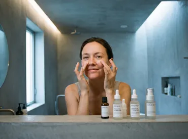
Daily Prevention and Skincare Strategies
Preventing the formation of new broken capillaries on the nose and halting the progression of existing lesions requires a disciplined, science-backed approach to daily skin maintenance. The cornerstone of vascular preservation lies in barrier optimization, photoprotection, and systematic trigger avoidance.
Sun Protection and Barrier Repair
Ultraviolet radiation remains the most aggressive environmental catalyst for capillary dilation and dermal thinning. Applying a broad-spectrum sunscreen with a minimum SPF of 30 every morning, regardless of weather or indoor exposure, is non-negotiable. Mineral formulations containing zinc oxide or titanium dioxide offer immediate physical blockage and are inherently less irritating for vascularly sensitive skin. Chemical sunscreens provide elegant cosmetic finishes but may contain avobenzone or octocrylene, which occasionally provoke stinging in compromised skin barriers.
Beyond UV filtration, reinforcing the stratum corneum and dermal matrix strengthens capillary support. Ingredients such as niacinamide enhance ceramide synthesis, improve epidermal cohesion, and reduce transepidermal water loss. Ceramide-dominant moisturizers replenish lipid bilayers, while peptides stimulate fibroblast activity and collagen remodeling. Antioxidants like vitamin C, vitamin E, and green tea polyphenols neutralize free radicals generated by environmental stressors, preventing endothelial degradation. Always cleanse with lukewarm water, avoid abrasive physical scrubs or high-concentration acid peels, and pat the face dry rather than rubbing.
Trigger Management and Lifestyle Adjustments
Systemic and behavioral factors exert profound influence over facial vascular tone. Maintaining a detailed symptom diary can help identify personal triggers, whether they involve specific foods, environmental conditions, or emotional states. Limiting alcohol consumption and moderating spicy food intake significantly reduces episodic flushing. Implementing stress-reduction techniques such as mindfulness meditation, diaphragmatic breathing, or gentle yoga downregulates sympathetic nervous system activity and stabilizes cortisol levels.
Occupational modifications prove equally valuable. Individuals working in high-heat environments should utilize protective face coverings, schedule regular cooling breaks, and apply soothing thermal spring water mists throughout the day. Humidifiers mitigate indoor dryness that compromises the skin barrier, while cooling gel pillows reduce nocturnal facial vasodilation. Consistent sleep hygiene and balanced hydration support overall vascular compliance and tissue repair. By addressing broken capillaries on the nose through both clinical intervention and lifestyle optimization, patients achieve compounding benefits that extend far beyond surface aesthetics.
When to Consult a Dermatologist
While occasional facial redness or mild telangiectasias can often be managed with over-the-counter products and trigger modification, certain clinical signs warrant professional evaluation. Early dermatological consultation prevents irreversible tissue changes, ensures accurate diagnosis of underlying inflammatory conditions, and provides access to prescription-strength and procedural interventions unavailable in retail channels.
Warning Signs and Progressive Symptoms
You should schedule a comprehensive dermatology assessment if you notice persistent facial erythema that fails to fade after several hours, visible blood vessels that rapidly multiply in size or distribution, or acneiform papules and pustules that do not respond to standard anti-acne treatments. Approximately 50% of patients with cutaneous rosacea experience ocular manifestations, including chronic dryness, burning, foreign body sensation, blepharitis, or conjunctival redness. Ocular involvement requires prompt management to prevent corneal damage and visual impairment.
Additionally, any thickening, irregular nodularity, or progressive enlargement of the nasal architecture should be evaluated immediately. These signs indicate phymatous transformation, characterized by sebaceous gland hypertrophy and fibrous tissue proliferation that cannot be reversed with topical therapies alone. Dermatologists may employ specialized diagnostic tools, order systemic evaluations if hereditary syndromes are suspected, and coordinate care with ophthalmologists or immunologists when necessary. For reliable, peer-reviewed educational resources on vascular skin conditions, patients can reference Mayo Clinic — Rosacea: Symptoms and Causes, Harvard Health — Rosacea A to Z, and the comprehensive epidemiological data available on Wikipedia — Telangiectasia. The American Academy of Dermatology and National Rosacea Society also provide extensive patient education materials and specialist directories for those seeking board-certified care.
Frequently Asked Questions
Can broken capillaries on the nose be prevented?
Yes, prevention focuses on minimizing cumulative vascular stress. Consistent daily sun protection with broad-spectrum SPF 30+, avoiding extreme temperature exposures, managing dietary triggers like alcohol and spicy foods, and maintaining a gentle, barrier-repair-focused skincare routine significantly reduce the likelihood of new vessel formation and slow the progression of existing telangiectasias.
Do broken capillaries on the nose disappear on their own?
Once these superficial vessels become permanently dilated, they will not resolve spontaneously. The loss of structural elasticity and chronic endothelial remodeling means that targeted medical intervention, typically involving pulsed dye lasers, Nd:YAG technology, or intense pulsed light therapy, is required to physically collapse and clear the visible vascular networks.
Are broken capillaries on the nose dangerous?
Isolated facial telangiectasias are primarily a cosmetic concern and pose no direct health threat. However, when accompanied by persistent flushing, inflammatory papules, ocular irritation, or rapid progression, they frequently indicate underlying conditions like rosacea or, in rare instances, systemic genetic syndromes that require comprehensive medical evaluation and management.
How many laser treatments are needed for visible results?
Most patients achieve optimal clearance after one to three treatment sessions spaced four to six weeks apart. The precise number depends on vessel density, depth, skin phototype, and the specific laser wavelength utilized. Follow-up sessions may be necessary over time as new vessels develop due to ongoing environmental exposure or aging processes.
Is there a link between stress and facial blood vessels?
Absolutely. Chronic psychological and physiological stress triggers the release of cortisol, neuropeptides, and catecholamines that stimulate facial vasodilation and increase localized perfusion. Repeated stress-induced flushing weakens the perivascular connective tissue, contributing to the chronic dilation that ultimately manifests as visible broken capillaries on the nose.
Conclusion
Navigating the landscape of broken capillaries on the nose requires a clear understanding of their vascular origins, a recognition of the multifaceted triggers that accelerate their development, and a commitment to evidence-based intervention strategies. While the terminology suggests rupture, these delicate red lines are actually dilated microvessels that have lost their capacity to constrict, often driven by chronic sun exposure, inflammatory skin conditions like rosacea, temperature extremes, hormonal fluctuations, and genetic predisposition. Modern dermatology offers highly effective, precision-targeted treatments such as pulsed dye lasers, Nd:YAG systems, and intense pulsed light therapy that safely eliminate visible vessels while preserving healthy surrounding tissue. Equally important is the daily commitment to barrier repair, rigorous photoprotection, and systematic trigger management, which collectively prevent new vascular dilation and extend the longevity of clinical results. By combining professional guidance with consistent, dermatologist-backed self-care practices, individuals can restore facial clarity, reduce vascular reactivity, and maintain long-term skin health. Always consult a board-certified dermatologist to receive an accurate diagnosis, rule out underlying systemic conditions, and develop a personalized treatment plan tailored to your unique skin physiology and lifestyle needs.

About the author
Elena Vance, MD, is a double board-certified dermatologist and pediatric dermatologist. She is an assistant professor of dermatology at a leading medical university in California and is renowned for her research in autoimmune skin disorders.