Big Toe Joint Pain: Anatomy, Conditions, Diagnosis & Treatment Guide
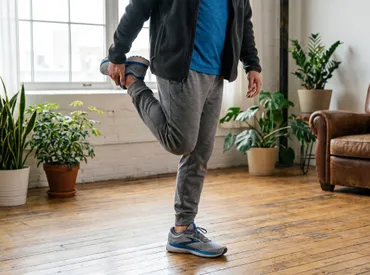
Every step you take relies heavily on a complex anatomical structure that many people never think about until something goes wrong. The big toe joint serves as the primary pivot point for forward propulsion, absorbing up to fifty percent of your body weight with each stride. When this critical mechanism functions properly, walking, running, and maintaining balance feel effortless. However, modern lifestyles, inappropriate footwear, repetitive microtrauma, and underlying metabolic conditions frequently place excessive strain on this vulnerable area. Understanding the intricate mechanics, common pathologies, and evidence-based management strategies for the big toe joint is essential for preserving long-term mobility and overall musculoskeletal health. This comprehensive guide explores everything from foundational anatomy to advanced treatment protocols, empowering you to recognize early warning signs, implement preventive measures, and collaborate effectively with healthcare professionals to maintain pain-free movement.
Anatomy and Function of the Big Toe Joint
The metatarsophalangeal (MTP) joint, commonly referred to as the big toe joint, operates as a highly specialized synovial hinge mechanism connecting the distal end of the first metatarsal bone to the proximal phalanx of the hallux per the Cleveland Clinic. Unlike simpler articulations found in the fingers or knees, this joint features a complex biomechanical architecture designed specifically to manage substantial ground reaction forces while facilitating smooth transition through the gait cycle. The joint capsule surrounds two small sesamoid bones embedded within the plantar plate tendon complex. These pea-shaped structures act as a mechanical pulley system, enhancing leverage for the flexor hallucis brevis muscle while distributing compressive loads away from vulnerable articular cartilage surfaces.
Free calculatorBMI CalculatorCalculate Body Mass IndexOpen the calculator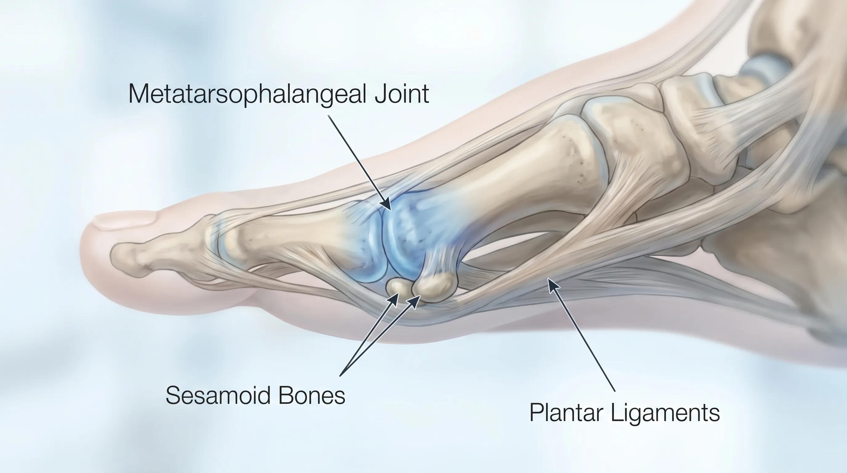
Articular hyaline cartilage lines both the metatarsal head and the base of the proximal phalanx, providing an ultra-low friction surface that permits approximately seventy degrees of dorsiflexion during toe-off. The surrounding synovial membrane continuously secretes viscous fluid that nourishes the avascular cartilage and reduces mechanical wear during dynamic activities. Supporting ligaments, including the medial and lateral collateral bands and the robust plantar plate, stabilize the joint against abnormal rotational forces and prevent hyperextension injuries. Proper biomechanical alignment ensures optimal weight distribution across the forefoot, but even minor deviations can initiate a cascade of compensatory movements that eventually compromise tissue integrity. When evaluating big toe joint dysfunction, clinicians carefully assess these interconnected structures to determine the precise source of pathology and develop targeted rehabilitation strategies.
The Metatarsophalangeal Joint Explained
During normal walking, the big toe joint remains relatively neutral until mid-stance, when the body weight shifts forward. As the heel lifts and the opposite foot swings forward, the joint must dorsiflex dramatically to allow proper clearance and forward momentum. This repetitive motion, occurring approximately ten thousand times daily for active individuals, places tremendous stress on the dorsal capsule, articular surfaces, and surrounding tendons. The joint's blood supply primarily originates from dorsal and plantar arterial networks, which become crucial during healing processes following injury or surgical intervention. Researchers studying gait biomechanics emphasize that inadequate joint mobility forces adjacent joints in the midfoot and ankle to compensate, often leading to secondary conditions like plantar fasciitis, Achilles tendinopathy, or knee pain.
Supporting Ligaments, Tendons, and Cartilage
The integrity of the big toe joint depends heavily on soft tissue health. The plantar plate, a thick fibrocartilaginous structure, anchors the flexor tendons and prevents dorsal migration of the phalanx during weight-bearing. Degeneration or partial tears in this structure frequently contribute to progressive deformities and chronic instability. Surrounding this framework, the extensor hallucis longus and brevis tendons provide dorsiflexion control, while intrinsic foot muscles maintain fine motor coordination. Synovial fluid viscosity and cartilage matrix composition rely on balanced nutritional delivery through diffusion, which explains why prolonged immobilization or chronic inflammation rapidly accelerates degenerative changes. Maintaining optimal tissue hydration through proper nutrition and targeted mobility exercises remains fundamental to preserving joint longevity.
Common Conditions Affecting the Big Toe Joint
Numerous pathological processes can compromise the structural and functional integrity of this essential articulation. Understanding the underlying mechanisms helps patients make informed decisions about conservative management and recognize when surgical consultation becomes necessary.
Hallux Rigidus and Osteoarthritis
Hallux rigidus represents a progressive degenerative condition characterized by cartilage breakdown, osteophyte formation, and restricted dorsiflexion. Initially presenting as stiffness and mild discomfort during push-off, the condition gradually advances to bone-on-bone articulation, significant pain with routine footwear, and visible swelling. The American Academy of Orthopaedic Surgeons classifies hallux rigidus using a grading system that evaluates joint space narrowing, osteophyte size, and range of motion limitation. Early intervention focuses on joint-sparing strategies, while advanced cases often require surgical reconstruction. Research indicates that genetic predisposition, previous trauma, and repetitive high-impact stress significantly accelerate disease progression according to the Mayo Clinic.
Gout and Inflammatory Arthritis
Metabolic disorders frequently target the big toe joint due to lower local tissue temperatures and slower circulatory turnover in distal extremities. Gout occurs when monosodium urate crystals precipitate within the joint space, triggering an intense autoimmune inflammatory response. Patients experience sudden, excruciating pain, pronounced erythema, and extreme sensitivity to light touch or bedding pressure. Unlike mechanical degeneration, inflammatory flares respond rapidly to targeted pharmacological interventions but require long-term uric acid management to prevent chronic arthropathy as detailed by the CDC. Rheumatoid arthritis and psoriatic arthritis can also affect the joint, typically presenting with symmetrical involvement, prolonged morning stiffness, and systemic symptoms that necessitate specialized immunomodulatory treatment.
Bunions (Hallux Valgus)
A bunion develops when biomechanical imbalances cause the first metatarsal to drift medially while the proximal phalanx angulates laterally. The resulting prominence creates friction against restrictive footwear, leading to adaptive bursitis, callus formation, and joint subluxation. Over time, the altered load distribution accelerates cartilage wear and destabilizes the entire forefoot complex. Contributing factors include inherited foot structure, hypermobility syndromes, and chronic use of narrow-toed shoes. Conservative management emphasizes pressure redistribution through orthotic intervention, while surgical correction addresses structural realignment and soft tissue balancing per clinical guidelines from Mayo Clinic.

Sprains, Fractures, and Turf Toe
Acute trauma frequently damages the joint capsule, collateral ligaments, or sesamoid apparatus. Turf toe occurs when the big toe joint experiences forced hyperextension during athletic activities, commonly tearing the plantar plate and surrounding ligaments. Athletes experience immediate pain, swelling, and diminished pushing power. Sesamoid fractures, often resulting from direct impact or repetitive stress in ballet dancers and runners, present with localized plantar tenderness and pain during toe-off. Proper acute management following the RICE protocol, combined with protected weight-bearing and progressive rehabilitation, typically restores function. However, neglected injuries frequently progress to chronic instability, avascular necrosis, or secondary osteoarthritis requiring surgical reconstruction.
Recognizing Symptoms and Warning Signs
Early identification of joint dysfunction dramatically improves treatment outcomes and prevents irreversible structural damage. Patients should monitor subtle changes in mobility patterns, pain localization, and gait mechanics.
Early Indicators vs. Advanced Symptoms
Initial warning signs often include mild morning stiffness lasting less than fifteen minutes, occasional sharp pain during toe-off, and subtle reduction in upward bending range. Shoes may feel tighter or cause friction over the dorsal joint surface. As pathology progresses, symptoms evolve into persistent aching pain unrelieved by rest, pronounced swelling that interferes with standard footwear, audible crepitus or grinding sensations during movement, and visible deformity or bony enlargement. Advanced stages frequently cause compensatory gait alterations, shifting weight laterally to avoid the painful joint. This altered biomechanics increases stress on the lesser toes, midfoot arches, and lower extremity joints, creating secondary conditions that complicate recovery.
When to Seek Professional Medical Evaluation
Consulting a qualified healthcare provider becomes essential when pain persists beyond two weeks despite conservative measures, when swelling restricts normal shoe wear, or when noticeable deformity develops. Red flag symptoms requiring immediate evaluation include sudden severe pain with joint redness and fever, inability to bear weight, visible open wounds or ulceration over prominent bony areas, and progressive neurological symptoms like numbness or tingling extending into the foot. A podiatrist or orthopedic specialist can perform comprehensive diagnostic testing to differentiate between mechanical degeneration, inflammatory processes, and acute structural damage.
Diagnosis and Medical Assessment
Accurate diagnosis relies on systematic clinical evaluation combined with advanced imaging modalities. Healthcare professionals integrate patient history, physical examination findings, and objective diagnostic data to formulate precise treatment pathways.
Clinical Examination and Patient History
A thorough assessment begins with detailed questioning regarding symptom onset, aggravating activities, footwear habits, previous injuries, and family medical history. Clinicians evaluate gait patterns, observing weight distribution, toe clearance, and compensatory movements. Manual palpation identifies precise tenderness localization, swelling distribution, and temperature variations. Range of motion testing measures both active and passive dorsiflexion and plantarflexion, comparing bilateral function. Specialized stress tests assess ligamentous stability, while muscle strength evaluations identify weakness patterns that contribute to joint overload. This comprehensive clinical picture guides subsequent diagnostic decisions.
Imaging Techniques and Laboratory Tests
Weight-bearing radiographs serve as the gold standard for evaluating joint space narrowing, osteophyte formation, bone alignment, and structural deformity. Standard anteroposterior, lateral, and oblique views provide multi-dimensional assessment capabilities. Advanced cases may require magnetic resonance imaging (MRI) or diagnostic ultrasound to visualize cartilage integrity, plantar plate tears, tendon pathology, or subtle stress fractures. Laboratory tests become crucial when inflammatory or metabolic conditions are suspected. Serum uric acid levels, complete blood counts, C-reactive protein, erythrocyte sedimentation rate, and rheumatoid factor testing help differentiate gout, rheumatoid arthritis, infection, or degenerative wear. Joint aspiration may be performed to analyze synovial fluid crystals or rule out septic arthritis.
Comprehensive Treatment Options
Management strategies span conservative interventions to advanced surgical procedures. Treatment selection depends on symptom severity, functional limitations, patient age, activity goals, and underlying pathology.
Conservative and Non-Surgical Approaches
First-line management emphasizes reducing inflammation, restoring mobility, and optimizing biomechanics following NIH osteoarthritis management guidelines. Over-the-counter or prescription nonsteroidal anti-inflammatory drugs (NSAIDs) effectively manage acute pain and swelling. Topical diclofenac provides localized relief with fewer systemic side effects. Physical therapy protocols incorporate manual joint mobilizations, soft tissue myofascial release, targeted stretching for gastrocnemius-soleus complex, and progressive strengthening of intrinsic foot muscles. Custom prescription orthotics with rigid forefoot posting or rocker-bottom designs limit excessive joint dorsiflexion during gait. Corticosteroid injections deliver potent anti-inflammatory medication directly into the joint space, providing months of relief for acute flare-ups. Hyaluronic acid viscosupplementation and platelet-rich plasma (PRP) therapies show promising results in stimulating cartilage metabolism and reducing synovial inflammation.
For patients managing gout, dietary modifications, adequate hydration, and urate-lowering medications prevent crystal deposition and recurrent attacks. Activity modification during acute phases protects healing tissues while maintaining cardiovascular fitness through low-impact alternatives. Comprehensive conservative care successfully manages symptoms in the majority of early to moderate cases, delaying or eliminating the need for invasive procedures.
Surgical Interventions and Recovery
When conservative measures fail to restore functional mobility or structural deformity progresses significantly, surgical consultation becomes appropriate. Arthroscopic debridement removes loose cartilage fragments and trims obstructive osteophytes, offering minimally invasive symptom relief for mild to moderate stiffness. Cheilectomy excises dorsal bone spurs and restores dorsiflexion range, preserving natural joint mechanics while alleviating impingement pain. For advanced hallux rigidus, arthrodesis permanently fuses the metatarsal and proximal phalanx at an optimal functional angle. Despite eliminating joint motion, patients consistently report high satisfaction rates, return to most activities, and durable pain relief. Total joint arthroplasty replaces damaged articular surfaces with prosthetic components, attempting to preserve mobility. However, implant longevity and potential complications require careful patient selection.
Postoperative rehabilitation follows structured protocols beginning with protected weight-bearing in surgical shoes or boots, transitioning to progressive range of motion exercises, strengthening programs, and gradual return to functional footwear. Adherence to postoperative guidelines significantly influences long-term outcomes and complication rates.
Prevention and Daily Management Strategies
Proactive joint preservation reduces disease incidence and extends functional longevity. Integrating evidence-based preventive measures into daily routines protects vulnerable structures from unnecessary wear and tear as recommended by the World Health Organization for healthy aging and musculoskeletal care.
Footwear Selection and Orthotics
Appropriate footwear represents the most impactful daily intervention for big toe joint health. Shoes must feature adequate length (typically one-half to one inch longer than the longest toe), generous width at the metatarsal heads, and minimal toe spring to allow natural movement during gait. Stiff-soled athletic shoes or hiking boots reduce repetitive dorsiflexion stress, while cushioned rocker-bottom designs facilitate smoother transitions for individuals experiencing early degeneration. Custom orthotics provide individualized arch support, control excessive pronation, and incorporate metatarsal pads that offload the forefoot. Over-the-counter inserts with rigid carbon fiber shanks offer effective stiffness for mild symptoms. Patients should rotate footwear daily, avoid worn-out shoes with compromised midsoles, and replace athletic shoes every three hundred to five hundred miles.

Exercises for Joint Mobility and Strength
Targeted exercises maintain synovial fluid circulation, prevent capsular contractures, and strengthen stabilizing musculature. Daily mobility routines should include gentle toe spreads, towel scrunches for intrinsic muscle activation, and seated dorsiflexion stretches against a wall to improve upward bending range. The short foot exercise, involving active arch elevation without toe curling, enhances neuromuscular control and reduces joint loading. Yoga-inspired toe poses and barefoot balance training improve proprioception and coordination. Consistency matters more than intensity; dedicating ten minutes daily yields superior results compared to sporadic intense sessions.
Nutritional and Lifestyle Considerations
Optimal joint health extends beyond mechanical interventions. Maintaining a healthy body weight reduces compressive forces transmitted through the foot during weight-bearing activities. Anti-inflammatory diets rich in omega-3 fatty acids, leafy greens, berries, and turmeric provide natural pain modulation and tissue repair support. Adequate hydration maintains synovial fluid viscosity and cartilage pliability. Smoking cessation improves peripheral circulation, enhancing tissue oxygenation and accelerating healing responses. Incorporating low-impact cross-training into fitness routines diversifies joint loading patterns, preventing repetitive stress accumulation. Periodic professional foot evaluations identify biomechanical deviations before they progress into symptomatic conditions.
| Treatment Modality | Primary Indications | Expected Recovery Timeline | Long-Term Efficacy |
|---|---|---|---|
| Physical Therapy & Orthotics | Early stiffness, mild pain, bunion management | 6-12 weeks | Moderate to High (maintenance required) |
| Corticosteroid Injection | Acute inflammatory flare, severe swelling | 1-2 weeks (relief lasts 3-6 months) | Temporary symptom control |
| Cheilectomy Surgery | Dorsal osteophyte impingement, preserved cartilage | 4-8 weeks | High (may progress over 5-10 years) |
| Arthrodesis Fusion | End-stage arthritis, severe deformity, chronic pain | 3-6 months | Very High (permanent, durable solution) |
| PRP/Biologic Therapy | Early degeneration, active patients seeking alternatives | 8-12 weeks | Variable (emerging evidence, promising) |
This comparative framework illustrates how clinical decision-making integrates symptom severity, functional goals, and tissue integrity to optimize patient outcomes.
Frequently Asked Questions
How do I know if my big toe pain is arthritis or a bunion?
Arthritis in the big toe joint typically presents as deep aching pain, stiffness (especially in the morning), and reduced range of motion when bending the toe upward. A bunion involves a visible bony protrusion on the inner side of the joint, often accompanied by redness, skin irritation from shoes, and progressive outward deviation of the toe. Imaging studies like weight-bearing X-rays provide definitive differentiation.
Can a damaged big toe joint heal without surgery?
Many conditions affecting the joint can be managed conservatively. While cartilage degeneration from osteoarthritis cannot be reversed, symptoms often improve significantly with targeted physical therapy, custom orthotics, anti-inflammatory protocols, and activity modification. Surgical intervention is typically reserved for severe cases where conservative care fails to restore functional mobility.
What footwear modifications help protect the big toe joint?
Shoes with a wide, rounded toe box prevent compression and allow natural toe splay. A stiff or rocker-bottom sole reduces excessive bending at the joint during toe-off. Low heels (under two inches) maintain proper foot alignment, while avoiding narrow, pointed styles minimizes pressure on vulnerable joint structures and surrounding soft tissues.
Is it safe to exercise when experiencing big toe joint pain?
Low-impact activities like swimming, cycling, and seated resistance training are generally safe and help maintain joint circulation and muscle support. However, high-impact exercises, hill running, or movements requiring extreme toe dorsiflexion should be temporarily modified or avoided during acute flare-ups to prevent further tissue irritation.
How long does recovery take after big toe joint surgery?
Recovery timelines vary by procedure. Minimally invasive soft tissue corrections often allow a return to regular footwear within four to six weeks. Arthrodesis or joint replacement surgeries typically require six to eight weeks of protected weight-bearing in a boot, followed by progressive rehabilitation. Full functional recovery and optimal gait mechanics may take three to six months.
Conclusion
The big toe joint plays an indispensable role in human locomotion, bearing substantial mechanical loads while facilitating smooth forward progression. When this intricate structure becomes compromised, the resulting pain and mobility restrictions significantly impact daily function, athletic performance, and overall quality of life. Early recognition of warning signs, proactive biomechanical optimization, and evidence-based conservative management often prevent disease progression and delay invasive interventions. Patients experiencing persistent symptoms should seek evaluation from qualified foot and ankle specialists who can perform comprehensive diagnostics and develop individualized treatment pathways. Whether utilizing targeted physical therapy, implementing footwear modifications, exploring advanced injection therapies, or considering surgical reconstruction, modern medicine offers numerous effective strategies to restore comfort and preserve function. Commitment to consistent joint care, lifestyle adjustments, and regular preventive monitoring empowers individuals to maintain active, pain-free mobility for decades to come. Prioritizing foot health is not merely about treating discomfort; it is an investment in long-term musculoskeletal resilience and sustained vitality.
How we work
Researched and drafted with AI assistance, then reviewed by a human editor against the cited sources.
This article is general health information, not medical advice. Consult a qualified healthcare professional about your own situation.