What Is the Purpose of the Gallbladder? Anatomy, Function, and Digestive Health Guide

The human digestive system operates as a remarkably coordinated network of organs, enzymes, and regulatory hormones designed to extract essential nutrients from the food we consume. Among these specialized structures sits a small, pear-shaped organ that plays a disproportionately vital role in lipid metabolism and nutrient assimilation. Understanding what is the purpose of the gallbladder reveals much about how our bodies process dietary fats, regulate cholesterol balance, and maintain long-term gastrointestinal harmony. Despite its modest size, this organ influences everything from energy extraction to fat-soluble vitamin absorption. When functioning optimally, it operates silently and efficiently, rarely drawing attention until a disruption occurs. However, its absence from daily conversation often masks the profound physiological contributions it makes to metabolic health. This guide explores the intricate anatomy, biochemical processes, and clinical significance of the biliary system, offering evidence-based insights into how this organ works, what happens when it malfunctions, and how you can actively support its function through targeted lifestyle interventions. By demystifying its physiological role, readers gain a comprehensive understanding of why digestive wellness depends on maintaining a healthy, well-functioning biliary tract.
Understanding the Gallbladder’s Anatomy and Location
The gallbladder is a hollow, sac-like structure approximately three to four inches in length, nestled against the visceral surface of the right lobe of the liver. Its anatomical positioning is not incidental; it sits directly within the biliary fossa, allowing for efficient communication with the hepatic ducts and the cystic duct. Measuring roughly the size of a small pear or a folded peach, it possesses a flexible, muscular wall capable of expanding to store up to fifty milliliters of concentrated bile. The organ's structure comprises three distinct histological layers: the mucosa, the muscularis, and the serosa. The innermost mucosal layer features numerous epithelial folds that dramatically increase the surface area available for fluid absorption. Beneath this lies a robust muscular layer responsible for generating the contractile force needed to expel bile into the common bile duct during digestion. The outermost serosal layer provides structural integrity and reduces friction against adjacent abdominal organs.
Free calculatorWater Intake CalculatorCalculate daily water needsOpen the calculatorPhysical Structure and Size
While often perceived as a simple storage vessel, the gallbladder's architecture is highly specialized for fluid dynamics and chemical modification. The mucosal lining contains tightly packed absorptive cells that actively transport sodium, chloride, and water from the incoming bile back into the bloodstream. This process transforms dilute, hepatic bile into a highly concentrated solution that is approximately ten times more potent. The organ's neck contains spiral valves known as the spiral valves of Heister, which prevent premature bile reflux and help maintain directional flow during contraction. Understanding what is the purpose of the gallbladder becomes clearer when examining this microarchitecture. The precise cellular arrangement ensures that bile remains stable, sterile, and primed for immediate deployment when chyme enters the duodenum. Variations in gallbladder size and shape are clinically normal, though significant anatomical anomalies may predispose individuals to biliary stasis or congenital ductal obstructions.
Relationship with Liver and Bile Ducts
The gallbladder does not operate in isolation; it functions as an integral component of the hepatobiliary system. Bile production begins in the hepatocytes, the primary functional cells of the liver, where it is continuously secreted into microscopic bile canaliculi. From there, bile flows into the right and left hepatic ducts, which merge to form the common hepatic duct. This duct then joins the cystic duct, creating the common bile duct that ultimately delivers bile to the small intestine. The junction where these pathways meet is regulated by a sophisticated network of sphincters, most notably the sphincter of Oddi, which prevents intestinal backflow and coordinates the precise timing of bile release. When asking what is the purpose of the gallbladder, it is essential to recognize its role as a dynamic buffer within this system. During fasting periods, the sphincter of Oddi remains closed, forcing bile backward into the gallbladder for storage. When food consumption triggers hormonal signals, the sphincter relaxes while the gallbladder contracts, creating a unidirectional flow that optimizes nutrient breakdown. This intricate anatomical relationship underscores why gallbladder dysfunction frequently manifests as hepatic or intestinal symptoms.
The Core Functions: What Exactly Does the Gallbladder Do?
At a fundamental level, this organ serves as a biological reservoir and chemical concentrator. Its primary physiological mandate revolves around the efficient management of bile, a complex digestive fluid synthesized by the liver. Without this regulatory structure, bile would continuously drain into the duodenum, diluting rapidly and losing its emulsifying potency. The organ ensures that highly concentrated digestive enzymes and bile salts are available precisely when they are most needed: immediately after fat-containing meals. By modulating bile storage, concentration, and targeted release, it transforms a continuous hepatic output into a meal-responsive digestive tool. This mechanism significantly improves metabolic efficiency, allowing the body to extract maximum nutritional value from dietary lipids while minimizing waste. Recognizing what is the purpose of the gallbladder highlights its indispensable role in maintaining digestive homeostasis and metabolic balance.
Bile Storage and Concentration
During periods between meals, the biliary system shifts into conservation mode. The liver continuously produces bile, but rather than allowing it to pass directly into the intestines where it would be ineffective, the cystic duct valves remain closed. This creates a pressure gradient that diverts hepatic secretions into the gallbladder. Once inside, the epithelial lining actively absorbs water and electrolytes through sodium-potassium ATPase pumps and aquaporin channels. This dehydration process increases the concentration of bile acids, phospholipids, cholesterol, and bilirubin by up to tenfold. The resulting fluid is thicker, darker, and far more chemically aggressive than its hepatic precursor. This concentration step is crucial for emulsification efficiency, as highly concentrated bile can rapidly disperse large lipid globules into microscopic micelles, vastly increasing the surface area available for pancreatic lipase action. When considering what is the purpose of the gallbladder, the storage and concentration phase represents its most defining biochemical contribution.
Regulated Release into the Small Intestine
The transition from fasting to feeding triggers a sophisticated neurohormonal cascade that precisely controls bile delivery. As partially digested food (chyme) exits the stomach and enters the duodenum, specialized enteroendocrine cells detect the presence of fats and amino acids. In response, these cells release cholecystokinin (CCK) and secretin into the bloodstream. CCK acts as the primary stimulant, binding to receptors on the gallbladder's smooth muscle layer and inducing forceful, rhythmic contractions. Simultaneously, it prompts the relaxation of the sphincter of Oddi, opening the pathway into the small intestine. Secretin further stimulates bicarbonate-rich pancreatic juice secretion, neutralizing gastric acid and creating an alkaline environment optimal for bile function. This synchronized release ensures that concentrated bile mixes precisely with incoming chyme, maximizing lipid breakdown without irritating the intestinal mucosa. The temporal precision of this mechanism demonstrates why the organ functions as a true regulatory valve rather than a passive pouch.
Facilitating Fat Digestion and Nutrient Absorption
Dietary triglycerides are hydrophobic molecules that naturally coalesce into large, indigestible droplets in aqueous environments. The gallbladder's concentrated bile contains amphipathic molecules called bile salts that act as biological detergents. These molecules possess both hydrophilic (water-loving) and hydrophobic (fat-loving) regions, allowing them to surround fat droplets and break them into minute micelles. This emulsification process is a prerequisite for enzymatic digestion, as pancreatic lipase can only access triglyceride molecules at the surface of these microscopic droplets. Once lipids are broken down into fatty acids and monoglycerides, they combine with bile salts to form mixed micelles that can traverse the unstirred water layer adjacent to the intestinal epithelium. From there, fats are absorbed into enterocytes, reassembled into chylomicrons, and transported via the lymphatic system. Additionally, this same mechanism facilitates the absorption of fat-soluble vitamins (A, D, E, and K), which rely on micellar transport for intestinal uptake. Without understanding what is the purpose of the gallbladder, one cannot fully appreciate how critical it is for converting inert dietary fats into usable metabolic fuel and essential cofactors.
Bile Composition and How the Gallbladder Modifies It
Bile is far more than a simple digestive juice; it is a complex biochemical solution composed of approximately eighty-five percent water, alongside bile acids, cholesterol, phospholipids, electrolytes, bilirubin, and various organic and inorganic compounds. The liver synthesizes primary bile acids from cholesterol through a tightly regulated metabolic pathway involving enzymes like cholesterol 7-alpha-hydroxylase. Once secreted, these compounds undergo significant modification within the gallbladder, where water absorption alters their solubility, concentration, and chemical behavior. The balance between these components determines whether bile remains in solution or precipitates into pathological formations. Monitoring this equilibrium is essential for maintaining long-term biliary health.
Water, Electrolytes, and Bile Salts
The gallbladder's mucosal epithelium is highly permeable to water and actively transports ions. Sodium and chloride ions are pumped out of the lumen, followed passively by water through osmotic gradients. This process reduces the volume of stored bile while preserving the total mass of bile acids and phospholipids. The resulting increase in bile acid concentration enhances their critical micellar concentration (CMC), the threshold at which they spontaneously form micelles. Bile salts themselves are steroid derivatives that remain chemically stable during fasting but become highly reactive upon contact with dietary fats. They also serve as signaling molecules, interacting with nuclear receptors like FXR and membrane receptors like TGR5 to regulate glucose metabolism, energy expenditure, and gut microbiota composition. This multifaceted role extends beyond simple digestion into systemic metabolic regulation.
Cholesterol and Bilirubin Management
Bile serves as the body's primary route for cholesterol excretion. The gallbladder must maintain cholesterol solubility within the bile solution, which requires a precise ratio of bile salts and phospholipids to keep cholesterol molecules dispersed in mixed micelles. When this balance tips, cholesterol can crystallize, forming the foundation for gallstones. Similarly, bilirubin, a breakdown product of senescent red blood cells, is conjugated in the liver and excreted via bile. The gallbladder's concentration process increases bilirubin density, which can occasionally precipitate as pigment stones if bacterial overgrowth or hemolysis occurs. These biochemical realities illustrate why the organ's function directly impacts systemic detoxification pathways and hematological waste management. Understanding what is the purpose of the gallbladder requires acknowledging its role as a metabolic filter that prevents lipid and pigment accumulation in peripheral tissues.
The Concentration Process
The concentration mechanism relies on specialized transporter proteins embedded in the apical and basolateral membranes of the gallbladder epithelium. Sodium-proton exchangers and chloride-bicarbonate antiporters work in tandem to establish electrochemical gradients that drive water reabsorption. Simultaneously, the epithelial barrier prevents the back-diffusion of concentrated bile acids into the tissue, protecting against chemical injury. The resulting hyperosmolar environment ensures that bile remains highly potent, ready to trigger rapid emulsification upon release. This process also contributes to bile viscosity management; overly dilute bile lacks emulsifying power, while overly thick bile risks stasis and sludge formation. Maintaining this physiological sweet spot depends on consistent hydration, balanced mineral intake, and healthy mucosal integrity.
| Component | Liver Secretion | Gallbladder Storage/Modification | Small Intestine Function |
|---|---|---|---|
| Water | High volume, dilute | Actively reabsorbed, concentrated | Aids chyme hydration |
| Bile Acids | Primary (cholic, chenodeoxycholic) | Concentrated up to 10x | Emulsify fats, form micelles |
| Cholesterol | Continuously secreted | Maintained in solubilized state | Absorbed or excreted |
| Bilirubin | Conjugated form | Density increases | Gives stool brown color |
| Electrolytes | Na+, Cl-, HCO3- | Reabsorbed via transporters | Maintains osmotic balance |
Common Gallbladder Conditions and Their Impact on Function
Despite its robust design, the gallbladder is susceptible to several pathological conditions that disrupt its normal physiology. Biliary stasis, chemical irritation, and bacterial colonization can all interfere with storage, concentration, or release mechanisms. These disorders frequently present with overlapping symptoms, making accurate diagnosis essential. Left unaddressed, chronic biliary dysfunction can cascade into systemic metabolic disturbances, nutritional deficiencies, and acute abdominal emergencies.
Gallstones (Cholelithiasis)
Gallstones represent the most prevalent biliary disorder, affecting millions globally. They form when the delicate solubility balance of bile is disrupted, leading to crystallization of cholesterol, bilirubin, or calcium salts. Supersaturated cholesterol bile, rapid weight loss, pregnancy, and genetic predispositions significantly increase risk. Stones can remain asymptomatic (silent stones) for years, or they can lodge in the cystic duct or common bile duct, causing intense colicky pain known as biliary colic. When a stone obstructs the cystic duct, gallbladder contraction increases intraluminal pressure, triggering ischemic pain and mucosal inflammation. Understanding what is the purpose of the gallbladder clarifies why these obstructions are so debilitating: they halt the organ's primary function and trigger compensatory spasms that amplify discomfort. For comprehensive clinical guidance on gallstone management, refer to Mayo Clinic's Gallstone Overview.
Inflammation (Cholecystitis)
Acute or chronic inflammation typically follows prolonged ductal obstruction, leading to mucosal edema, bacterial overgrowth, and compromised blood supply. Patients often experience prolonged right upper quadrant pain, fever, leukocytosis, and positive Murphy's sign upon clinical examination. If untreated, cholecystitis can progress to gangrenous tissue changes, perforation, or secondary peritonitis. The inflammatory cascade also impairs the gallbladder's contractile responsiveness, creating a vicious cycle of bile stasis and further irritation. Chronic inflammation may lead to porcelain gallbladder, a calcified wall that carries increased oncogenic potential and typically requires prophylactic removal. Early intervention with antibiotics, fasting protocols, and surgical evaluation remains the standard of care for acute presentations.
Biliary Dyskinesia and Gallbladder Sludge
Biliary dyskinesia refers to functional impairment without structural obstruction. The gallbladder exhibits reduced ejection fraction during hepatobiliary iminodiacetic acid (HIDA) scans, indicating poor contractility despite normal anatomy. Patients experience classic biliary symptoms—postprandial pain, nausea, bloating—yet imaging reveals no stones. Gallbladder sludge, conversely, represents a pre-stone condition where concentrated bile thickens into particulate matter. Often transient, sludge can progress to calculi if left unmanaged. Both conditions highlight the importance of neuromuscular coordination in biliary function. Dietary modifications, hydration optimization, and targeted prokinetic agents often provide symptomatic relief before considering surgical intervention. For deeper insights into biliary motility disorders, consult Cleveland Clinic's Gallbladder Function Guide.
Living Without a Gallbladder: Adaptation and Long-Term Health
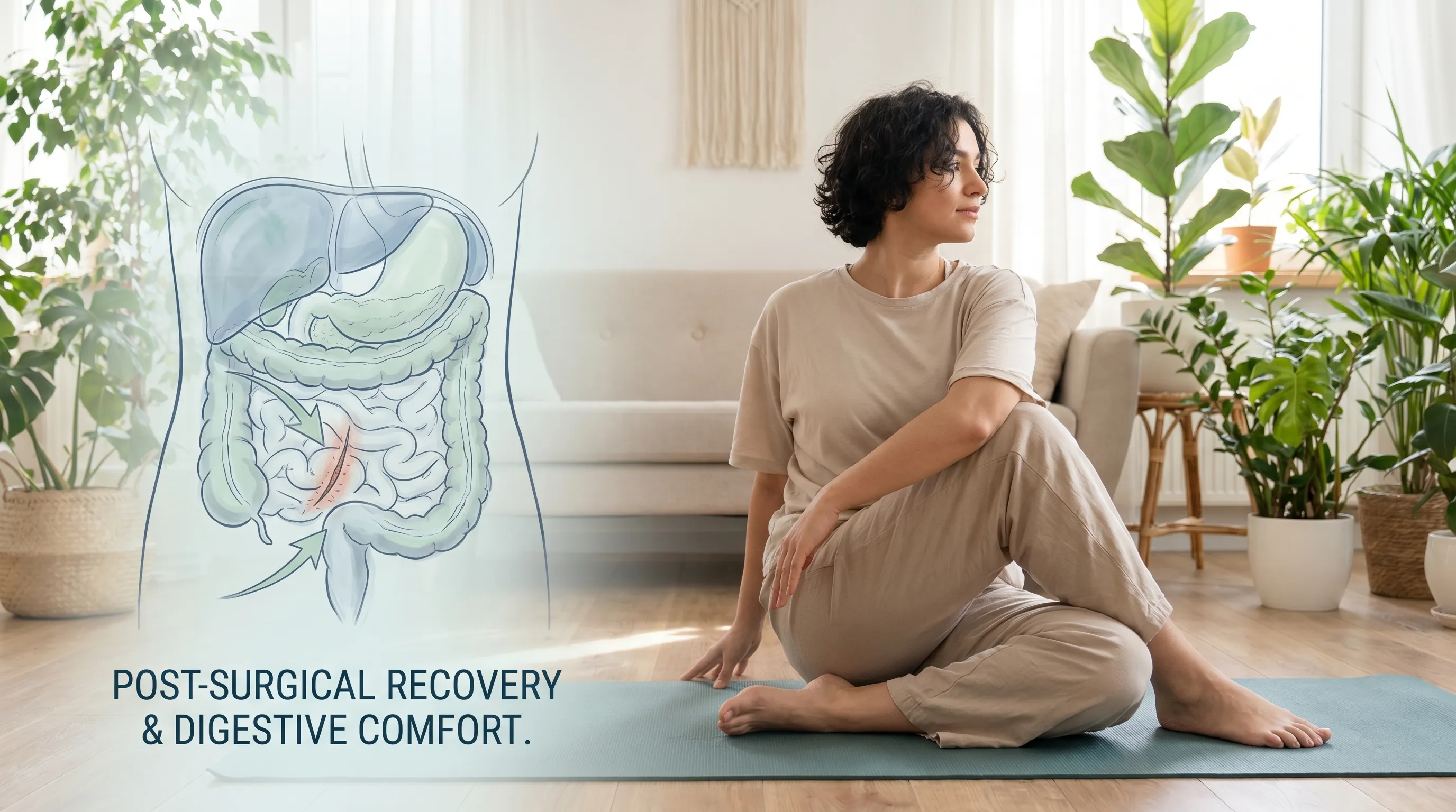
Surgical removal (cholecystectomy) is among the most common gastrointestinal procedures worldwide. While the organ is not essential for survival, its absence requires physiological adaptation. The liver continues producing bile, but instead of being stored and concentrated, it flows continuously into the duodenum. This baseline drip can initially overwhelm the small intestine, leading to bile acid diarrhea or fat malabsorption. However, the body gradually compensates through upregulation of intestinal bile acid transporters and microbiome adjustments.
How the Digestive System Compensates
Following surgery, the bile ducts undergo mild dilation, serving as secondary reservoirs. Over several months, the sphincter of Oddi develops adaptive tone regulation, preventing excessive reflux into the pancreas while allowing steady bile passage. The intestinal mucosa increases expression of sodium-dependent bile acid cotransporters, improving reabsorption efficiency during enterohepatic circulation. The gut microbiota also shifts, favoring strains that metabolize bile acids into secondary forms with varying solubility profiles. These compensatory mechanisms illustrate the remarkable plasticity of the hepatobiliary-gastrointestinal axis. Most patients report full symptom resolution within six to twelve weeks post-operation. For evidence-based post-surgical care protocols, review NHS Gallbladder Recovery Guidelines.
Dietary Adjustments Post-Cholecystectomy
Immediate post-operative nutrition focuses on reducing biliary demand. Patients are advised to consume small, frequent meals rich in soluble fiber, lean proteins, and low-fat plant sources. Gradual reintroduction of healthy fats (avocado, olive oil, nuts) helps the digestive system rebuild lipid tolerance. Avoiding large, high-fat meals prevents overwhelming the continuous bile drip, which can cause osmotic diarrhea. Hydration remains critical, as adequate water intake supports bile fluidity and intestinal transit. Long-term, the Mediterranean diet pattern consistently demonstrates protective effects against post-cholecystectomy syndrome, offering balanced lipid profiles and anti-inflammatory phytonutrients. Registered dietitians often recommend tracking food intolerances using elimination journals during the transition phase.
Monitoring Digestive Changes
Not all patients adapt seamlessly. Post-cholecystectomy syndrome affects approximately ten to twenty percent of individuals, presenting with persistent abdominal pain, bloating, or altered bowel habits. Differential diagnoses include retained common bile duct stones, sphincter of Oddi dysfunction, irritable bowel syndrome exacerbation, or pancreatic insufficiency. Monitoring stool consistency, frequency, and nutritional markers (vitamin D, K, B12) helps identify malabsorption early. If bile acid diarrhea persists, sequestrants like cholestyramine can bind excess intestinal bile acids, reducing colonic irritation. Regular follow-up with a gastroenterologist ensures that functional adaptations remain on track and that secondary complications are addressed promptly.

Practical Strategies to Support Gallbladder Health
Proactive biliary care revolves around maintaining optimal bile composition, supporting smooth muscle function, and preventing stasis. Evidence-based lifestyle interventions significantly reduce the risk of calculi formation, inflammation, and functional impairment. By aligning dietary habits with physiological demands, individuals can preserve this organ's efficiency well into advanced age.
Diet and Hydration Guidelines
Nutritional balance directly impacts bile chemistry. Diets high in refined sugars and trans fats promote hepatic cholesterol overproduction and bile supersaturation. Conversely, diets rich in soluble fiber (oats, legumes, flaxseed) bind intestinal bile acids, prompting the liver to synthesize fresh bile from circulating cholesterol, thereby reducing biliary cholesterol saturation. Hydration ensures adequate bile volume, preventing sludge formation. Aim for at least two to three liters of water daily, adjusting for activity level and climate. Incorporating bitter foods (dandelion greens, arugula, artichoke) stimulates cephalic and gastric phases of digestion, priming biliary release. Coffee consumption in moderate amounts has been epidemiologically linked to reduced gallstone risk, likely due to caffeine's cholecystokinin-releasing properties. Always pair dietary fats with fiber to moderate absorption rates and reduce acute biliary strain.
Weight Management and Physical Activity
Rapid weight loss remains a primary trigger for gallstone development due to mobilized adipose tissue cholesterol and decreased gallbladder motility from caloric restriction. Gradual, sustainable fat loss (one to two pounds weekly) preserves biliary rhythm while improving metabolic markers. Aerobic exercise enhances splanchnic blood flow and stimulates gastrointestinal motility, reducing fasting intervals where bile stagnates. Strength training supports basal metabolic rate, improving insulin sensitivity and reducing hepatic lipid synthesis. Combining cardiovascular conditioning with resistance work creates an optimal physiological environment for bile synthesis and gallbladder emptying. Consistency matters more than intensity; daily movement patterns prevent prolonged fasting states that favor stone nucleation.
When to Seek Medical Evaluation
Certain symptoms warrant immediate clinical assessment. Persistent right upper quadrant pain radiating to the right shoulder or back, especially after meals, suggests biliary obstruction or inflammation. Jaundice, dark urine, pale stools, or fever indicate possible common bile duct involvement or cholangitis. Nausea, vomiting, or unexplained weight loss alongside digestive discomfort may signal advanced pathology. Diagnostic workups typically include abdominal ultrasound, liver function panels, and HIDA scans. Early consultation prevents complications like pancreatitis or hepatic impairment. Understanding what is the purpose of the gallbladder empowers patients to recognize dysfunction early, seek appropriate imaging, and collaborate with clinicians on personalized management strategies. For detailed symptom differentiation, explore NCBI's Biliary Disease Pathophysiology.
Frequently Asked Questions
Can you live a normal life without a gallbladder?
Yes, the vast majority of individuals adapt successfully to cholecystectomy and maintain normal digestive function. While initial dietary modifications are necessary to accommodate continuous bile flow, the body compensates through ductal dilation and enhanced intestinal bile acid reabsorption. Most patients resume full physical and social activities within four to six weeks, requiring only long-term awareness of fat tolerance levels.
What happens if gallbladder function is compromised?
Impaired function typically manifests as biliary stasis, reduced contractility, or ductal obstruction. Symptoms include postprandial pain, bloating, nausea, and fat intolerance. Over time, poor emptying promotes sludge and stone formation, increasing inflammation risk. Functional impairment may also contribute to nutrient malabsorption, particularly fat-soluble vitamins, and alter gut microbiome composition.
How quickly does digestion adapt after gallbladder removal?
Physiological adaptation begins immediately post-surgery, with the most noticeable adjustments occurring within the first two to four weeks. Complete stabilization of bile acid metabolism and intestinal motility usually requires six to eight weeks. During this period, patients should prioritize small, low-fat meals and gradually expand dietary variety as tolerance improves.
Does the gallbladder produce bile?
No. Bile synthesis occurs exclusively in the liver, where hepatocytes convert cholesterol into primary bile acids. The gallbladder's sole function is storage, concentration, and regulated release. This distinction is critical when considering what is the purpose of the gallbladder, as it operates as a reservoir and concentrator rather than a synthetic organ.
What foods should I avoid to protect my gallbladder?
Limit highly refined carbohydrates, trans fats, excessive saturated animal fats, fried foods, and heavily processed snacks. These ingredients promote hepatic cholesterol overproduction, bile supersaturation, and delayed gastric emptying. Instead, emphasize soluble fiber, plant sterols, lean proteins, and adequate hydration to maintain bile solubility and support smooth muscular contractions.
Conclusion
The biliary system represents one of the most elegant examples of physiological efficiency in human biology. By answering what is the purpose of the gallbladder, we uncover an organ meticulously designed to store, concentrate, and strategically release bile, transforming continuous hepatic output into a meal-responsive digestive catalyst. Its role in emulsifying dietary fats, enabling fat-soluble vitamin absorption, and maintaining cholesterol homeostasis underscores its irreplaceable contribution to metabolic health. While gallbladder dysfunction is common, understanding its mechanisms empowers individuals to implement preventive strategies, recognize early warning signs, and navigate post-operative adaptation with confidence. Prioritizing balanced nutrition, consistent hydration, mindful weight management, and regular medical evaluation ensures that this small but mighty organ continues to support digestive harmony throughout life. Whether functioning optimally or surgically removed, the body's ability to maintain nutritional equilibrium highlights the remarkable resilience of our digestive architecture. Embracing evidence-based wellness practices not only protects biliary health but enhances overall systemic vitality, proving that digestive care remains a cornerstone of long-term well-being.
How we work
Researched and drafted with AI assistance, then reviewed by a human editor against the cited sources.
This article is general health information, not medical advice. Consult a qualified healthcare professional about your own situation.