Understanding TMJ Tinnitus: Causes, Symptoms, and Evidence-Based Relief Strategies

The persistent ringing, buzzing, or humming in your ears may not originate from the inner ear at all. For millions of adults experiencing unexplained auditory symptoms, the true source lies much closer to the jawline. The connection between temporomandibular joint dysfunction and phantom ear noises is increasingly recognized by otolaryngologists, neurologists, and dental specialists alike. Understanding how tmj tinnitus develops requires a deep dive into craniofacial anatomy, neuromuscular pathways, and the complex way our nervous system processes sensory information. When the jaw becomes inflamed, misaligned, or chronically overworked, the tension radiates outward, disrupting the delicate structures responsible for hearing and balance. This phenomenon, while frustrating and often distressing, is highly manageable when approached with a structured, evidence-based treatment plan. By addressing the root musculoskeletal imbalances, integrating targeted therapies, and adopting sustainable daily habits, most individuals experience meaningful relief and reclaim their auditory comfort. The journey toward resolution begins with accurate recognition, professional evaluation, and a commitment to consistent, informed self-care.
Understanding the Anatomical and Neurological Link
The Temporomandibular Joint: Structure and Function
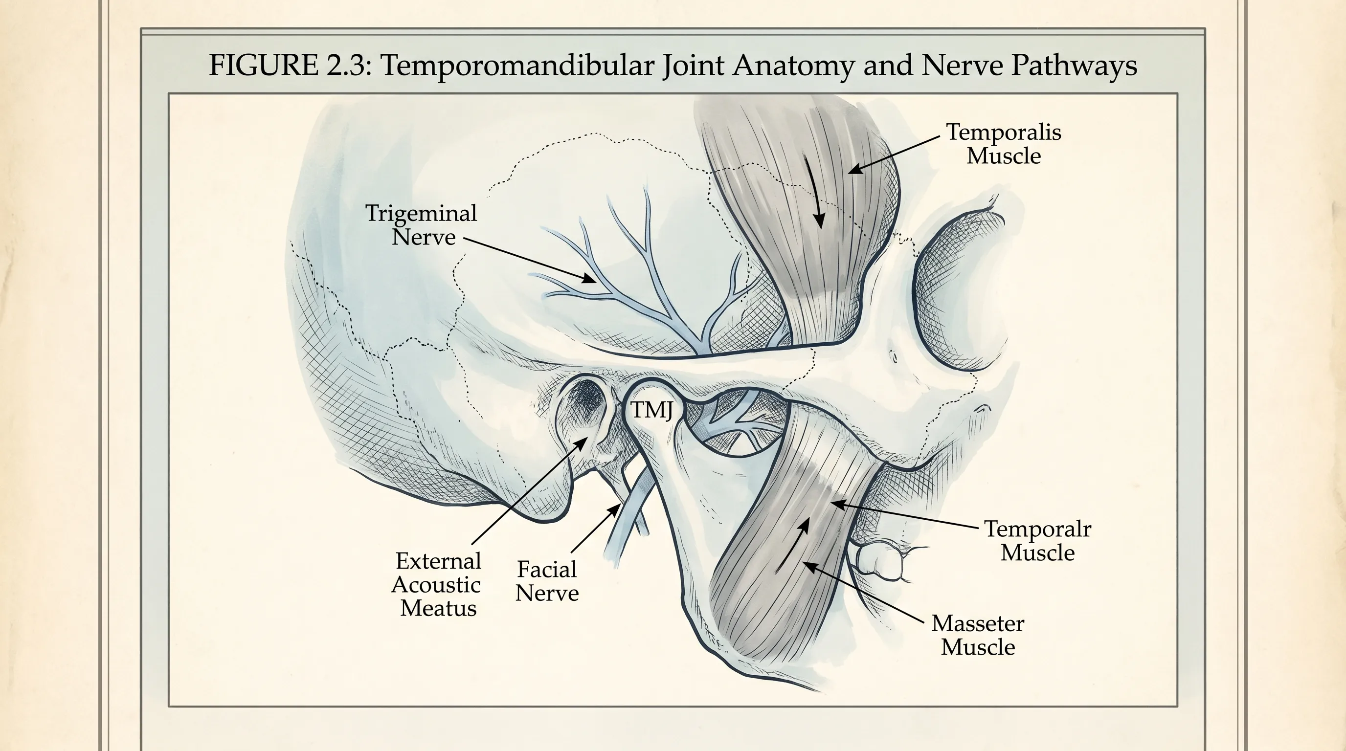
The temporomandibular joint serves as a highly complex, bilateral articulation connecting the mandible to the temporal bone of the skull. Unlike simpler hinge joints found in the extremities, this structure operates through a combination of rotational and translational movements, allowing for essential functions such as chewing, speaking, swallowing, and yawning. Each joint features a fibrocartilaginous disc that acts as a cushion, preventing bone-on-bone friction and ensuring smooth gliding motions. Surrounding the joint are powerful masticatory muscles, including the masseter, temporalis, medial pterygoid, and lateral pterygoid, which coordinate with extreme precision to control bite force and jaw trajectory. The region is densely innervated by the trigeminal nerve, the largest cranial nerve, which carries sensory information from the face, jaw, and oral cavity directly to the brainstem. Because of this intricate anatomical arrangement, any disruption to joint alignment, disc positioning, or muscular balance sends neurological signals that can easily misfire. The proximity of the joint to the middle ear cavity and the auditory nerve pathways explains why dysfunction frequently manifests as auditory disturbances rather than purely localized jaw discomfort. When inflammatory mediators, muscle spasms, or mechanical compression alter normal neural transmission, the brain may interpret these aberrant signals as continuous ringing, clicking, or pressure.
Free calculatorSleep CalculatorCalculate sleep cyclesOpen the calculatorHow Jaw Misalignment Affects the Auditory System
When the mandible shifts out of its optimal resting position, even by fractions of a millimeter, it creates a cascade of compensatory adaptations throughout the craniofacial skeleton. The altered biomechanics place excessive strain on the ligaments supporting the temporomandibular joint, while simultaneously pulling on the tensor tympani and tensor veli palatini muscles, both of which attach to the ear and play crucial roles in regulating middle ear pressure and protecting the eardrum from sudden loud noises. Chronic hyperactivity or spasm in these muscles can cause the eardrum to tense abnormally, producing a low-frequency rumbling or high-pitched whine. Additionally, misalignment often leads to uneven distribution of bite forces, triggering localized inflammation and joint effusion. The resulting swelling compresses adjacent blood vessels, reducing oxygen delivery to surrounding tissues and promoting further neuromuscular irritation. This compromised vascular supply, combined with persistent muscular tension, creates a feedback loop that sustains the tmj tinnitus cycle. Over time, the central auditory cortex adapts to this constant low-level noise, effectively turning it into a perceived baseline sound. Understanding this physiological chain reaction is essential for selecting interventions that address both the structural cause and the neurological response, rather than merely masking the symptom with temporary noise cancellation.
Neurological Pathways and Referred Sensory Symptoms
The human nervous system operates through highly interconnected referral networks, meaning that stimulation in one region can generate perceptual experiences in another. The trigeminal nerve shares anatomical and functional pathways with the vestibulocochlear nerve, which governs hearing and equilibrium. This convergence occurs in the trigeminal nucleus caudalis, located in the brainstem, where sensory signals from the jaw, face, and ears integrate and relay to higher cortical centers. When joint receptors, muscle spindles, or pain fibers in the temporomandibular region fire abnormally due to injury, inflammation, or chronic tension, the brainstem can misinterpret these signals as originating from the cochlea. This phenomenon, known as somatic tinnitus, is well-documented in peer-reviewed audiology and neurology literature. Patients frequently report that their ear ringing fluctuates with jaw movement, clenching, or head positioning, confirming its musculoskeletal origin. Furthermore, the cervical spine shares overlapping innervation patterns with the cranial nerves. Forward head posture and upper cervical dysfunction amplify tension in the suboccipital and trapezius muscles, which in turn increases baseline nervous system arousal and lowers the threshold for phantom auditory perception. Addressing these neurological crossovers requires a holistic approach that combines joint mobilization, muscle relaxation, and central nervous system desensitization to restore normal sensory filtering.
Recognizing and Differentiating TMJ-Induced Ear Ringing
Auditory Characteristics and Symptom Patterns
The acoustic presentation of tmj tinnitus varies significantly among individuals, but several hallmark characteristics distinguish it from purely auditory disorders. Most patients describe the sound as a constant, unilateral ringing, buzzing, humming, or clicking that fluctuates in intensity throughout the day. Unlike age-related hearing loss or acoustic neuroma-related ringing, which typically remains stable regardless of physical activity, tmj-related symptoms often intensify during jaw use, such as chewing tough foods, talking for extended periods, or clenching during periods of concentration. Some individuals experience a pulsating quality synchronized with their bite force, while others notice a sudden onset of noise after prolonged dental procedures or orthodontic adjustments. The volume can range from barely perceptible background static to overwhelming frequencies that interfere with concentration and sleep. Importantly, the sound frequently changes or temporarily disappears when patients gently reposition their mandible, apply heat, or massage the surrounding musculature. This direct mechanical correlation provides valuable clinical clues and helps differentiate musculoskeletal ringing from inner ear pathology. Keeping a detailed symptom diary that tracks jaw activity, stress levels, dietary habits, and auditory fluctuations can assist clinicians in identifying specific triggers and tailoring personalized management protocols.
Co-Occurring Musculoskeletal and Neurological Signs
Auditory disturbances rarely present in isolation when stemming from craniofacial dysfunction. A comprehensive clinical picture typically includes multiple secondary manifestations that reinforce the diagnosis. Chronic facial pain, particularly along the zygomatic arch and near the earlobe, is highly prevalent. Patients frequently report tenderness upon palpation of the masseter and temporalis muscles, accompanied by restricted mouth opening or deviation during jaw movement. Joint noises, such as clicking, popping, or grinding, often accompany mastication and indicate disc displacement or cartilage wear. Headaches, especially those localized to the temples, forehead, or base of the skull, frequently accompany the condition due to shared muscular attachments and nerve pathways. Dental symptoms, including worn enamel, fractured fillings, and heightened tooth sensitivity, suggest prolonged bruxism and abnormal occlusal loading. Additionally, many individuals experience neck stiffness, shoulder tension, and reduced cervical range of motion, highlighting the interconnected nature of cranial posture and thoracic mechanics. Recognizing this cluster of co-occurring signs enables earlier intervention and prevents unnecessary auditory testing when the primary driver is clearly musculoskeletal.
Distinguishing from Sensorineural and Conductive Conditions
Accurate differentiation is critical for directing appropriate care. Sensorineural tinnitus typically arises from damage to the hair cells within the cochlea, often triggered by noise exposure, ototoxic medications, or presbycusis. It is frequently accompanied by measurable hearing loss on pure-tone audiometry and lacks the direct mechanical fluctuation associated with jaw movement. Conductive tinnitus, conversely, results from physical obstructions in the outer or middle ear, such as cerumen impaction, otitis media, or Eustachian tube dysfunction. This form often produces a booming or roaring quality and improves temporarily when patients perform the Valsalva maneuver or swallow. tmj tinnitus, however, presents with normal or near-normal audiograms in most cases, and the perceived noise strongly correlates with masticatory activity, clenching, or postural shifts. Imaging of the temporomandibular joint via cone-beam computed tomography or magnetic resonance imaging frequently reveals disc displacement, joint effusion, or degenerative changes absent in primary otologic conditions. Collaborative evaluation between audiologists and maxillofacial specialists ensures that treatment targets the correct anatomical system, avoiding prolonged ineffective interventions and accelerating functional recovery.
Comprehensive Diagnostic Evaluation and Clinical Protocols
Initial Clinical Assessment and Patient History
A thorough diagnostic process begins with an extensive medical and dental history, capturing onset patterns, symptom progression, previous trauma, dental work history, and lifestyle factors. Clinicians utilize standardized questionnaires such as the Jaw Functional Limitation Scale and Tinnitus Functional Index to quantify symptom severity and track treatment response. Physical examination involves systematic palpation of the temporomandibular joints and surrounding musculature, assessing for tenderness, asymmetry, and range of motion limitations. Bite alignment analysis using articulating paper reveals uneven occlusal contacts that contribute to joint stress. Dynamic assessment during jaw opening, lateral excursions, and protrusion identifies joint noises, deviations, or locking episodes. Clinicians also evaluate cervical spine alignment, scapular positioning, and forward head posture to determine whether upper quadrant biomechanics are exacerbating craniofacial tension. Patient-reported data regarding sleep quality, bruxism awareness, stress triggers, and dietary habits provides essential context for designing a multidimensional management plan. This comprehensive baseline ensures that subsequent diagnostic steps remain targeted and clinically relevant.
Advanced Imaging and Specialized Diagnostic Tools
When conservative assessment indicates structural involvement or when symptoms persist despite initial therapy, advanced imaging becomes necessary. Magnetic resonance imaging delivers high-resolution visualization of the articular disc position, joint fluid accumulation, and surrounding soft tissue integrity, making it the gold standard for evaluating internal derangements. Cone-beam computed tomography provides detailed bony architecture assessment, identifying condylar resorption, osteophyte formation, or erosive changes indicative of degenerative joint disease. Panoramic radiography offers a broad overview of dental and maxillofacial structures, useful for detecting occlusal imbalances, impacted teeth, or asymmetrical growth patterns. Electromyography may be utilized in complex cases to measure muscle activity levels, identify hyperactive chewing patterns, and objectively quantify tension before and after therapeutic interventions. Hearing tests, including tympanometry and otoacoustic emissions, confirm cochlear function and rule out independent otologic pathology. Integrating these diagnostic modalities creates a precise anatomical and functional map, enabling clinicians to select the most appropriate therapeutic pathway and establish realistic recovery timelines.
Building an Interdisciplinary Care Team
Effective management of tmj tinnitus rarely relies on a single practitioner. Successful outcomes emerge from coordinated collaboration among dentists specializing in orofacial pain, physical therapists trained in craniofacial rehabilitation, otolaryngologists ruling out primary ear pathology, and sometimes pain management specialists or cognitive behavioral therapists addressing the psychological burden of chronic symptoms. Clear communication protocols ensure that imaging results, treatment responses, and patient progress are shared seamlessly across disciplines. Regular interdisciplinary meetings facilitate unified care plans that address structural alignment, neuromuscular reeducation, and lifestyle modifications simultaneously. Patients benefit from having a central care coordinator who synthesizes recommendations and prioritizes interventions based on symptom severity and functional impact. This collaborative model minimizes fragmented care, reduces redundant testing, and accelerates meaningful recovery by addressing the condition from every relevant physiological and behavioral angle.
Evidence-Based Medical and Therapeutic Interventions
Customized Occlusal Splints and Dental Appliances
Oral appliances represent one of the most widely studied and clinically validated interventions for tmj tinnitus. Unlike over-the-counter boil-and-bite guards, which can exacerbate joint stress by encouraging uncontrolled clenching, custom-fabricated occlusal splints are designed to redistribute bite forces evenly, prevent tooth grinding, and stabilize the mandible in a physiologically optimal position. Stabilization splints, covering all maxillary or mandibary teeth, are typically worn during sleep to mitigate nocturnal bruxism and allow inflamed joint tissues to rest. Anterior repositioning appliances may be indicated for patients with documented disc displacement, temporarily guiding the condyle into a more favorable alignment to reduce muscle tension and auditory referral. Progressive adjustment ensures that the splint promotes neuromuscular adaptation without creating permanent bite changes. Clinical studies demonstrate significant symptom reduction in over 70 percent of compliant users within three months of consistent wear. Regular dental follow-ups monitor appliance fit, track occlusal changes, and adjust therapy parameters as joint mechanics improve.
Targeted Physical Therapy and Rehabilitation Regimens
Physical therapy serves as the cornerstone of functional restoration, combining manual techniques, neuromuscular reeducation, and progressive exercise protocols to restore normal jaw mechanics and reduce auditory referral. Trained therapists utilize intraoral and extraoral myofascial release techniques to break up adhesions, decrease muscle hypertonicity, and improve local circulation. Gentle joint mobilization and traction techniques enhance capsular elasticity and promote disc repositioning when appropriate. Therapeutic exercise programs focus on controlled mandibular opening, lateral stretching, isometric strengthening, and cervical stabilization to correct postural imbalances contributing to craniofacial tension. Therapists frequently incorporate modalities such as low-level laser therapy, transcutaneous electrical nerve stimulation, and ultrasound to reduce inflammation and accelerate tissue healing. Patient education regarding proper ergonomics, breathing mechanics, and stress management reinforces clinical gains and empowers individuals to maintain progress independently. Consistent adherence to a structured rehabilitation program typically yields measurable improvements in pain scores, range of motion, and tinnitus intensity within 6 to 8 weeks.
Pharmacological Management and Pain Relief Strategies
Medications do not resolve underlying structural dysfunction but can play a vital adjunctive role in breaking the pain-tension-inflammation cycle. Short-term use of nonsteroidal anti-inflammatory drugs helps reduce joint effusion and muscular pain, creating a more favorable environment for physical therapy. Muscle relaxants, prescribed cautiously and for limited durations, can interrupt severe spasm cycles that perpetuate nerve irritation and auditory symptoms. In cases of significant central sensitization or comorbid chronic pain conditions, clinicians may consider neuromodulators such as low-dose tricyclic antidepressants or gabapentinoids to lower nervous system hyperexcitability and reduce phantom sound perception. Topical analgesic gels applied to the masseter and temporal regions provide localized relief without systemic side effects. All pharmacological interventions require careful monitoring, periodic reassessment, and gradual tapering to avoid dependency and ensure that medication supports rather than replaces active rehabilitation and lifestyle modification.
Holistic Self-Care and Daily Lifestyle Modifications
Dietary Adjustments and Chewing Habit Optimization
What you consume directly impacts joint loading and muscular recovery. Transitioning to a soft-food diet during acute flare-ups minimizes mechanical stress on compromised structures, allowing inflammation to subside and tissues to heal. Avoiding excessively hard, chewy, or sticky foods prevents sudden torque and excessive condylar pressure. Cutting food into small, manageable pieces reduces the required bite force and promotes symmetrical jaw movement. Chewing slowly and alternating sides distributes load evenly, preventing unilateral overload. Eliminating habitual gum chewing, ice crunching, and nail biting removes repetitive microtrauma that sustains muscular hyperactivity. Adequate hydration maintains tissue elasticity and synovial fluid viscosity, supporting smooth joint mechanics. Anti-inflammatory nutrition, emphasizing omega-3 fatty acids, leafy greens, turmeric, and lean proteins, complements clinical treatment by reducing systemic inflammatory markers and promoting tissue regeneration.
Stress Reduction, Mindfulness, and Jaw Relaxation
Psychological tension directly manifests as physical clenching, particularly during sleep or high-focus activities. Implementing daily mindfulness practices, such as diaphragmatic breathing, progressive muscle relaxation, and guided imagery, lowers baseline sympathetic nervous system arousal and reduces involuntary jaw tightening. Progressive jaw relaxation exercises involve consciously placing the tongue against the roof of the mouth, allowing teeth to separate slightly, and focusing on releasing facial tension with each exhale. Journaling, regular light exercise, and digital detox periods before bedtime improve sleep architecture, which is crucial for tissue repair and nervous system reset. Patients often benefit from biofeedback training, which provides real-time auditory or visual cues when clenching occurs, enabling conscious behavioral modification. Integrating stress management into daily routines creates a sustainable buffer against flare-ups and supports long-term auditory comfort.
Postural Alignment, Sleep Hygiene, and Ergonomic Adjustments
The head weighs approximately 10 to 12 pounds in neutral alignment. For every inch of forward displacement, the effective load on the cervical spine and jaw doubles, dramatically increasing muscular tension and joint compression. Ergonomic workstation setups should position screens at eye level, support lumbar curvature, and keep shoulders relaxed and retracted. Regular micro-breaks featuring gentle neck rolls, chin tucks, and shoulder blade squeezes interrupt prolonged static postures that strain the craniofacial complex. Sleep position significantly influences jaw alignment and nocturnal clenching. Side sleepers should avoid resting the jaw directly on the pillow, instead using a supportive cervical pillow that maintains neutral head and neck posture. Back sleeping with a contoured pillow reduces rotational torque on the temporomandibular joint. Limiting screen time before bed, maintaining consistent sleep schedules, and creating a cool, dark environment enhance restorative sleep quality, which directly correlates with symptom severity and recovery trajectory.
| Self-Care Strategy | Implementation Guidelines | Expected Timeframe for Noticeable Improvement | Key Precautions |
|---|---|---|---|
| Soft Diet Transition | Replace tough meats, nuts, raw vegetables with steamed alternatives; cut food into small pieces | 1–3 weeks | Ensure adequate protein and nutrient intake to support tissue healing |
| Occlusal Splint Wear | Use only custom-fitted devices; clean daily with mild soap and cool water; store in ventilated case | 4–8 weeks | Avoid over-the-counter boil-and-bite guards; report bite changes promptly |
| Jaw Stretching Routine | Perform 5–10 gentle controlled openings and lateral movements twice daily; stop if pain exceeds 3/10 | 6–12 weeks | Do not force beyond natural range; maintain upright posture during exercises |
| Postural Ergonomics | Align monitor at eye level; use lumbar support; take 2-minute stretch breaks every 45 minutes | 2–4 weeks | Adjust workspace gradually to prevent sudden muscular strain |
| Mindfulness Relaxation | Practice 10 minutes diaphragmatic breathing and facial tension scanning daily before sleep | 3–6 weeks | Consistency outweighs duration; pair with digital wind-down routine |

Frequently Asked Questions
Can a misaligned bite really cause ringing in the ears?
Yes. Malocclusion and abnormal jaw positioning can compress muscles, ligaments, and nerve pathways near the ear canal, creating phantom auditory signals. Correcting the bite through dental appliances often reduces or eliminates these symptoms.
How long does it take for TMJ tinnitus treatment to show results?
Timeline varies based on severity. Many patients experience noticeable relief within 4 to 6 weeks of consistent physical therapy, occlusal splint usage, and stress management. Chronic cases may require 3 to 6 months of multidisciplinary care.
Are there specific exercises that help reduce jaw-related ear ringing?
Yes. Controlled jaw opening, lateral jaw stretches, and resisted opening exercises strengthen and relax the masticatory muscles. Cervical stretching and postural corrections also reduce tension transmitted to the auditory system.
Should I see a dentist or an ENT specialist first?
Start with a healthcare provider to rule out primary ear pathology. If hearing tests are normal, an ENT often coordinates with a TMJ-focused dentist or maxillofacial specialist for a comprehensive musculoskeletal and occlusal evaluation.
Does stress make TMJ tinnitus worse?
Absolutely. Psychological stress increases cortisol and adrenaline, which heightens muscle tension, jaw clenching, and central nervous system sensitivity. Mindfulness, breathing techniques, and sleep optimization significantly reduce symptom intensity.
Conclusion
The persistent challenge of tmj tinnitus demands a comprehensive, multidimensional approach that addresses both structural dysfunction and neurological adaptation. By understanding the intricate relationship between the temporomandibular joint, craniofacial musculature, and auditory pathways, patients and clinicians can move beyond temporary masking strategies toward sustainable, root-cause resolution. Customized dental appliances, targeted physical therapy, strategic pharmacological support, and consistent lifestyle modifications work synergistically to restore normal joint mechanics, reduce muscular hyperactivity, and desensitize the central nervous system to phantom sounds. Success hinges on patience, professional guidance, and daily commitment to postural alignment, stress management, and ergonomic optimization. With accurate diagnosis, coordinated interdisciplinary care, and evidence-based self-care practices, most individuals experience significant symptom reduction, improved jaw function, and lasting relief from auditory disturbances. Prioritizing craniofacial health not only resolves the immediate discomfort of ringing ears but also enhances overall quality of life, enabling clearer focus, deeper rest, and greater physical comfort. Taking informed, proactive steps today lays the foundation for enduring wellness tomorrow.
How we work
Researched and drafted with AI assistance, then reviewed by a human editor against the cited sources.
This article is general health information, not medical advice. Consult a qualified healthcare professional about your own situation.