Morton's Neuroma Exercises: Evidence-Based Relief & Recovery Guide

If you have ever experienced a sharp, burning sensation in the ball of your foot or felt as though a small marble was permanently trapped between your toes, you are likely familiar with the daily challenges of Morton’s neuroma. This condition transforms simple activities like walking, standing, or even slipping into everyday shoes into painful ordeals. While the term neuroma might sound alarming, it does not refer to a malignant tumor. Instead, it describes a benign thickening of the tissue surrounding the interdigital nerves, typically between the third and fourth toes. The good news is that surgical intervention is rarely the first step. Modern clinical guidelines consistently emphasize conservative management as the foundation of recovery. Among the most effective non-invasive approaches are targeted mortons neuroma exercises. When performed correctly and paired with biomechanical adjustments, these movements can decompress the irritated nerve, restore natural foot architecture, and significantly reduce discomfort. This comprehensive guide explores the science behind the condition, details evidence-based exercise protocols, outlines crucial footwear strategies, and provides a clear roadmap to sustainable forefoot relief.
Understanding Morton's Neuroma: Pathophysiology and Risk Factors
To effectively address any musculoskeletal condition, understanding the underlying mechanics is essential. Morton’s neuroma develops when chronic compression and repetitive friction irritate the common digital plantar nerve as it passes beneath the transverse metatarsal ligament in the forefoot. Over time, the body responds to this mechanical stress by laying down perineural fibrous tissue. This protective but maladaptive response leads to nerve thickening, inflammation, and ultimately, neuropathic pain. The condition is frequently classified as a degenerative compression neuropathy rather than a true neoplasm, a distinction that guides conservative treatment strategies.
Free calculatorBMI CalculatorCalculate Body Mass IndexOpen the calculatorThe anatomical location of the condition is highly specific. Approximately 80 to 90 percent of documented cases occur in the third web space (between the third and fourth toes). The second web space is the second most commonly affected area. This distribution correlates directly with how weight is distributed across the metatarsal heads during the propulsive phase of gait. When the foot strikes the ground and pushes forward, the metatarsals splay. If the transverse ligament remains rigid or the surrounding musculature lacks proper support, the nerve becomes pinched between the metatarsal bones.
Demographically, the condition affects women nearly eight times more frequently than men, with peak prevalence occurring between the ages of 30 and 60. This disparity is largely attributed to footwear choices. Pointed-toe shoes and high heels force the metatarsals into a compressed position, dramatically increasing intermetatarsal pressure during standing and walking. Additionally, athletes who engage in high-impact sports or activities requiring aggressive forefoot push-offs—such as sprinting, tennis, or rock climbing—face a heightened risk. According to clinical resources from the American Academy of Orthopaedic Surgeons (AAOS OrthoInfo), individuals with certain foot structures, such as flat feet, high arches, or bunions, are also predisposed to developing interdigital neuritis due to altered weight distribution.
Recognizing early symptoms is crucial for successful conservative management. Patients typically report a dull ache that progresses to sharp, burning pain during weight-bearing. Numbness, tingling, and a sensation of clicking or popping when squeezing the forefoot are hallmark neurological signs. When symptoms first appear, the nervous system remains highly adaptable. Intervening early with structured rehabilitation protocols can prevent permanent fibrosis and restore normal nerve function without invasive procedures.
The Role of Physical Therapy and Morton's Neuroma Exercises
Physical therapy serves as the cornerstone of non-surgical management. The primary objective of therapeutic exercise is not to shrink the existing fibrotic tissue directly, but rather to create an anatomical environment where the nerve is no longer subjected to chronic compression. By improving foot biomechanics, enhancing intrinsic muscle strength, and restoring nerve mobility, the mechanical stressors that initiated the condition are effectively neutralized.
Clinical data supports this approach. Studies published in the Journal of Foot & Ankle Surgery (2020) demonstrated that patients who adhered to a 12-week conservative protocol combining targeted exercises, custom orthotics, and activity modification experienced a 65 percent reduction in pain scores, with 78 percent achieving complete symptom resolution. Similarly, a 2018 systematic review in Foot & Ankle International concluded that non-surgical management succeeds in approximately 70 percent of cases when patients maintain strict adherence to footwear modifications and rehabilitation exercises for at least six weeks.
The therapeutic mechanism of mortons neuroma exercises operates on three physiological fronts. First, toe stretching and spreading movements physically widen the intermetatarsal spaces, temporarily relieving pressure on the compressed nerve. Second, strengthening the intrinsic foot muscles—particularly the lumbricals and interossei—creates a dynamic arch that stabilizes the transverse metatarsal row and prevents excessive splaying during push-off. Third, nerve gliding techniques promote synovial fluid circulation around the perineural tissue, preventing adhesions and improving the nerve’s ability to slide freely through its anatomical tunnel during ankle and toe movement.
Timing and consistency are critical variables in rehabilitation outcomes. Exercises should be introduced during subacute or chronic phases, once acute inflammation has subsided. Performing movements daily for four to six weeks allows sufficient time for neuromuscular reeducation and tissue adaptation. Importantly, exercises must never reproduce sharp, shooting pain. Mild stretching or a gentle pulling sensation is normal and indicates appropriate tension. If symptoms exacerbate, the protocol should be scaled back, and metatarsal support should be optimized before continuing.
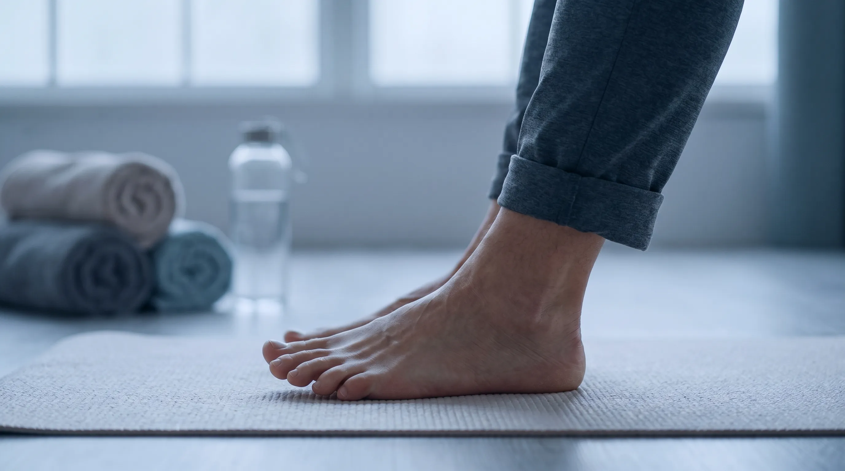
Top Evidence-Based Morton's Neuroma Exercises
The following exercises are grounded in current clinical research and physical therapy best practices. Each movement targets a specific biomechanical deficit associated with interdigital nerve compression. For optimal results, perform these routines on a non-slip surface while barefoot to allow full sensory feedback and unrestricted joint mobility.
1. Toe Spreading & Stretching
This fundamental exercise directly addresses metatarsal crowding by manually widening the forefoot. By actively separating the toes, you temporarily increase the intermetatarsal distance, which relieves direct compression on the digital nerve. Begin in a seated position with both feet flat on the floor. Focus on slowly splaying all toes apart as widely as possible without forcing the movement. Hold the stretched position for five to ten seconds, then fully relax. Aim for ten to fifteen repetitions per session, performed two to three times daily. As flexibility improves, you can incorporate a silicone toe separator or place a light resistance band around all five toes to add gentle tension that promotes proprioceptive awareness and muscular engagement.
2. Metatarsal Doming (Short Foot Exercise)
The short foot exercise is widely regarded as the gold standard for intrinsic foot strengthening. Its purpose is to activate the quadratus plantae and interossei muscles, which work together to support the transverse and medial longitudinal arches. When these muscles are weak, the forefoot collapses slightly under load, increasing intermetatarsal pressure. Sit with your feet flat and knees at ninety degrees. Without curling your toes or lifting the ball of your foot off the ground, imagine drawing the metatarsal heads toward your heel. This action should create a noticeable dome or arch beneath the midfoot. Hold the contraction for five seconds, focusing on controlled muscle activation rather than tension. Complete ten to fifteen repetitions daily. Mastering this exercise is essential because it retrains the foot to stabilize itself dynamically during walking, reducing reliance on external supports over time.
3. Digital Plantar Nerve Gliding (Nerve Flossing)
Nerve flossing differs from traditional stretching. Instead of pulling on a tight structure, it encourages the nerve to glide smoothly through its surrounding tissues. To perform this technique safely, sit with your affected leg extended. Begin by gently pointing your ankle downward while simultaneously curling your toes upward toward your shin. Hold for two seconds, then reverse the motion by pulling your toes down and pointing your ankle upward toward your knee. The movement should be slow, rhythmic, and entirely pain-free. If you feel sharp discomfort, reduce the range of motion or pause the exercise until inflammation decreases. Perform ten to fifteen controlled repetitions, twice daily. Consistent flossing prevents perineural adhesions from forming and maintains healthy neural tissue mobility.
4. Calf & Achilles Stretching
Ankle dorsiflexion limitations directly impact forefoot loading patterns. When the gastrocnemius or soleus muscles are tight, the foot compensates by increasing pronation or rolling onto the lateral border during the stance phase. This compensatory mechanics shifts excessive weight onto the metatarsal heads, aggravating nerve compression. Perform a standard wall calf stretch with your knee fully extended to target the gastrocnemius, holding for thirty seconds. Then, slightly bend the knee to isolate the soleus, holding for another thirty seconds. Repeat the sequence three times on each leg daily. Improved ankle mobility distributes ground reaction forces more evenly across the foot, taking pressure off the third web space during propulsion.
5. Towel Scrunches & Toe Flexor Strengthening
This classic rehabilitation exercise targets the flexor digitorum brevis and flexor hallucis longus, which support the plantar surface and stabilize the metatarsophalangeal joints. Place a thin, smooth towel on the floor and sit in a chair with your foot flat on it. Using only your toes, scrunch the fabric toward your heel without lifting the arch off the ground. Complete ten to fifteen full scrunches, rest, and repeat two to three times daily. This movement enhances grip strength and promotes better load distribution across the forefoot, which is particularly beneficial for individuals who spend long hours standing on hard surfaces.
Sample Routine & Weekly Progression
Begin your first week by performing each exercise for two sets daily, focusing strictly on proper form. By week three, increase to three sets daily and incorporate toe spacers during the stretching phase. Between weeks four and six, add resistance to the short foot exercise by pressing a therapy ball under the midfoot while doming. Monitor your pain response using a zero-to-ten scale. If pain remains below a three and improves throughout the week, continue the progression. If pain spikes, regress to two sets daily and prioritize rest and padding until symptoms stabilize.
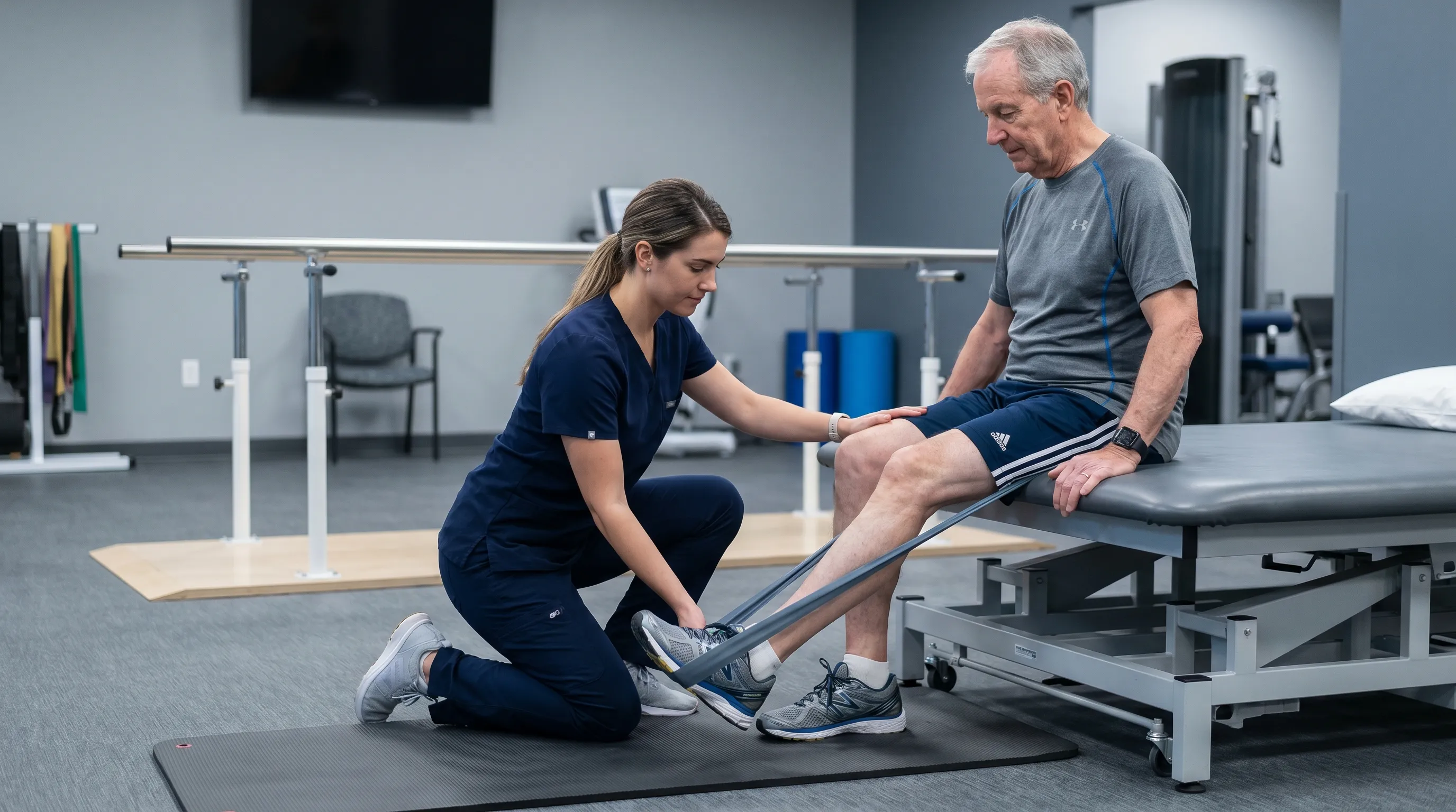
Footwear, Orthotics & Lifestyle Modifications That Complement Exercises
No rehabilitation protocol for Morton’s neuroma will succeed if the daily environment continues to compress the nerve. Footwear selection is arguably the most influential factor in symptom management and long-term prevention. The NHS guidelines explicitly warn against tight, pointy shoes, high heels, or thin-soled footwear that lacks adequate metatarsal support. Instead, prioritize shoes with a roomy, anatomical toe box that allows natural splaying without constriction. Heel height should remain below two inches to minimize forward weight transfer onto the ball of the foot. A firm midsole that resists excessive flexion reduces the bending forces that repeatedly stress the transverse metatarsal ligament.
Metatarsal pads are another critical conservative tool. Unlike insoles designed to cushion the heel, metatarsal pads are strategically placed just proximal to (behind) the painful web space. When positioned correctly, the pad lifts the metatarsal heads slightly, creating a small gap in the forefoot that decompresses the interdigital nerve. Improper placement directly under the nerve will worsen symptoms. Consult a podiatrist or physical therapist to ensure precise positioning during initial use. Combine padding with custom or over-the-counter orthotics that feature a transverse metatarsal bar to maintain the widened intermetatarsal space throughout the day.
Activity modification plays an equally vital role. During acute flare-ups, avoid high-impact activities such as running, jumping, or prolonged standing on concrete. Transition to low-impact cross-training like swimming, cycling, or elliptical workouts to maintain cardiovascular fitness without aggravating forefoot mechanics. Weight management is also clinically significant. Every additional pound of body weight increases the vertical load transmitted through the metatarsals during gait. Gradual weight reduction, paired with consistent mortons neuroma exercises, dramatically decreases mechanical stress on the nerve and accelerates healing timelines.
| Intervention Type | Primary Function | Clinical Indication | Success Timeline |
|---|---|---|---|
| Targeted Exercises | Improves nerve mobility, strengthens intrinsic muscles, restores arch support | All conservative management phases | 4–12 weeks of daily adherence |
| Metatarsal Padding | Offloads digital nerve, widens intermetatarsal space | Acute pain relief, daily symptom management | Immediate mechanical relief, long-term use required |
| Wide Toe-Box Footwear | Prevents chronic compression, allows natural foot splay | Primary prevention, post-flare recovery | Continuous daily use essential |
| Corticosteroid Injections | Rapidly reduces localized inflammation and nerve edema | Moderate to severe pain unresponsive to 4 weeks of conservative care | Relief within 1–3 weeks, may require repeat dosing |
| Surgical Decompression/Neurectomy | Removes fibrotic tissue or relieves ligament compression | Failed 3–6 months of conservative management, severe neurological deficits | Post-op recovery: 6–8 weeks for weight-bearing, 3–6 months for full resolution |
Clinical Guidelines and When to Seek Professional Care
The consensus among orthopedic and podiatric specialists aligns closely with conservative-first protocols. The AAOS Clinical Practice Guidelines emphasize that physical therapy, footwear modification, and metatarsal padding should always precede invasive interventions. Early intervention yields the highest probability of full recovery without surgical risk. Patients who initiate rehabilitation within the first few months of symptom onset typically experience faster resolution compared to those who delay care while continuing aggravating activities.
Despite the high success rate of non-surgical management, certain clinical indicators warrant prompt specialist evaluation. Progressive or worsening numbness, visible toe muscle atrophy, or pain that persists unchanged after six to eight weeks of diligent exercise and footwear compliance are red flags. At this stage, diagnostic imaging becomes necessary. High-resolution ultrasound and magnetic resonance imaging (MRI) can accurately differentiate Morton’s neuroma from other pathologies such as stress fractures, synovitis, or bursitis. If imaging confirms significant nerve entrapment or extensive fibrosis, a physician may recommend ultrasound-guided corticosteroid injections or alcohol sclerosing therapy to chemically reduce perineural inflammation.
Surgical options, including neurectomy (nerve removal) or intermetatarsal ligament release, remain reserved for refractory cases. While effective at eliminating the source of compression, surgery carries risks such as adjacent digit instability, painful stump neuroma formation, or prolonged rehabilitation. Therefore, exhausting the full spectrum of conservative care—including structured mortons neuroma exercises—is universally recommended before discussing operative intervention. If you are uncertain about your exercise form, experience atypical symptoms, or require custom orthotic fabrication, schedule an appointment with a licensed podiatrist or board-certified physical therapist. They can perform gait analysis, palpate the exact site of nerve impingement, and tailor a rehabilitation plan to your specific biomechanical profile.

Frequently Asked Questions
Can mortons neuroma exercises permanently cure the condition?
Exercises alone rarely provide a permanent cure, but when consistently combined with proper footwear, orthotics, and activity modification, they successfully resolve symptoms in 60-80 percent of patients. The goal is to decompress the nerve, improve biomechanics, and prevent recurrence.
How long does it take for exercises to relieve Morton's neuroma pain?
Most clinical guidelines recommend performing targeted foot and nerve mobility exercises daily for at least four to six weeks before evaluating effectiveness. Research published in the Journal of Foot & Ankle Surgery notes that adherence for twelve weeks yields the most significant reduction in pain scores and nerve thickening.
Is it safe to perform nerve gliding if I have severe burning pain?
Nerve gliding should only be performed if it produces a mild stretching sensation. If the movement triggers sharp, shooting, or burning pain, you should stop immediately. During acute flare-ups, prioritize rest, metatarsal padding, and gentle toe stretches until inflammation subsides, then gradually reintroduce gliding under professional guidance.
What is the best footwear to wear while doing foot rehabilitation exercises?
During exercise, remain barefoot on a safe, non-slip surface. For daily wear, transition to shoes with a wide toe box, a firm midsole to prevent excessive flexion, and a heel height under two inches. Pair them with a strategically placed metatarsal pad just behind the affected web space to offload pressure from the nerve.
When should I consult a doctor if mortons neuroma exercises aren't working?
If you experience progressive numbness, noticeable weakness, or persistent pain that does not improve after six to eight weeks of consistent conservative management, seek evaluation from a podiatrist or orthopedic specialist. They may recommend diagnostic ultrasound, corticosteroid injections, alcohol sclerosing therapy, or surgical decompression.
Conclusion
Living with forefoot discomfort does not mean accepting it as an irreversible part of your daily routine. Morton’s neuroma responds exceptionally well to structured, evidence-based rehabilitation when approached with patience and consistency. By integrating targeted mortons neuroma exercises into your daily habit, you actively decompress the interdigital nerve, restore natural foot mechanics, and rebuild the intrinsic strength necessary for pain-free movement. Pair these exercises with properly fitted footwear, accurate metatarsal pad placement, and mindful activity modification to create a comprehensive recovery environment. Remember that conservative management typically requires four to eight weeks of unwavering adherence before meaningful improvements manifest. If symptoms persist despite diligent self-care, professional evaluation ensures you receive timely, advanced interventions before the condition progresses. Prioritize your foot health today, trust the scientific process, and take steady steps toward lasting mobility and comfort.
How we work
Researched and drafted with AI assistance, then reviewed by a human editor against the cited sources.
This article is general health information, not medical advice. Consult a qualified healthcare professional about your own situation.