How to Tell If You Have Flat Feet: A Complete Medical Guide to Self-Assessment & Management

Understanding the structure and function of your feet is essential for maintaining mobility, preventing injury, and ensuring long-term musculoskeletal health. The human foot is a marvel of biological engineering, containing twenty-six bones, thirty-three joints, and over a hundred muscles, tendons, and ligaments working in precise harmony. At the core of this intricate system lies the medial longitudinal arch, a crucial shock-absorbing structure that dictates how force is distributed across the lower body. When this arch flattens, either partially or completely, it can alter your entire biomechanical chain. Many individuals navigate daily life unaware of their foot type, while others experience chronic discomfort, fatigue, or recurring injuries. If you have been wondering how to tell if u have flat feet, this comprehensive guide will walk you through evidence-based self-assessment techniques, clinical diagnostic procedures, symptom recognition, and scientifically validated management strategies. Whether you are an active runner, a parent monitoring your child's development, or someone experiencing unexplained ankle pain, learning how to tell if u have flat feet is the first critical step toward reclaiming pain-free movement and optimizing your overall wellness journey.
Understanding the Anatomy of Your Foot Arches
To accurately assess your foot structure and understand the clinical significance of a flattened arch, it is vital to first grasp the fundamental biomechanics of the human foot. The foot does not sit flat against the ground in a perfectly planar orientation during optimal function. Instead, it features three distinct arches: the medial longitudinal arch (the one most people refer to), the lateral longitudinal arch, and the transverse arch. The medial arch, located along the inside edge of the foot, is the highest and most dynamic. It is supported by the posterior tibial tendon, the spring ligament, the plantar fascia, and a network of intrinsic foot muscles. These structures work synergistically to act as a natural spring, absorbing impact forces during heel strike and converting them into propulsive energy during toe-off.
When weight is applied during standing or walking, a healthy arch compresses slightly to absorb shock, then recoils to provide rigidity for forward propulsion. In flat feet, also known clinically as pes planus or fallen arches, this structural integrity is compromised (Cleveland Clinic). The arch collapses under load, allowing the medial aspect of the foot to make complete or near-complete contact with the ground. This altered geometry triggers a cascade of compensatory movements. Excessive inward rolling, medically termed overpronation, can pull on the Achilles tendon, rotate the tibia internally, and misalign the patella. Over time, this kinetic chain disruption increases stress on the plantar fascia, medial ankle ligaments, knee meniscus, and lumbar spine. Recognizing how to tell if u have flat feet requires observing how your foot interacts with the ground under weight-bearing conditions, not just examining it in a relaxed, seated position.
How to Tell If You Have Flat Feet: Step-by-Step Self-Assessment
Learning how to tell if u have flat feet does not require immediate medical imaging or expensive clinical equipment. Several reliable, evidence-backed self-evaluation methods allow you to accurately identify your arch profile at home. These techniques have been validated by podiatric and orthopedic specialists and are routinely recommended in clinical diagnostic guidelines. By combining visual observation with functional testing, you can determine whether your foot structure aligns with normal anatomy or exhibits characteristic flattening.
The Standing Visual Inspection
The most straightforward method begins with a simple weight-bearing examination. Remove your shoes and socks, then stand naturally on a hard, level surface with your feet shoulder-width apart. Distribute your weight evenly and look straight down at the medial aspects of both feet. In a foot with a healthy arch, you will observe a distinct upward curve along the inner edge. There should be a visible gap between the ground and the arch area. If you have flat feet, this curve will be absent or severely diminished, causing the entire sole to appear flat against the floor.
To enhance accuracy, ask a partner to take photographs from multiple angles, including the front, side, and directly overhead. Comparing the medial height of your arch against the floor provides immediate visual confirmation. You can also observe the angle of your heel. A healthy foot typically shows a straight vertical alignment of the calcaneus, while flat feet often display a valgus deformity, where the heel tilts outward. This heel eversion is a classic indicator of compensatory pronation and reinforces the visual findings of the standing test.
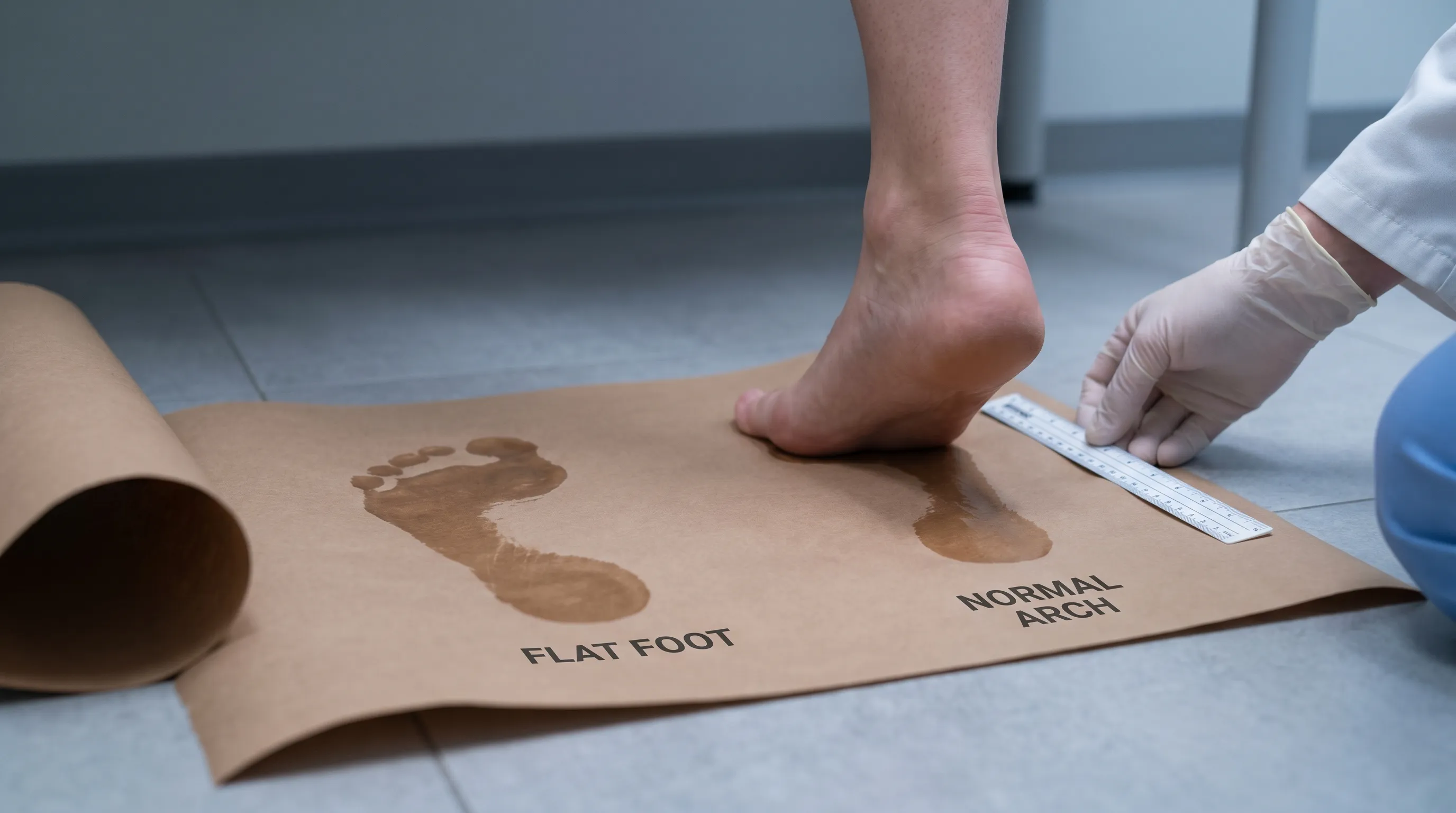
The Classic Wet Footprint Test
For a more tangible and highly accurate assessment, the wet footprint test remains a cornerstone of home diagnosis. Fill a shallow basin or sink with water, ensuring it is not too deep to avoid splashing. Carefully step into the water, saturating the soles of your feet, then immediately step onto a piece of heavy cardboard, construction paper, or a flat, absorbent mat. Stand in a relaxed posture for three to five seconds before stepping off. Allow the footprint to dry completely for analysis.
Interpreting the results is straightforward. A normal footprint displays a clear inward curve along the medial side, typically leaving only the forefoot, heel, and a narrow band connecting them. If your entire foot outline is visible, with minimal or no concave curve on the inner edge, this strongly indicates flat feet. The footprint test eliminates subjective guesswork and provides a permanent record you can reference over time or share with a healthcare provider during consultations. This method is particularly useful when evaluating children, as the fat pads on a toddler's feet can mask arch development until age three or four.
The "Too Many Toes" Sign Explained
A highly specific clinical indicator that you can easily replicate at home is the "too many toes" sign. Stand in your natural posture while a partner or family member observes your feet from directly behind you at ground level. Focus on the lateral (outer) edge of each foot. In a healthy foot alignment, you should see only the fifth toe (little toe) protruding slightly beyond the outer contour of the foot. If you can clearly see the fourth toe or even the third toe visible past the lateral margin, this is a positive "too many toes" sign.
This phenomenon occurs because flat feet cause the forefoot to abduct and the hindfoot to evert, rotating the entire foot outward. As the arch collapses, the foot splays laterally, exposing more of the lesser toes from a posterior viewpoint. While not definitive on its own, this sign strongly correlates with pes planus and compensatory lower extremity malalignment. Combining this observation with the standing visual inspection and footprint test creates a robust, multi-angle self-assessment protocol that accurately answers the question of how to tell if u have flat feet.
Differentiating Flexible vs. Rigid Flatfoot
Determining whether your flat feet are flexible or rigid is crucial for understanding prognosis and guiding management strategies. To perform this differentiation, sit comfortably and allow your feet to rest completely unweighted. Look for the presence of an arch. Next, stand on your tiptoes or have someone gently lift your big toe while keeping the heel planted. If an arch reappears when you are seated, standing on tiptoes, or during the toe-raising maneuver, you have flexible flatfoot. This is the most common type and typically responds well to conservative treatments like orthotics, stretching, and supportive footwear.
Conversely, if the arch remains completely flat regardless of position, weight-bearing status, or muscle contraction, you may be experiencing rigid flatfoot. Rigid arch collapse often indicates underlying structural anomalies, tarsal coalitions, joint degeneration, or neurological dysfunction. Rigid flatfoot rarely improves with over-the-counter inserts and usually requires professional imaging and specialized medical intervention. Recognizing this distinction early helps prevent unnecessary trial-and-error with self-care measures and ensures you pursue the most appropriate therapeutic pathway.
Recognizing the Symptoms Beyond a Flat Appearance
While structural observation provides the initial answer to how to tell if u have flat feet, clinical reality dictates that form must be evaluated alongside function. Importantly, a significant portion of individuals with flat feet experience absolutely no pain or functional limitations (NIH MedlinePlus). The human body possesses remarkable adaptive capacity, and many people live active, symptom-free lives with naturally low arches. However, when biomechanical compensation exceeds physiological thresholds, symptoms emerge. Understanding these warning signs helps differentiate a benign anatomical variant from a progressive pathological condition.
Pain Patterns and Activity-Related Discomfort
Symptomatic flat feet typically manifest as localized discomfort along the medial foot and ankle region. The pain is often described as a deep, aching sensation that intensifies after prolonged standing, walking, or high-impact exercise. Because the collapsed arch loses its natural shock-absorption properties, repetitive impact forces transmit directly to the plantar fascia, tibialis posterior tendon, and navicular bone. You may notice sharp pain during the push-off phase of walking, particularly when transitioning from flat surfaces to inclined terrain.
Activity-related worsening is a hallmark feature. Pain that consistently flares after specific movements, such as running, jumping, or wearing minimalist shoes, strongly suggests that your foot's supportive structures are being overstressed. Unlike acute injuries that cause sudden, localized swelling, flat foot pain often builds gradually throughout the day and eases with rest or elevation. This chronic, low-grade discomfort can subtly alter your gait, causing you to shift weight to the outer foot or shorten your stride to avoid medial ankle strain.
Swelling, Balance Issues, and Shoe Wear Clues
Beyond pain, secondary symptoms provide additional diagnostic clues. Medial ankle swelling is extremely common in progressive flat foot deformity. As the posterior tibial tendon stretches or degenerates, inflammatory fluid accumulates around the medial malleolus. You may notice puffiness that pits slightly when pressed or feels warm to the touch after long days on your feet. This swelling often accompanies a feeling of instability or clumsiness. When the arch collapses, the subtalar joint loses its stable platform, impairing proprioceptive feedback and increasing the risk of ankle sprains.
Footwear degradation patterns offer objective, long-term evidence of abnormal loading. Examine the soles of your frequently worn shoes. Normal wear distributes relatively evenly across the forefoot and heel, with slight outer heel erosion. In flat feet, you will observe pronounced medial wear along the inside edge of the midsole and outsole. The upper fabric may also stretch outward near the toe box, and the heel counter often leans inward due to persistent overpronation. Uneven shoe wear is not merely a cosmetic issue; it is a clear biomechanical indicator that your gait requires corrective support.
When Asymptomatic Flat Feet Still Warrant Attention
Even without pain, flat feet can silently contribute to systemic musculoskeletal strain. Altered foot mechanics shift load vectors upward through the kinetic chain, potentially accelerating cartilage wear in the knees and hips. Runners, athletes, and individuals who stand for occupational requirements should monitor subtle changes in endurance, recovery time, and joint stiffness. Periodic self-assessment of your foot structure ensures that early interventions, such as targeted strengthening or footwear upgrades, are implemented before compensatory patterns become entrenched. Learning how to tell if u have flat feet proactively empowers you to maintain optimal lower extremity alignment and prevent secondary overuse injuries.
Types of Flat Feet and Their Distinct Characteristics
Flat feet are not a monolithic condition. Medical literature categorizes them into distinct etiological and structural subtypes, each carrying unique clinical implications, progression trajectories, and management protocols. Accurately identifying your specific type is essential for tailoring interventions that address the root cause rather than merely masking symptoms.
Flexible Flatfoot: The Pediatric Norm
Flexible flatfoot dominates the pediatric population and represents a normal developmental stage in early childhood. Infants and toddlers are born with flat feet due to ligamentous laxity, a thick protective fat pad in the sole, and immature neuromuscular control. As children begin walking and their musculoskeletal system matures, the arch typically develops between ages three and ten. In flexible flatfoot, the arch is clearly visible when the child is seated or standing on tiptoes but disappears when weight is applied.
This variant is overwhelmingly painless and rarely restricts physical activity. Children with flexible flat feet participate normally in sports, running, and playground activities without increased injury risk. Clinical guidelines universally recommend observation and reassurance rather than aggressive intervention. Braces, rigid orthotics, or specialized footwear have not been proven to accelerate arch development or prevent future problems in asymptomatic children. Most cases resolve spontaneously as intrinsic foot muscles strengthen and skeletal growth stabilizes.
Adult-Acquired Flatfoot: Progressive Collapse
Adult-acquired flatfoot, frequently termed progressive collapsing foot deformity, presents a stark contrast to the benign pediatric form. This condition emerges in previously normal arches, typically between ages forty and sixty, though it can occur earlier in athletes or individuals with high physical demands. The primary driver is posterior tibial tendon dysfunction (PTTD). The posterior tibial tendon acts as the main dynamic stabilizer of the medial arch. When subjected to chronic overuse, microtrauma, degenerative changes, or acute rupture, it loses its tensile strength.
Without adequate tendon support, the arch gradually descends, the heel bone shifts outward, and the forefoot abducts. This progression occurs in stages. Stage one involves tendon inflammation with a normal arch. Stage two features flexible deformity with tendon elongation but preserved joint mobility. Stage three introduces rigid joint contracture and arthritic changes. Stage four extends deformity to the ankle joint itself. Early recognition of how to tell if u have flat feet in this progressive phase is critical, as conservative interventions like bracing, anti-inflammatory protocols, and physical therapy are most effective before irreversible structural changes occur.
Rigid Flatfoot: Structural and Neurological Origins
Rigid flatfoot represents the most complex clinical variant, characterized by complete absence of an arch across all weight-bearing and non-weight-bearing states. Unlike flexible deformities, rigid flatfoot does not respond to positional changes or muscle contraction. The underlying pathology is often congenital or structural, involving abnormal bone fusion, joint malformation, or neurological impairment. Tarsal coalition, a rare condition where two or more foot bones fuse abnormally during development, severely restricts subtalar joint motion and forces the foot into a permanently pronated position.
Neurological disorders such as cerebral palsy, spina bifida, or Charcot-Marie-Tooth disease can also produce rigid flatfoot through muscle imbalance, spasticity, or loss of proprioceptive control. Additionally, severe arthritis in the midfoot or hindfoot can fuse joints in a collapsed position. Because rigid flatfoot resists conservative biomechanical correction, it demands comprehensive imaging, often including CT or MRI scans, to delineate the exact anatomical disruption. Management typically involves specialized orthotics, custom bracing, and frequently surgical reconstruction to restore joint alignment and function.
| Feature | Flexible Flatfoot | Adult-Acquired Flatfoot (PTTD) | Rigid Flatfoot |
|---|---|---|---|
| Arch Visibility | Present when seated/on tiptoes, absent when standing | Progressively absent; initially flexible, later fixed | Absent in all positions and weights |
| Primary Cause | Normal development, ligamentous laxity | Posterior tibial tendon degeneration/tear | Tarsal coalition, arthritis, neurological conditions |
| Pain Level | Usually painless | Moderate to severe, worsens with activity | Chronic, often severe, constant discomfort |
| Joint Mobility | Fully mobile | Mobile in early stages, becomes restricted later | Severely restricted or fused |
| Typical Treatment | Observation, supportive shoes | Physical therapy, orthotics, ankle braces | Surgical reconstruction, rigid orthotics, fusion |
Causes, Risk Factors, and Underlying Conditions
Understanding why arches flatten illuminates how to tell if u have flat feet in context and guides preventive strategies. Arch collapse rarely occurs in isolation; it typically results from a convergence of genetic predisposition, mechanical stress, tissue degeneration, and systemic health factors. Recognizing these contributors helps individuals modify lifestyle habits and mitigate progression risks.
Congenital and Developmental Factors
Genetics play a substantial role in foot architecture. If your parents or siblings have flat feet, you inherit a higher probability of similar ligamentous laxity and bone morphology. Some individuals are born with hypoplastic arches due to variations in navicular bone positioning or Achilles tendon length. Developmentally, premature arch collapse in children can stem from rapid growth spurts outpacing muscular strengthening, or prolonged use of unsupportive footwear during critical formative years. While congenital factors cannot be altered, understanding hereditary patterns prompts earlier monitoring and proactive biomechanical support during athletic or occupational activities.
Tendon Dysfunction, Trauma, and Degeneration
The posterior tibial tendon is the linchpin of medial arch stability. Repetitive microtrauma from high-impact sports, prolonged occupational standing, or sudden ankle sprains can cause microscopic tendon tears. Over time, collagen degeneration and reduced vascularity impair healing capacity, leading to elongation and functional failure. Trauma, including ankle fractures or midfoot ligament ruptures, can instantly destabilize the arch structure. Degenerative arthritis in the talonavicular or calcaneocuboid joints further compromises the osseous scaffolding that supports the arch. These mechanical failures explain why adult-acquired flatfoot often emerges after years of seemingly healthy activity, suddenly manifesting as progressive arch collapse and medial ankle pain.
Lifestyle and Systemic Risk Modifiers
Several modifiable and non-modifiable factors accelerate flat foot development. Obesity places excessive compressive load on the foot's supporting ligaments and plantar fascia, gradually stretching them beyond their elastic limit. Diabetes and rheumatoid arthritis induce systemic inflammation and tissue breakdown, weakening ligamentous integrity and tendon collagen (CDC). Pregnancy temporarily increases relaxin hormone levels, softening pelvic and foot ligaments to prepare for childbirth; this can induce transient flat foot deformity that may persist postpartum if not properly rehabilitated. Aging naturally reduces muscle mass, tendon elasticity, and fat pad cushioning, diminishing the foot's shock-absorption capacity. Regularly wearing unsupportive, flat-soled footwear without arch reinforcement compounds these stresses, making mindful shoe selection a critical preventive measure.
When Self-Checks Aren't Enough: Professional Diagnosis
While home assessments effectively answer how to tell if u have flat feet, clinical evaluation provides definitive diagnosis, precise staging, and personalized treatment planning. Healthcare professionals utilize advanced observational techniques, specialized imaging, and functional assessments to map your exact biomechanical profile and rule out mimicking conditions.
What to Expect During a Clinical Examination
A thorough podiatric or orthopedic evaluation begins with a detailed medical history, exploring pain patterns, activity levels, prior injuries, and family history. The physical examination involves observing your feet both seated and standing. Clinicians assess arch height, heel alignment, forefoot positioning, and ankle range of motion. They frequently perform the single-leg heel-rise test, asking you to balance on one foot and lift your heel repeatedly. Inability to perform multiple heel rises on the affected side strongly indicates posterior tibial tendon insufficiency.
Shoe wear analysis remains a valuable diagnostic tool, revealing chronic gait deviations. Practitioners palpate the medial ankle, arch, and plantar fascia for tenderness, swelling, or structural irregularities. Tightness in the gastrocnemius-soleus complex is evaluated using the Silfverskiöld test, as Achilles contracture exacerbates arch strain. Neurological screening checks sensation and reflexes to exclude peripheral neuropathy or spinal radiculopathy contributing to altered foot mechanics. This comprehensive examination establishes a baseline for tracking progression and measuring treatment efficacy.
Imaging and Advanced Diagnostic Tools
When structural damage or rigid deformity is suspected, imaging becomes essential. Weight-bearing X-rays are the gold standard for evaluating bone alignment, joint spacing, and arch height under physiological load. They reveal talonavicular coverage angles, calcaneal inclination, and signs of osteoarthritis. If soft tissue pathology is suspected, ultrasound dynamically visualizes tendon thickness, integrity, and fluid accumulation in real time during movement. Magnetic resonance imaging (MRI) provides detailed cross-sectional views of tendons, ligaments, bone marrow edema, and cartilage degeneration, particularly valuable for surgical planning in advanced posterior tibial tendon dysfunction.
Gait analysis laboratories use pressure-sensitive walkways and high-speed cameras to quantify foot strike patterns, pronation velocity, and ground reaction forces. This technology precisely identifies compensatory mechanisms and customizes orthotic prescriptions to your unique movement signature. By integrating clinical examination with advanced diagnostics, medical professionals deliver accurate, individualized care that addresses the full spectrum of flat foot pathology.
Managing Flat Feet: Treatment and Preventative Care
Discovering how to tell if u have flat feet is only the beginning; effective management transforms structural awareness into sustainable pain relief and enhanced mobility. Treatment strategies are strictly guided by symptom severity, deformity flexibility, and functional impact. Asymptomatic flat feet require no medical intervention, focusing instead on preventive maintenance and monitoring.
Footwear Modifications and Orthotic Solutions
Footwear serves as the first line of defense against flat foot progression. Ideal shoes feature a firm heel counter, wide toe box, and stable midsole with built-in arch support. Avoid completely flat, unsupportive shoes like flip-flops or thin-soled fashion sneakers, which eliminate shock absorption and exacerbate medial strain. For moderate symptoms, over-the-counter arch supports or prefabricated insoles made from cork, EVA foam, or gel provide immediate load redistribution and comfort.
Custom-molded orthotics, prescribed by a podiatrist, are fabricated from precise foot casts or digital scans. They correct abnormal alignment, control excessive pronation, and cushion high-pressure zones. Orthotics must be gradually introduced to allow tissue adaptation, typically wearing them two hours on day one, increasing by one hour daily until full-day use is achieved. Proper footwear combined with orthotics often resolves pain, improves balance, and restores functional capacity within weeks.
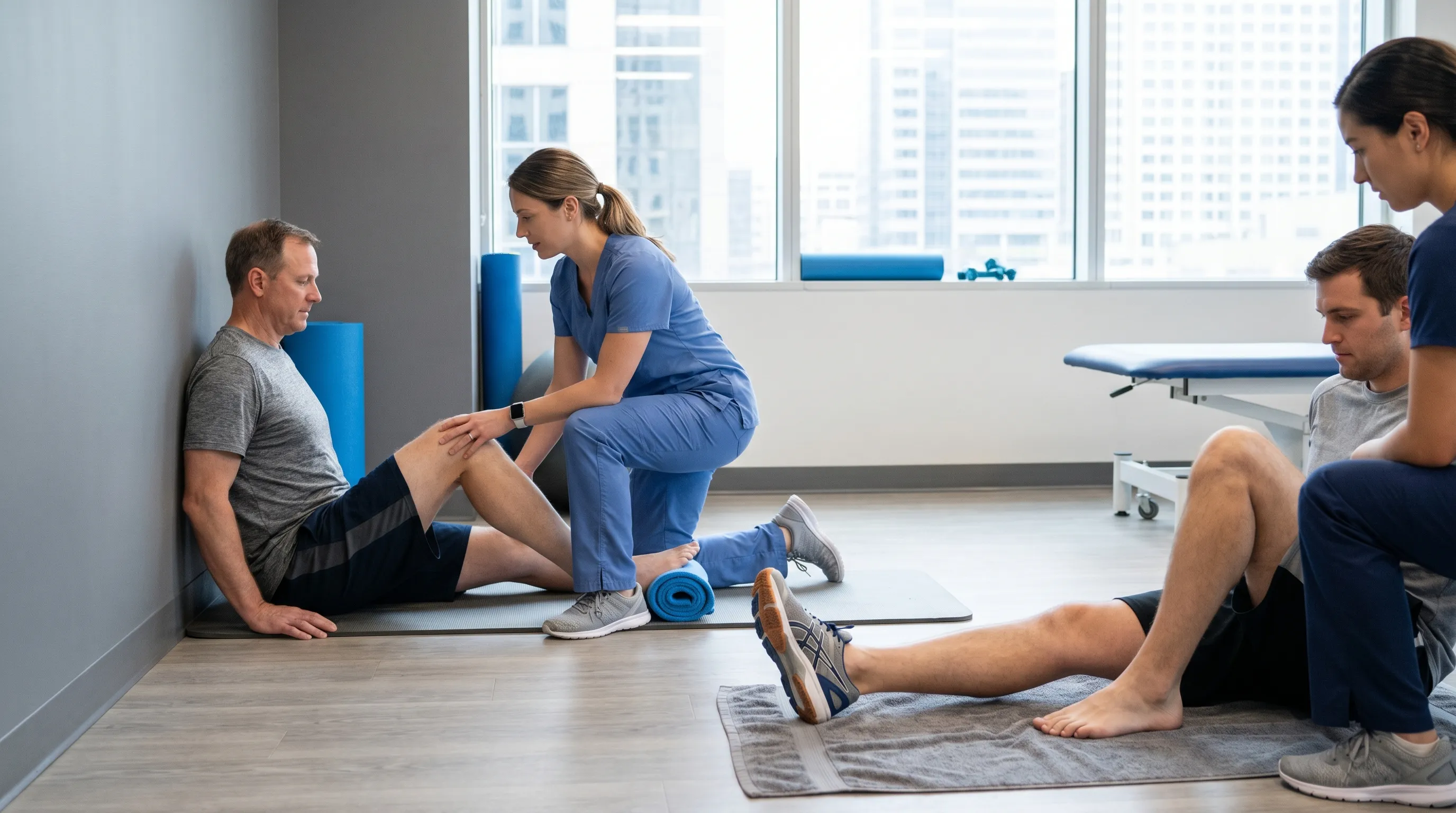
Physical Therapy, Stretching, and Strengthening
Targeted rehabilitation addresses the muscular deficits and fascial restrictions that accompany flat feet. The Achilles tendon and calf muscles frequently become tight, pulling the heel upward and increasing forefoot pressure. Daily gastrocnemius and soleus stretching using a wall lean or slanted board restores ankle dorsiflexion and reduces compensatory strain on the plantar fascia. Strengthening exercises rebuild intrinsic foot muscle endurance, creating a natural muscular arch.
The towel curl exercise improves toe flexor strength: sit barefoot, place a small towel under your foot, and use your toes to scrunch it toward your heel. Marble pickups enhance fine motor control and arch activation. Heel raises strengthen the posterior tibial tendon and calf complex; perform them slowly on both legs, progressing to single-leg repetitions as tolerance improves. Short foot exercises, where you contract the sole to lift the arch without curling the toes, retrain proprioceptive control and improve static stability. Consistency is paramount; daily practice for ten to fifteen minutes yields measurable improvements in arch height, pain reduction, and gait efficiency over eight to twelve weeks.
Surgical Interventions for Severe Cases
When conservative measures fail after six to twelve months, and deformity progresses to rigid collapse or debilitating pain, surgical intervention becomes necessary. Procedures are tailored to the specific anatomical failure. Tendon transfers, often relocating a healthy tendon to replace the degenerated posterior tibial tendon, restore dynamic support. Osteotomies involve cutting and realigning bones to correct heel valgus and restore arch geometry. In advanced arthritis or rigid coalition, joint fusion (arthrodesis) eliminates painful motion and stabilizes the foot structure. Recovery involves prolonged non-weight-bearing periods, intensive physical therapy, and gradual return to weight-bearing activities. While surgery carries inherent risks, modern techniques and postoperative protocols achieve high success rates in restoring pain-free ambulation and preventing long-term joint deterioration.
Frequently Asked Questions
Can flat feet develop suddenly in adulthood?
Yes, adult-acquired flatfoot can develop gradually or suddenly due to posterior tibial tendon dysfunction, arthritis, trauma, or neurological conditions. If you notice a sudden flattening of your arch, especially accompanied by pain or swelling along the inside of the ankle, consult a healthcare provider promptly.
Is it normal for toddlers to have flat feet?
Absolutely. Flexible flat feet are completely normal in infants and toddlers. The arch typically begins to develop between ages 3 and 10 as foot muscles strengthen and the fat pad under the sole naturally diminishes. Unless the child experiences persistent pain or gait abnormalities, observation is usually sufficient.
Do flat feet cause knee or back pain?
Flat feet can alter your lower extremity alignment, leading to overpronation that stresses the knees, hips, and lower back. This kinetic chain disruption is well-documented in biomechanical studies. Proper footwear, custom orthotics, and targeted strengthening often alleviate secondary joint discomfort.
How do I know if I need custom orthotics or just arch supports?
Over-the-counter arch supports work well for mild, flexible flatfeet with minimal pain. Custom orthotics are recommended for rigid flatfoot, significant asymmetry, progressive tendon dysfunction, or when OTC inserts fail to resolve symptoms. A podiatrist or physical therapist can perform a gait analysis to determine the appropriate intervention.
Will running or high-impact exercise worsen flat feet?
Exercise itself does not cause arches to collapse, but high-impact activities without proper footwear can exacerbate symptoms or accelerate tendon wear. Gradual progression, strength training for foot intrinsics, and choosing stability or motion-control shoes allow most individuals with flat feet to run safely and comfortably.
Key Takeaways
Learning how to tell if u have flat feet empowers you to take proactive control of your lower extremity health. Simple, reliable home tests like the standing visual inspection, wet footprint analysis, and "too many toes" observation provide immediate structural insight. Distinguishing between flexible, acquired, and rigid flatfoot guides appropriate management pathways and prevents unnecessary interventions. Remember that flat feet are often asymptomatic and entirely manageable through supportive footwear, targeted orthotics, and consistent physical therapy. Early recognition of pain patterns, swelling, or balance issues ensures timely professional evaluation before progressive deformity compromises mobility. By integrating biomechanical awareness with evidence-based self-care, you can maintain pain-free movement, optimize athletic performance, and protect the intricate architecture that carries you through life. If structural changes are accompanied by persistent discomfort, gait instability, or sudden onset flattening, seek evaluation from a podiatrist or orthopedic specialist for personalized diagnostics and long-term treatment planning. Prioritizing foot health today prevents compensatory joint degeneration and preserves lifelong mobility and vitality.

About the author
Leo Martinez, DPT, is a board-certified orthopedic physical therapist specializing in sports medicine and post-surgical rehabilitation. He is the founder of a sports therapy clinic in Miami, Florida that works with collegiate and professional athletes.