Male Chest Health: Anatomy, Training, and Medical Awareness

The male chest represents far more than a focal point for aesthetic development; it is a complex anatomical region integral to respiratory function, upper body biomechanics, and overall physiological health. While fitness culture frequently emphasizes pectoral hypertrophy and sculpted definition, a comprehensive understanding of this area extends well beyond muscle growth. The interplay of muscular architecture, glandular tissue, subcutaneous fat distribution, and underlying skeletal structures determines both functional capacity and long-term wellness. Unfortunately, many individuals overlook the medical significance of this region until discomfort, asymmetry, or noticeable tissue changes prompt concern. From postural adaptations caused by sedentary lifestyles to hormonal fluctuations that alter tissue composition, the male chest serves as a barometer for broader systemic health. Recognizing the intricate balance between strength training, nutritional support, and clinical vigilance empowers men to optimize both appearance and physiological resilience. This guide explores the foundational anatomy, prevalent conditions, evidence-based training methodologies, nutritional strategies, and clinical warning signs that every man should understand. By integrating scientific insights with actionable lifestyle modifications, readers can cultivate a healthier, stronger, and more functional torso while maintaining proactive awareness of potential medical concerns.
Understanding the Anatomy of the Male Chest
The structural foundation of the upper torso involves multiple interconnected systems that facilitate breathing, upper limb movement, and postural stability. Comprehending these components provides critical context for both exercise programming and clinical awareness. The thoracic region houses vital organs, yet its external presentation is primarily governed by the superficial musculature, connective tissue networks, and adipose distribution patterns (Cleveland Clinic Chest Anatomy Guide).
Free calculatorBMI CalculatorCalculate Body Mass IndexOpen the calculatorMuscular Structure and Pectoral Muscles
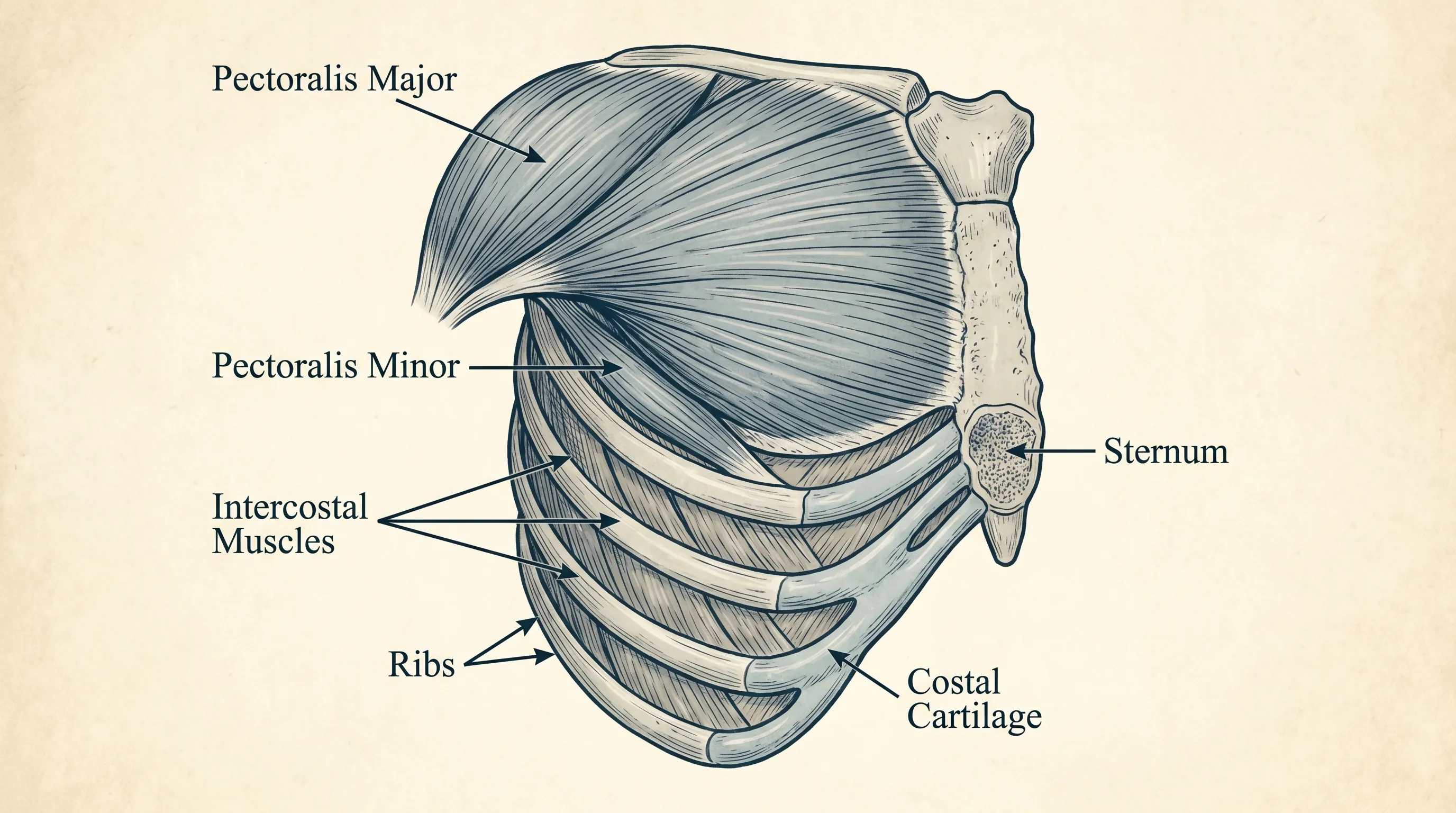
The pectoralis major represents the most prominent superficial muscle of the male chest. This fan-shaped muscle originates along the medial half of the clavicle, the sternum, and the cartilages of the first six ribs. Its fibers converge laterally to insert onto the humerus, enabling powerful adduction, internal rotation, and flexion of the shoulder joint. Functionally, the pectoralis major is divided into clavicular, sternal, and costal heads, each responding uniquely to different movement planes and resistance angles.
Beneath the pectoralis major lies the pectoralis minor, a thinner triangular muscle that attaches to the third through fifth ribs and the coracoid process of the scapula. While smaller in mass, the pectoralis minor plays an essential role in scapular stabilization, downward rotation, and protraction. Chronic tension in this region frequently contributes to rounded shoulders and restricted overhead mobility.
Supporting these primary movers are the serratus anterior, intercostals, and subclavius muscles. The serratus anterior stabilizes the scapula against the thoracic wall, while the intercostal muscles facilitate rib cage expansion and contraction during respiration. Understanding this muscular synergy explains why isolated bench press movements alone rarely optimize thoracic health or functional strength.
Fat Distribution and Tissue Composition
Adipose tissue in the thoracic region varies significantly based on genetics, age, metabolic health, and endocrine balance. Subcutaneous fat deposits over the pectoral muscles follow standard androgen-mediated distribution patterns, typically accumulating along the lower outer quadrants and inframammary crease. Unlike true glandular tissue, subcutaneous fat remains compressible and lacks the fibrous density characteristic of breast development.
Men also possess residual mammary ductal structures beneath the nipple-areolar complex. During puberty, androgen dominance suppresses further ductal proliferation, resulting in minimal glandular tissue. However, shifts in the testosterone-to-estrogen ratio can reactivate these pathways, leading to noticeable tissue enlargement. This physiological baseline underscores why comprehensive male chest health requires attention to both muscular development and systemic hormonal regulation (NIH Endocrine & Hormonal Health).
Common Conditions Affecting the Male Chest
The upper torso is susceptible to a diverse range of structural, hormonal, and inflammatory conditions. Recognizing early indicators enables timely intervention and prevents long-term complications. Medical literature consistently emphasizes the importance of differentiating between benign tissue changes, musculoskeletal dysfunction, and serious pathological developments.
Gynecomastia vs. Pseudogynecomastia
Gynecomastia represents a clinical condition characterized by benign proliferation of male breast glandular tissue. It frequently emerges during infancy, puberty, and late adulthood due to transient or sustained hormonal fluctuations. Pubertal cases often resolve spontaneously within twelve to thirty-six months as endocrine equilibrium stabilizes. Adult-onset presentations typically require diagnostic evaluation to identify underlying triggers such as medication side effects, hepatic impairment, hyperthyroidism, or substance-induced endocrine disruption (Mayo Clinic: Gynecomastia).
In contrast, pseudogynecomastia involves isolated adipose accumulation without glandular proliferation. It commonly develops in individuals experiencing weight gain, metabolic slowdown, or chronic sedentary behavior. Distinguishing between these conditions relies on physical palpation, imaging assessments, and laboratory hormone panels. Treatment approaches differ substantially; pseudogynecomastia responds to caloric restriction and cardiovascular training, whereas true gynecomastia may necessitate pharmacological management or surgical reduction.
Male Breast Cancer: Rare but Serious
Although representing less than one percent of all breast malignancies, male breast cancer demands serious clinical attention. Risk factors include inherited BRCA1 and BRCA2 mutations, familial predisposition, radiation exposure, chronic liver disease, Klinefelter syndrome, and advanced age. Unlike female presentations, male tumors frequently manifest as a painless subareolar mass, nipple retraction, skin dimpling, or spontaneous unilateral nipple discharge.
Early detection significantly improves prognostic outcomes. Clinical breast examinations, diagnostic mammography, ultrasound imaging, and biopsy protocols follow established oncological guidelines. Men with strong family histories or genetic risk factors should discuss surveillance strategies with specialized healthcare providers (Mayo Clinic: Male Breast Cancer). Awareness remains the most powerful preventive tool, as delayed consultation frequently correlates with advanced staging at diagnosis.
Posture-Related Issues and Rounded Shoulders
Modern occupational habits heavily influence thoracic musculoskeletal alignment. Prolonged computer use, mobile device handling, and asymmetric lifting patterns frequently promote forward head posture, scapular winging, and internal shoulder rotation. The resulting muscular imbalances place excessive tension on the pectoralis minor, shorten the anterior chest wall, and weaken the rhomboids and lower trapezius.
Chronic postural deviation restricts diaphragmatic breathing, reduces thoracic extension, and predisposes individuals to impingement syndromes and rotator cuff tendinopathy. Corrective interventions require comprehensive mobility protocols, targeted strengthening of posterior chain musculature, and ergonomic workspace optimization (CDC Workplace Ergonomics). Consistent postural retraining restores neutral spinal alignment and optimizes mechanical efficiency during daily movements and athletic activities.
Building a Strong, Healthy Male Chest Through Exercise
Effective pectoral development extends beyond repetitive pressing movements. A scientifically grounded training framework prioritizes movement variety, progressive overload, recovery optimization, and injury prevention. Implementing evidence-based programming ensures sustainable muscle growth, joint integrity, and functional strength.
Foundational Strength Training Principles
Progressive overload remains the cornerstone of hypertrophy and strength adaptation. Gradually increasing mechanical tension through load increments, volume accumulation, or tempo variations stimulates muscle protein synthesis and myofibrillar expansion. Research demonstrates that training within six to twelve repetition ranges optimizes pectoral hypertrophy, while three to five rep ranges maximize neural drive and maximal force production, aligning with federal physical activity guidelines.
Training frequency plays an equally critical role. Targeting the male chest two to three times weekly allows sufficient stimulus while preserving recovery capacity. Compound movements should form the foundation of each session, supplemented by isolation exercises to address structural imbalances or weak points. Rest intervals of ninety to one hundred twenty seconds between heavy sets optimize adenosine triphosphate regeneration, while shorter pauses during hypertrophy-focused blocks enhance metabolic stress.
Targeted Pectoral Workouts
A balanced pectoral routine incorporates movements across multiple planes to stimulate all fiber bundles comprehensively. The barbell bench press remains the gold standard for overall sternal and costal head activation. Incline dumbbell pressing preferentially recruits clavicular fibers, addressing upper chest development often neglected in traditional programming. Decline variations and weighted dips emphasize lower sternal recruitment while minimizing anterior shoulder stress.
Cable crossovers, pec deck machines, and dumbbell flyes isolate horizontal adduction, creating continuous tension throughout the range of motion. Incorporating time-under-tension techniques such as three-second eccentrics and one-second pauses at maximal stretch significantly enhances fascial adaptation and muscular endurance. Implementing these methodologies ensures comprehensive development and reduces compensatory loading on the anterior deltoids and triceps.
Stretching and Mobility for Optimal Function
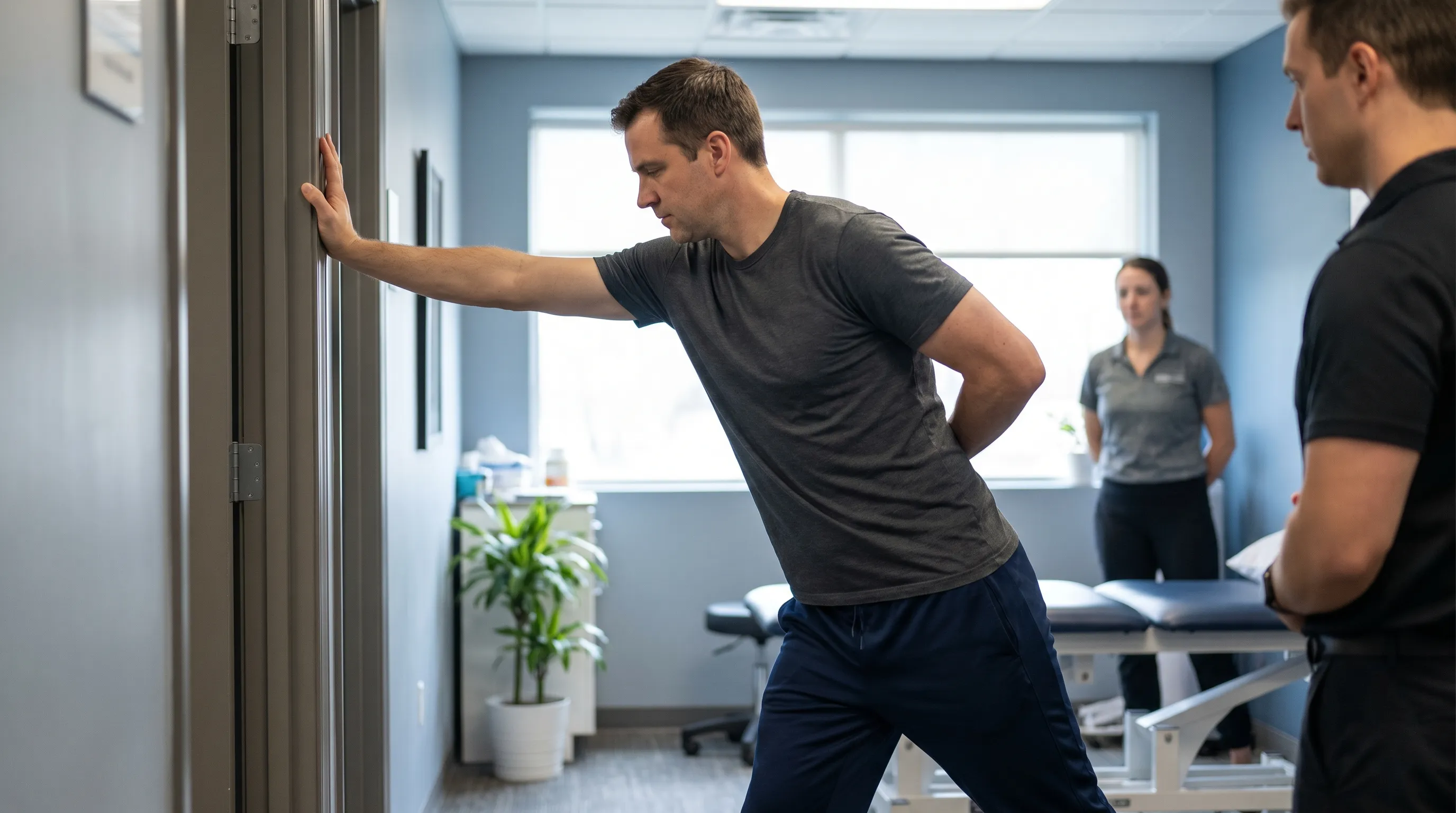
Flexibility and joint mobility directly influence training longevity and movement quality. Chronic tightness in the anterior chest restricts thoracic extension, compromises breathing mechanics, and increases injury risk during overhead activities. Daily mobility protocols should prioritize myofascial release using foam rollers or lacrosse balls, dynamic scapular mobilization drills, and controlled stretching techniques.
Doorway pec stretches, sleeper stretches, and band pull-aparts effectively counteract anterior dominance. Integrating thoracic spine rotations and quadruped cat-camel movements restores vertebral segmental motion and rib cage elasticity. Performing these mobility routines before resistance training and during recovery days maintains tissue pliability, supports proper biomechanics, and accelerates neuromuscular adaptation.
Nutrition and Hormones, and Lifestyle Factors
Dietary patterns and lifestyle choices exert profound influence over thoracic tissue composition, recovery capacity, and endocrine equilibrium. Optimizing these variables enhances both structural development and systemic resilience.
Dietary Strategies to Support Muscle Growth and Recovery
Protein synthesis depends on consistent amino acid availability. Consuming 1.6 to 2.2 grams of protein per kilogram of body weight daily provides optimal substrate for pectoral repair and hypertrophic adaptation. Distributing protein across four to six meals ensures sustained muscle protein synthesis rates throughout waking hours. Prioritizing complete protein sources such as lean poultry, fatty fish, eggs, and legumes guarantees comprehensive essential amino acid profiles (NIH Nutrition & Dietary Research).
Carbohydrate timing directly impacts training performance and glycogen replenishment. Complex carbohydrates including oats, sweet potatoes, and brown rice supply sustained energy for high-volume pectoral sessions. Strategic post-workout carbohydrate consumption restores hepatic and muscular glycogen stores while facilitating nutrient transport to fatigued tissue. Healthy fats from avocados, olive oil, nuts, and omega-3 rich fish support cellular membrane integrity and hormonal synthesis pathways essential for long-term male chest development.
Hormonal Balance and Weight Management
Endocrine regulation dictates tissue distribution, recovery capacity, and metabolic rate. Chronic caloric deficits or extreme dieting suppress testosterone production, impair muscle preservation, and increase cortisol-mediated fat accumulation in the thoracic region. Conversely, sustained caloric surpluses without adequate resistance training promote adipose storage rather than lean tissue accretion.
Maintaining a moderate energy surplus of two hundred to three hundred calories during muscle-building phases and a controlled deficit of three hundred to five hundred calories during cutting phases optimizes body composition shifts. Prioritizing seven to nine hours of quality sleep nightly restores endocrine function, enhances growth hormone secretion, and mitigates systemic inflammation. Reducing alcohol consumption, managing chronic stress through mindfulness practices, and maintaining regular physical activity collectively stabilize hormonal environments conducive to healthy pectoral development (NIH Hormone Health Overview).

When to Seek Medical Evaluation
While most upper torso variations stem from benign physiological processes, specific symptoms warrant immediate clinical attention. Understanding warning signs and diagnostic pathways prevents delayed treatment and ensures appropriate medical management.
Red Flag Symptoms and Pain Assessment
Acute, crushing, or radiating chest discomfort requires emergency evaluation to exclude myocardial infarction, pulmonary embolism, or aortic dissection. Symptoms accompanied by diaphoresis, dyspnea, syncope, or left arm radiation should never be ignored (CDC Heart Disease & Heart Attack Signs). Musculoskeletal pain typically presents as localized tenderness exacerbated by palpation, shoulder movement, or deep breathing. Differentiating between cardiac emergencies and benign thoracic discomfort relies on clinical presentation and diagnostic testing.
Persistent subareolar lumps, unilateral nipple discharge, skin ulceration, or rapid asymmetrical enlargement require prompt physician assessment. Pain that interferes with daily function, worsens over weeks despite conservative management, or presents with systemic symptoms such as unexplained weight loss or fever necessitates comprehensive clinical evaluation. Early intervention significantly improves therapeutic outcomes across all diagnostic categories.
Diagnostic Procedures and Specialist Consultations
Initial evaluation typically involves thorough physical examination, hormone panel testing, liver function assessment, and thyroid screening. Ultrasound imaging differentiates cystic formations, solid masses, and adipose tissue with high diagnostic accuracy. Mammography and magnetic resonance imaging provide detailed structural visualization when ultrasound results remain inconclusive or when malignancy suspicion exists (Mayo Clinic Breast Cancer Diagnosis).
Biopsy protocols, endocrine specialist consultations, and surgical referrals guide definitive treatment planning. Men experiencing medication-induced tissue changes should consult prescribing physicians regarding alternative therapies or dosage adjustments. Maintaining open communication with primary care providers ensures coordinated management and minimizes unnecessary anxiety regarding benign thoracic variations. Regular self-examinations combined with annual physicals establish baseline awareness and facilitate early detection of clinically significant changes.
| Feature | Gynecomastia | Pseudogynecomastia | Male Breast Cancer |
|---|---|---|---|
| Tissue Type | Firm, rubbery glandular tissue beneath areola | Soft, diffuse subcutaneous fat | Hard, irregular, often painless subareolar mass |
| Onset Pattern | Gradual enlargement over weeks to months | Progressive weight gain related expansion | Sudden or progressive unilateral growth |
| Nipple Changes | Possible slight protrusion, typically symmetrical | None | Retraction, discharge, scaling, or ulceration |
| Pain Level | Often tender during early development | Generally painless | Usually painless, occasionally aching |
| Diagnostic Focus | Hormone panels, ultrasound, medication review | Body composition analysis, caloric assessment | Mammography, biopsy, genetic screening |
Frequently Asked Questions
What causes gynecomastia in adult men?
Gynecomastia typically develops due to an imbalance between estrogen and testosterone levels. Triggers include natural aging, certain medications like antidepressants or blood pressure drugs, liver or kidney dysfunction, thyroid disorders, recreational substance use, and underlying endocrine conditions. A thorough medical evaluation is necessary to identify the precise etiology (Mayo Clinic Gynecomastia Causes).
How can I differentiate between chest fat and true glandular tissue?
Excess chest fat (pseudogynecomastia) feels soft and diffuse, whereas true glandular tissue is typically firm, rubbery, and located concentrically beneath the areola. Fat distribution often decreases with caloric deficits and targeted cardiovascular exercise, while glandular enlargement usually persists without medical intervention or surgical consultation.
What is the optimal training frequency for pectoral development?
Research indicates that training the chest two to three times weekly with at least 48 hours of recovery between sessions maximizes muscle protein synthesis and structural adaptation. Beginners should focus on mastering form with moderate volume, while advanced lifters may benefit from periodized intensity cycling and strategic deload weeks (CDC Physical Activity Guidelines).
Are chest pains always related to cardiovascular issues?
Not necessarily. Chest discomfort can originate from musculoskeletal strain, costochondritis, gastrointestinal reflux, anxiety, or respiratory conditions. However, any new, severe, radiating, or persistent chest pain requires immediate clinical evaluation to rule out cardiac events. Never self-diagnose acute thoracic symptoms without professional medical assessment (Mayo Clinic Chest Pain Overview).
Does diet significantly influence male chest composition?
Yes. Body fat percentage directly impacts the visibility and shape of the pectoral muscles, while adequate protein intake supports tissue repair and hypertrophy. Chronic caloric surpluses, excessive alcohol consumption, and diets high in processed foods can alter hormonal profiles and promote adipose accumulation, particularly in the thoracic region (NIH Diet & Body Composition Research).
How long does it take to see noticeable changes in chest development?
With consistent resistance training, optimized protein intake, and adequate recovery, most men observe measurable strength improvements within four to six weeks. Visible muscular definition typically requires eight to twelve weeks of dedicated programming, while substantial fat loss to reveal underlying pectoral structure often spans three to six months depending on baseline composition and metabolic rate.
Conclusion
Cultivating optimal health and function within the upper torso requires a balanced integration of evidence-based training, strategic nutrition, postural awareness, and proactive medical vigilance. The male chest serves as both a powerhouse for upper body strength and a clinical indicator of systemic wellness. Understanding anatomical foundations enables more effective exercise programming, while recognizing pathological variations ensures timely professional intervention when necessary. By implementing structured resistance protocols, prioritizing mobility and recovery, maintaining endocrine-friendly lifestyle habits, and remaining attentive to abnormal tissue changes, men can sustain long-term thoracic resilience and functional capacity. Whether pursuing aesthetic enhancement, athletic performance optimization, or preventative health maintenance, a comprehensive approach yields sustainable results. Consult qualified fitness professionals for programming adjustments and seek clinical evaluation for persistent discomfort or structural abnormalities. Empowered by scientific insight and consistent practice, individuals can confidently support a stronger, healthier, and more balanced physique for years to come.
How we work
Researched and drafted with AI assistance, then reviewed by a human editor against the cited sources.
This article is general health information, not medical advice. Consult a qualified healthcare professional about your own situation.