Loose Adult Tooth: Causes, Treatments, and How to Stabilize It Naturally
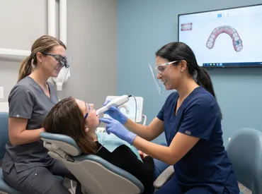
Experiencing the unsettling sensation of a loose adult tooth can trigger immediate concern, especially when we have been conditioned since childhood to associate tooth mobility exclusively with normal dental development. While primary teeth naturally shed to make room for permanent successors, adult teeth are designed to remain securely anchored for a lifetime. When an adult tooth begins to wiggle, shift, or feel unstable, it serves as a critical biological warning sign that the supporting structures of your dentition are compromised. This symptom should never be dismissed as a temporary anomaly or ignored out of fear. Early intervention is the single most important factor in preserving natural dentition, preventing irreversible bone loss, and maintaining optimal oral function. Modern dentistry offers a wide array of diagnostic tools, conservative therapies, and advanced restorative techniques specifically engineered to stabilize mobility and restore periodontal health. By understanding the underlying mechanisms, recognizing the clinical progression, and implementing evidence-based treatment protocols, patients can effectively address a loose adult tooth and safeguard their long-term dental vitality. This comprehensive guide will walk you through the anatomical foundations, pathological triggers, diagnostic criteria, and proven management strategies to help you navigate this concerning dental condition with confidence and informed decision-making.
Understanding the Anatomy and Physiology of Tooth Stability
To comprehend why a tooth becomes mobile, one must first understand the intricate biological architecture responsible for securing it in the oral cavity. Unlike artificial implants that fuse directly with bone through osseointegration, natural teeth rely on a sophisticated suspension system that allows for microscopic movement during chewing while maintaining structural integrity. This system is composed of multiple specialized tissues that work in perfect synchrony to distribute mechanical forces, absorb occlusal shock, and protect the underlying nerve and vascular supply.
Free calculatorHeadache Location ChartIdentify headache types by locationOpen the calculatorThe Role of the Periodontal Ligament and Alveolar Bone
The periodontal ligament (PDL) is a highly specialized connective tissue situated between the cementum of the tooth root and the alveolar bone of the jaw. This ligament contains thousands of collagen fiber bundles arranged in distinct orientations that function like microscopic shock absorbers. When you bite down, the PDL compresses slightly, allowing the tooth to move imperceptibly before transmitting the force evenly to the surrounding bone. This physiological mobility is essential for preventing root fractures and enabling proprioceptive feedback to the jaw muscles. However, when inflammatory processes or traumatic forces disrupt the PDL, the collagen matrix degrades, and the ligament widens pathologically. This degradation directly translates to clinical mobility. The alveolar bone, which houses the tooth roots, relies on continuous remodeling stimulated by healthy periodontal ligament activity. Chronic inflammation accelerates bone resorption through the activation of osteoclasts, gradually reducing the depth and density of the bony socket. As the alveolar ridge diminishes, the foundation supporting the tooth weakens, resulting in progressive instability. For deeper clinical insights into bone and ligament interaction, visit the National Institute of Dental and Craniofacial Research (NIDCR) guide to periodontal structures.
How Gum Tissue Supports Dental Integrity
The gingival tissue, commonly referred to as the gums, serves as a biological seal that protects the periodontal ligament and underlying bone from bacterial invasion. Healthy gingiva is firmly attached to the cementum just below the crown margin, forming a tight epithelial barrier. When periodontal disease takes hold, this attachment apparatus breaks down, leading to the formation of pathological pockets. These pockets create an anaerobic environment where pathogenic bacteria thrive, producing endotoxins that further stimulate inflammatory cascades. The progressive destruction of the gingival attachment not only exposes the root surfaces to decay and sensitivity but also removes the primary soft-tissue barrier that stabilizes the tooth within the jaw. Without this supportive tissue matrix, even minor functional forces can cause noticeable shifting or wobbling.
The Natural Limits of Tooth Mobility
Clinicians classify physiological mobility as a microscopic movement measuring less than 0.2 millimeters. This slight movement is completely imperceptible to patients and is a normal functional characteristic. Pathological mobility becomes clinically significant once it exceeds 0.5 millimeters or becomes detectable to the tongue or fingers. Understanding this threshold is crucial because many individuals mistakenly believe that any sensation of movement indicates inevitable tooth loss. In reality, the periodontium possesses a remarkable capacity for regeneration and stabilization if the destructive triggers are removed promptly. Recognizing the difference between normal physiological adaptation and pathological deterioration is the first step toward effective clinical management.
Primary and Secondary Causes of a Loose Adult Tooth
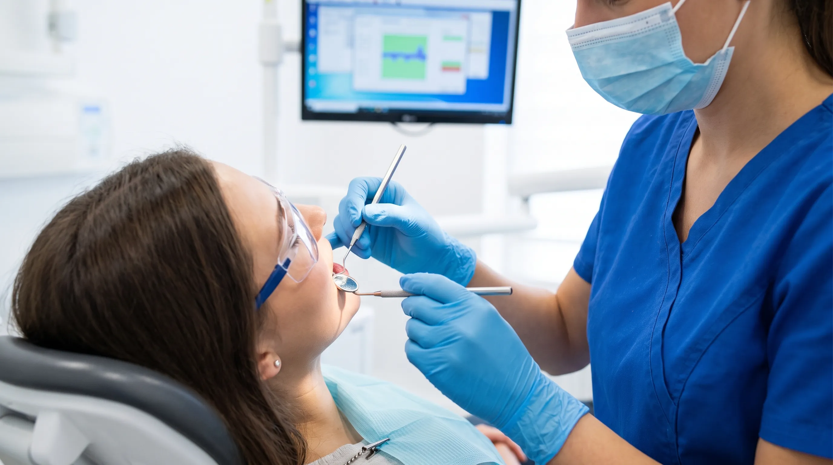
Identifying the root cause of dental mobility is essential for formulating an accurate treatment plan. While poor oral hygiene is the most frequently cited trigger, numerous systemic, mechanical, and pharmacological factors can independently or collectively contribute to periodontal breakdown and subsequent instability.
Advanced Periodontal Disease and Bone Loss
Periodontal disease remains the leading cause of a loose adult tooth globally. It begins as gingivitis, a reversible inflammation of the superficial gum tissue, but rapidly progresses to periodontitis when bacterial plaque mineralizes into calculus. The subgingival accumulation of pathogenic species such as Porphyromonas gingivalis, Tannerella forsythia, and Treponema denticola triggers a chronic immune response. The host’s inflammatory mediators, including interleukin-1β, tumor necrosis factor-alpha, and matrix metalloproteinases, inadvertently destroy the very connective tissues and bone they are attempting to protect. Over time, this relentless cycle leads to horizontal or vertical bone defects, pocket deepening, and irreversible attachment loss. Without professional intervention and rigorous home maintenance, periodontitis will continue to compromise the structural foundation until the tooth becomes severely mobile and ultimately non-restorable. The CDC provides extensive epidemiological data on periodontal disease progression.
Traumatic Injuries and Occlusal Stress
Acute dental trauma from falls, sports impacts, vehicular accidents, or altercations can fracture roots, luxate teeth, or severely bruise the periodontal ligament. Even when the crown appears intact, subgingival trauma can initiate inflammatory resorption and compromise long-term viability. Beyond acute injuries, chronic occlusal stress plays a significant role in tooth mobility. Malocclusions, premature contacts, high dental restorations, and parafunctional habits like clenching generate excessive lateral and vertical forces that exceed the adaptive capacity of the periodontium. This condition, known as primary occlusal trauma, does not directly cause periodontitis but can accelerate bone loss in already compromised tissues. When combined with untreated periodontal disease, the synergistic effect dramatically hastens the progression of mobility.
Systemic Health Factors and Hormonal Influences
The health of your dentition is intrinsically linked to your overall physiological status. Systemic conditions such as uncontrolled diabetes mellitus severely impair neutrophil function, reduce collagen synthesis, and exacerbate periodontal inflammation. Diabetic patients experience accelerated alveolar bone loss and are three times more likely to develop severe periodontitis. The Cleveland Clinic highlights the strong bidirectional relationship between diabetes and gum disease. Autoimmune disorders like rheumatoid arthritis and lupus involve systemic inflammatory pathways that similarly target periodontal tissues. Hormonal fluctuations during pregnancy, menstruation, and menopause can also temporarily increase vascular permeability and alter the tissue response to plaque, making gums more susceptible to swelling, bleeding, and transient mobility. Additionally, metabolic bone diseases like osteoporosis and osteopenia reduce the density of the mandibular and maxillary bones, weakening the alveolar housing and predisposing patients to tooth loss even with minimal periodontal challenge.
Medication-Induced and Lifestyle-Related Triggers
Certain pharmacological agents significantly impact periodontal health. Anticonvulsants like phenytoin, calcium channel blockers, and immunosuppressants such as cyclosporine can induce gingival overgrowth, creating plaque-retentive niches that harbor pathogenic bacteria and complicate oral hygiene. Long-term corticosteroid use suppresses immune responses, allowing opportunistic infections to flourish. Furthermore, tobacco use remains one of the most potent risk factors for periodontal breakdown. The World Health Organization (WHO) emphasizes that tobacco cessation is critical for periodontal recovery and overall oral health. Nicotine induces vasoconstriction, masking classic signs of inflammation like bleeding while simultaneously impairing fibroblast function, reducing blood flow, and accelerating alveolar bone resorption. Chronic heavy alcohol consumption and nutritional deficiencies in vitamins C, D, and calcium further compromise the body’s ability to maintain healthy periodontal architecture.
Recognizing Symptoms and Assessing Dental Emergencies
Early recognition of pathological mobility is paramount in preventing irreversible structural damage. Patients often misinterpret subtle shifts as normal aging, delaying professional consultation until extensive bone loss has occurred. Understanding the clinical progression and accompanying symptoms enables timely intervention and maximizes treatment success.
Grading Tooth Mobility: From Mild to Severe
Dental professionals utilize a standardized mobility grading scale to quantify instability and guide treatment decisions. Grade I mobility involves horizontal movement of approximately 1 millimeter in a buccolingual direction, often detectable only with a dental instrument. Grade II mobility presents with increased horizontal displacement between 1 and 2 millimeters, which patients can frequently feel with their tongue or fingers. Grade III mobility is characterized by horizontal movement exceeding 2 millimeters combined with vertical depressibility, indicating near-complete loss of periodontal support. In Grade IV, the tooth is non-functional, severely loose, and requires immediate extraction or splinting. Accurate grading helps clinicians determine whether conservative therapy, surgical intervention, or extraction is the most appropriate pathway for a loose adult tooth.
Accompanying Signs That Require Immediate Attention
Mobility rarely occurs in isolation. It is typically accompanied by other periodontal warning signs that warrant urgent evaluation. Persistent bleeding during brushing or flossing indicates active inflammation and ulcerated epithelial linings. Gum recession exposing root surfaces increases susceptibility to sensitivity and cervical caries. Suppuration, or pus discharge from the gingival sulcus, signals an active periodontal abscess that can rapidly destroy surrounding bone. Halitosis that persists despite rigorous hygiene often stems from anaerobic bacterial metabolism within deep pockets. Changes in bite alignment, spacing between previously tight teeth, or a sensation of teeth shifting forward are classic indicators of advanced attachment loss. When these symptoms coincide with noticeable mobility, delaying professional care significantly reduces the prognosis for natural tooth retention.
Differentiating Between Temporary Wiggle and Pathological Mobility
Not all tooth movement signifies irreversible disease. Following orthodontic treatment, minor transient mobility is common as the periodontal ligament remodels around the new tooth position. Similarly, mild mobility may occur after minor dental trauma or during severe sinus infections that exert pressure on the maxillary posterior teeth. These temporary conditions typically resolve within weeks as the tissues heal and stabilize. True pathological mobility, however, persists, progressively worsens, and correlates with radiographic evidence of bone loss. If movement does not improve within two weeks, worsens over time, or is accompanied by pain, swelling, or bleeding, it indicates active periodontal destruction requiring immediate professional assessment. For comprehensive symptom guidance, consult the Mayo Clinic's overview of bruxism and occlusal stress.
Evidence-Based Professional Treatments and Interventions
Modern periodontal therapy focuses on halting disease progression, eliminating pathogenic biofilms, and reconstructing the supportive architecture of the dentition. Treatment plans are highly individualized, progressing from non-invasive therapies to complex surgical procedures based on disease severity and patient response.
Comprehensive Periodontal Therapy and Deep Cleaning
The cornerstone of managing periodontal breakdown is the removal of etiologic factors. Scaling and root planing (SRP) is a meticulous non-surgical procedure that eliminates calculus, plaque, and bacterial toxins from root surfaces below the gumline. Ultrasonic instruments disrupt heavy deposits, while hand curettes smooth the cementum, creating a biocompatible surface that encourages periodontal reattachment. Local antimicrobial agents like minocycline microspheres or chlorhexidine chips may be placed directly into deep pockets to eradicate residual pathogens. Adjunctive systemic antibiotics such as amoxicillin-metronidazole combinations are prescribed for aggressive or refractory cases. Patients typically undergo a re-evaluation 4 to 6 weeks post-therapy to assess tissue response and determine if residual pockets require additional intervention.
Dental Splinting and Stabilization Techniques
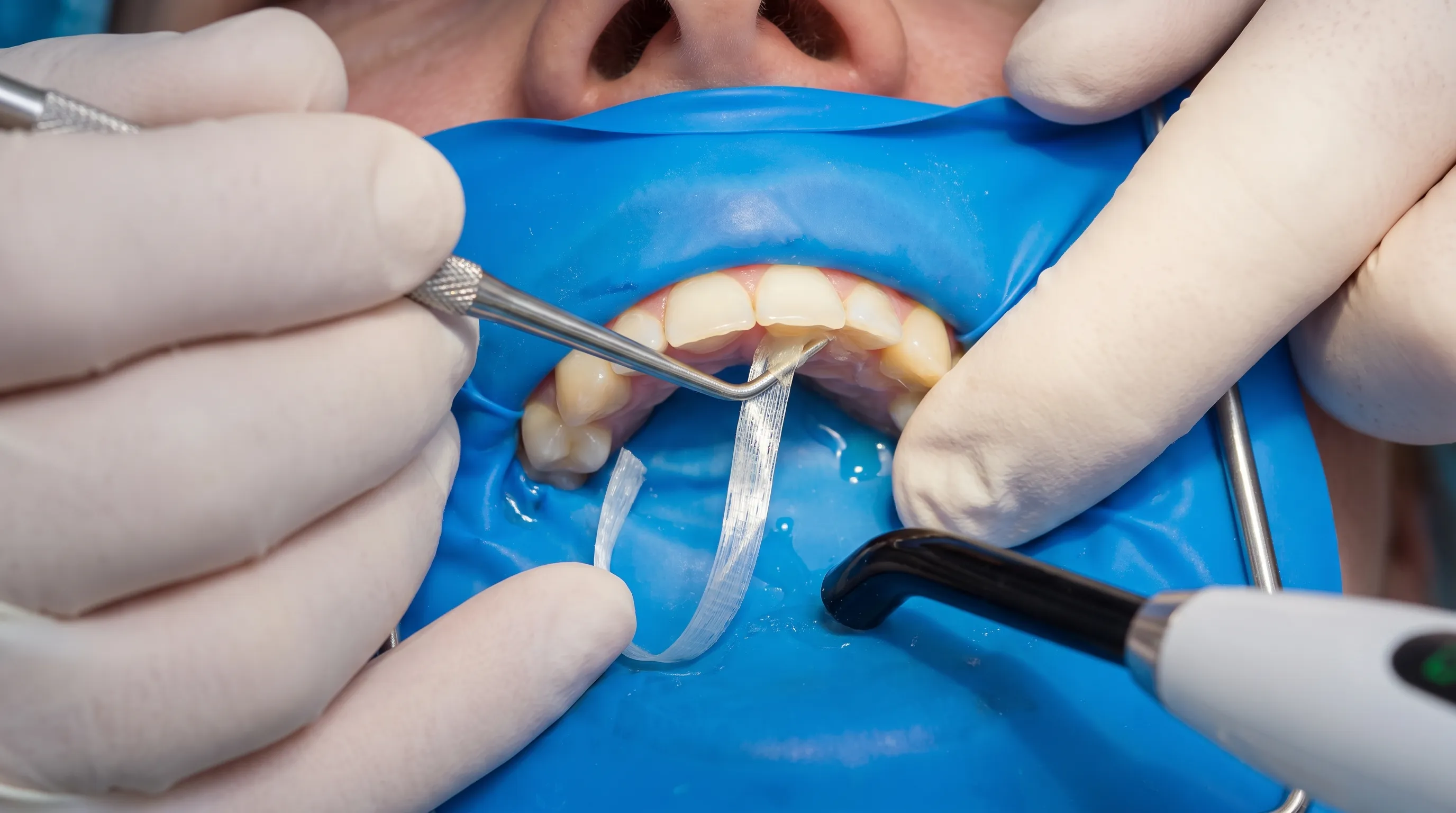
When a loose adult tooth exhibits functional mobility but retains adequate supporting structure, splinting provides immediate mechanical stabilization. Extracoronal splinting involves bonding adjacent teeth together using composite resin and fiber-reinforced materials, distributing occlusal forces across a broader arch segment. Intracoronal splints utilize metal or zirconia connectors embedded within the teeth, offering superior durability and aesthetic integration. For severely mobile teeth, temporary splinting with acrylic resin stabilizes the area while periodontal tissues heal and mature. Permanent splinting is considered when bone regeneration has stabilized the prognosis but residual mobility compromises chewing efficiency. Properly designed splints must allow for physiological cleaning and prevent secondary caries formation.
Surgical Options: Bone Grafting and Guided Tissue Regeneration
In cases of significant vertical bone defects or furcation involvement, non-surgical therapy alone cannot restore lost architecture. Regenerative periodontal surgery utilizes osteoconductive graft materials, barrier membranes, and biologic agents like enamel matrix derivatives to stimulate new bone, cementum, and ligament formation. Autografts harvested from the patient, allografts from tissue banks, or synthetic bone substitutes are meticulously packed into debrided defects. Resorbable or non-resorbable membranes isolate the surgical site from gingival epithelial downgrowth, allowing slower periodontal cells to repopulate the root surface. Guided tissue regeneration can achieve remarkable results, restoring up to 60 percent of lost periodontal support and transforming a hopeless prognosis into a long-term viable tooth.
When Extraction Becomes Necessary
Despite advanced therapeutic options, some teeth reach a point of structural compromise where retention becomes impractical. Indications for extraction include Grade III or IV mobility, hopeless prognosis with less than 10 percent bone support, severe root fractures extending subgingivally, uncontrollable infection threatening adjacent structures, or non-restorable carious destruction. In these scenarios, proactive extraction prevents further bone loss and eliminates chronic infection sources. Following extraction, implant therapy, fixed bridgework, or removable prosthetics can restore function and aesthetics. Modern implantology offers highly predictable long-term outcomes, and timely intervention prevents the devastating consequences of prolonged edentulism. Professional clinical guidelines can be reviewed in the American Dental Association's periodontal disease resources.
Safe Home Care, Prevention, and Lifestyle Modifications
Professional intervention establishes the foundation for stability, but long-term success hinges entirely on rigorous patient compliance and proactive lifestyle management. Implementing evidence-based self-care protocols transforms periodontal maintenance from a burden into a sustainable daily habit.
Optimizing Your Daily Oral Hygiene Routine
Effective plaque control requires technique refinement and appropriate tool selection. Manual brushing should utilize a soft-bristled brush with modified Bass technique, angling the bristles 45 degrees toward the gumline to disrupt subgingival biofilm. Power toothbrushes with oscillating-rotating or sonic technology demonstrate superior plaque removal and gingival health improvement in clinical studies. Interdental cleaning is non-negotiable; traditional floss, water flossers, or interdental brushes must be used daily to clean embrasure spaces inaccessible to brushes. Antimicrobial mouthwashes containing cetylpyridinium chloride or essential oils provide adjunctive bacterial reduction but should never replace mechanical debridement. For best practices in daily oral care, see the Mayo Clinic's recommendations on dental hygiene. Patients with mobility must exercise gentle pressure to avoid further periodontal ligament trauma while ensuring thorough biofilm disruption.
Dietary Adjustments and Nutritional Support
Nutritional intake profoundly influences periodontal tissue resilience and inflammatory regulation. Diets high in refined sugars and simple carbohydrates fuel pathogenic bacterial metabolism, accelerating plaque accumulation and acid production. Emphasizing anti-inflammatory foods rich in omega-3 fatty acids, antioxidants, and polyphenols modulates systemic inflammatory responses and support tissue healing. Vitamin C is essential for collagen cross-linking and gingival integrity, while vitamin D and calcium maintain alveolar bone mineral density. Magnesium and zinc play crucial roles in enzymatic bone formation and immune modulation. Hydration stimulates salivary flow, naturally cleansing the oral cavity and neutralizing acids. Avoiding hard, sticky, or excessively chewy foods during active mobility periods prevents exacerbating traumatic forces on compromised teeth.
Protective Measures Against Bruxism and Night Grinding
Parafunctional habits generate occlusal forces up to ten times normal chewing pressure, devastating already weakened periodontal structures. Nocturnal bruxism often occurs subconsciously, making self-awareness difficult. Custom-fabricated hard acrylic occlusal guards provide a protective buffer, redistributing destructive lateral forces and preventing enamel attrition, root microfractures, and ligament trauma. Stress management techniques including cognitive behavioral therapy, mindfulness meditation, progressive muscle relaxation, and regular aerobic exercise significantly reduce bruxism frequency by lowering sympathetic nervous system activation. Avoiding caffeine and alcohol before bedtime improves sleep architecture and reduces sleep-related grinding episodes. Regular guard inspections and professional adjustments ensure optimal fit and continued protective efficacy.
| Mobility Grade | Clinical Presentation | Movement Range | Prognosis | Recommended Intervention |
|---|---|---|---|---|
| Grade I | Slight horizontal movement | < 1 mm | Excellent | Enhanced hygiene, SRP, monitoring |
| Grade II | Noticeable horizontal movement | 1–2 mm | Good | Splinting, regenerative surgery, occlusal adjustment |
| Grade III | Severe horizontal + vertical depression | > 2 mm | Guarded | Aggressive regenerative therapy, extraction evaluation |
| Grade IV | Non-functional, extreme looseness | Variable | Poor/Hopeless | Extraction, implant or prosthetic planning |
Frequently Asked Questions
Can a loose adult tooth tighten up on its own without dental treatment?
In cases of minor trauma, transient hormonal changes, or temporary orthodontic forces, a tooth may spontaneously re-stabilize as the periodontal ligament heals and inflammation subsides. However, if the mobility stems from chronic periodontal disease, bacterial plaque accumulation, or progressive bone loss, it will not tighten on its own. Pathological mobility indicates irreversible attachment loss that requires professional intervention to halt disease progression. Delaying treatment allows continued bone resorption, ultimately transforming a salvageable condition into one requiring extraction. Professional evaluation, diagnostic imaging, and targeted periodontal therapy are essential to determine if natural stabilization is biologically possible.
Is it safe to wiggle a loose tooth with my tongue or fingers to check it?
While occasional tactile awareness is natural, repeatedly manipulating a mobile tooth with your tongue, fingers, or objects is strongly discouraged. Each wiggling motion stretches the compromised periodontal ligament fibers, disrupts the delicate healing process, and accelerates inflammatory mediator release. Constant mechanical stimulation prevents the collagen matrix from reattaching to the root surface and can widen the periodontal space. Furthermore, transferring oral bacteria from the hands or introducing external contaminants increases the risk of secondary infection. Clinicians recommend leaving the tooth completely undisturbed except during gentle, prescribed hygiene routines and professional evaluations to maximize the potential for stabilization.
What is the fastest way to stop a loose tooth from falling out?
There is no instant remedy for pathological mobility, as periodontal tissue regeneration is a biological process measured in weeks and months. The fastest pathway involves immediately eliminating the primary destructive factors: professional scaling and root planing to remove subgingival calculus, strict adherence to optimized home hygiene, complete cessation of tobacco use, and implementation of an occlusal guard if parafunctional habits exist. In advanced cases, rapid stabilization through dental splinting prevents further functional movement while underlying tissues respond to therapy. Emergency extraction may be necessary if infection threatens systemic health or if the tooth presents an aspiration risk. Consistent, disciplined follow-up care is the only evidence-based approach to preserving a loose adult tooth.
Can vitamin supplements reverse tooth mobility caused by gum disease?
While nutritional deficiencies can exacerbate periodontal inflammation and impair tissue healing, no vitamin or mineral supplement can regenerate lost alveolar bone or reattach severed periodontal ligament fibers once pathological mobility has developed. Supplements like vitamin C, D, calcium, and omega-3 fatty acids serve as crucial adjuncts that support systemic immunity, reduce inflammatory cytokines, and optimize the body’s capacity for tissue repair. They enhance the success of professional treatments and improve long-term maintenance outcomes but cannot replace mechanical debridement, antimicrobial therapy, or surgical regeneration. A balanced, nutrient-dense diet combined with evidence-based periodontal care remains the gold standard for addressing mobility.
How often should I see a periodontist if I have a history of loose teeth?
Patients with established periodontal disease or documented tooth mobility require lifelong supportive maintenance beyond standard biannual cleanings. Periodontal therapy typically involves recall intervals of every 3 to 4 months during the first year following active treatment, allowing clinicians to monitor pocket depths, assess bleeding indices, and detect early recurrence. Once periodontal parameters stabilize, intervals may gradually extend to 6 months, though many specialists recommend continuing quarterly visits indefinitely. These maintenance appointments enable early intervention before clinical mobility develops, preserve surgical and regenerative outcomes, and provide continuous reinforcement of optimal home care techniques tailored to your specific anatomical and biological needs.
Conclusion
Navigating the reality of a loose adult tooth requires a shift from reactive panic to proactive, evidence-based management. Tooth mobility is not a normal part of aging, nor is it an inevitable precursor to extraction. It is a clear biological signal that the periodontium is under siege from mechanical overload, bacterial pathogenicity, or systemic compromise. By understanding the anatomical foundations, recognizing the early clinical signs, and committing to comprehensive professional therapy alongside rigorous home care, patients can frequently stabilize compromised teeth and restore long-term functional harmony. The journey toward periodontal health demands partnership between clinician expertise and patient dedication, transforming a frightening diagnosis into an opportunity for renewed oral vitality. Prioritizing timely evaluation, embracing preventive lifestyle modifications, and maintaining consistent supportive care will safeguard your natural dentition, preserve your smile, and ensure that every tooth remains securely anchored for decades to come.
How we work
Researched and drafted with AI assistance, then reviewed by a human editor against the cited sources.
This article is general health information, not medical advice. Consult a qualified healthcare professional about your own situation.