Clicky Sore Knee: Causes, Treatments, and Expert Relief Strategies
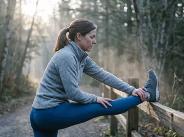
You are halfway through your morning walk when you hear it: a distinct pop, followed by a grinding sensation that shoots through your lower leg. The next step brings a dull ache that refuses to fade. If you have ever wondered why your joints suddenly announce your movements with audible friction and lingering discomfort, you are not alone. Millions of people navigate daily life while managing a clicky sore knee, often dismissing it as normal aging until the pain begins interfering with stairs, workouts, or even simple walking. Joint noise combined with tenderness is a classic clinical presentation that bridges biomechanical dysfunction and tissue-level inflammation. Understanding the difference between harmless physiological sounds and pathological joint stress is the first step toward reclaiming your mobility. In this comprehensive guide, we will explore the underlying anatomy, decode the most frequent triggers, and provide evidence-based strategies to calm inflammation, restore tracking alignment, and build resilient supporting musculature. Whether your symptoms emerged after a sudden misstep or have developed gradually over years of repetitive impact, targeted interventions exist to interrupt the pain cycle and promote long-term joint health. Let us break down exactly what is happening inside your joint space and how you can safely guide your recovery forward.
Understanding the Anatomy and Mechanics of a Noisy Knee
The human knee is a marvel of biomechanical engineering, functioning primarily as a hinge joint that must also accommodate subtle rotational movements. It connects three bones: the femur, tibia, and patella, relying on a complex network of ligaments, tendons, cartilage, and synovial fluid to maintain smooth articulation. When any of these components experience abnormal friction, misalignment, or fluid disruption, audible and tactile feedback emerges. Learning to interpret these signals helps you distinguish normal joint physiology from early warning signs of structural compromise.
Free calculatorKnee Pain Location ChartIdentify knee pain causes by locationOpen the calculatorThe Science Behind Joint Sounds (Crepitus)
Crepitus is the medical term for grating, cracking, or popping sensations within joints. Research published in orthopedic journals consistently shows that isolated clicking without pain is frequently benign. It typically occurs when nitrogen gas bubbles rapidly form and collapse in the synovial fluid, similar to cracking your knuckles. Another common mechanism involves tendons or ligaments sliding over bony prominences during flexion and extension. However, when crepitus couples with localized tenderness, swelling, or mechanical resistance, it shifts from physiological noise to pathological signaling. Persistent grinding sensations often reflect uneven cartilage wear, where the smooth hyaline layer that normally cushions bone ends becomes roughened. This roughened surface increases friction during weight-bearing activities, triggering inflammatory cascades that sensitize surrounding nerve endings. Understanding this transition helps individuals recognize when self-monitoring should evolve into structured rehabilitation or professional evaluation.
Tracking Misalignments and Biomechanical Stress
The patella glides along a specialized femoral groove during knee movement. Ideal tracking ensures even pressure distribution across the joint surface. When muscular imbalances develop—particularly weakness in the vastus medialis or tightness in the lateral structures—the kneecap deviates from its intended pathway. This lateral tracking forces the patella against the outer ridge of the femur, generating both audible clicking and concentrated stress points that become increasingly tender over time. Hip mechanics also play a crucial role; excessive femoral internal rotation or gluteal weakness alters the entire lower-limb kinetic chain, redirecting impact forces directly into the knee rather than absorbing them through proximal stabilizers. Addressing a clicky sore knee effectively requires evaluating movement patterns beyond the joint itself. Foot pronation, pelvic positioning, and core engagement all influence how cleanly your knee moves through its full range of motion. Corrective strategies that restore balanced muscle recruitment significantly reduce abnormal joint loading and prevent progressive tissue breakdown.
Common Causes Behind a Clicky Sore Knee
Pinpointing the root cause of your symptoms guides appropriate management and prevents unnecessary interventions. While multiple conditions share overlapping presentations, distinct clinical patterns emerge when you analyze onset timing, aggravating factors, and associated functional limitations.
Patellofemoral Pain Syndrome and Overuse
Patellofemoral pain syndrome (PFPS) represents one of the most prevalent explanations for anterior knee discomfort accompanied by audible crepitus. Frequently labeled as runner’s knee, this condition disproportionately affects individuals who engage in repetitive impact activities, sudden training spikes, or prolonged sitting with flexed knees. The hallmark symptom includes pain behind or around the kneecap that worsens with squatting, stair descent, or sitting for extended periods. Overuse disrupts the delicate balance of compressive forces within the patellofemoral compartment, irritating subchondral bone and sensitizing the synovial lining. Studies from Mayo Clinic emphasize that PFPS rarely stems from acute structural damage. Instead, it develops from cumulative microtrauma and muscular dysfunction that gradually degrade joint homeostasis. Managing this condition successfully requires load modification, progressive quadriceps strengthening, and hip stabilization work rather than passive treatments alone.
Degenerative Changes and Osteoarthritis
Age-related joint degeneration remains a leading contributor to persistent knee noise and tenderness. Osteoarthritis progressively breaks down protective cartilage, narrows joint space, and stimulates osteophyte formation. Unlike sudden traumatic injuries, degenerative changes unfold gradually, often producing a consistent grinding sensation that intensifies after periods of rest and temporarily eases with gentle movement. The synovial membrane becomes chronically irritated, producing excess inflammatory mediators that amplify pain perception. Clinical guidelines from NHS highlight that early intervention through weight management, targeted strengthening, and anti-inflammatory lifestyle practices can significantly slow progression. Maintaining joint nutrition through movement stimulates synovial fluid circulation, which remains essential for delivering oxygen and removing metabolic waste from avascular cartilage tissue. Ignoring early degenerative signals often accelerates functional decline, while proactive management preserves mobility well into later decades.
Soft Tissue Injuries: Meniscus and Tendons
The meniscus acts as a shock-absorbing wedge between the femur and tibia, while tendons transmit muscular force across the joint. Tears in the meniscus frequently produce audible clicks or catches during rotation or deep knee bending. Degenerative meniscal lesions commonly occur without dramatic trauma, particularly in individuals over forty, while acute tears usually follow sudden twisting motions under load. Tendinopathy affecting the patellar or quadriceps tendon creates localized soreness just below or above the kneecap, often worsening during eccentric loading phases like downhill walking or jumping. When tendon tissue experiences repetitive strain beyond its repair capacity, collagen fibers disorganize and neovascularization develops, sensitizing nociceptive pathways. Comprehensive rehabilitation for soft tissue injuries prioritizes gradual load progression rather than complete immobilization. Controlled mechanical stress actually stimulates tenocyte activity and promotes organized collagen remodeling, ultimately restoring tensile strength and reducing discomfort during daily activities.
Red Flags: When a Click Requires Immediate Medical Attention
Not every noisy knee demands urgent intervention, but certain clinical indicators signal underlying structural compromise that requires professional assessment. Recognizing these warning signs prevents delayed treatment and minimizes the risk of long-term joint instability.
Distinguishing Harmless Noise from Structural Damage
Painless crepitus during routine movement generally reflects normal physiological processes. However, when clicking synchronizes with sharp pain, sudden swelling, or mechanical locking, the clinical picture changes dramatically. Joint locking occurs when a displaced meniscal fragment or loose cartilage body physically blocks full extension or flexion. Instability sensations, where the knee feels like it might buckle or give way, indicate ligamentous insufficiency or significant quadriceps inhibition. Rapid-onset swelling within two hours of an incident suggests hemarthrosis or capsular disruption, whereas gradual fluid accumulation over days typically reflects inflammatory arthritis or chronic irritation. Differentiating these patterns guides appropriate triage decisions. If your clicky sore knee consistently produces catching, buckling, or persistent effusion despite rest and activity modification, scheduling a thorough orthopedic evaluation remains essential for accurate diagnosis and targeted treatment planning.
The Role of Inflammation and Effusion
Joint effusion represents your body’s protective response to internal stress or injury. Synovial fluid production increases to cushion irritated tissues and deliver immune mediators. While temporary swelling supports healing, chronic effusion indicates ongoing irritation that disrupts normal neuromuscular function. Excess fluid mechanically stretches the joint capsule, triggering reflexive inhibition of the quadriceps and reducing overall stability. This inhibition creates a vicious cycle where weakened supporting muscles place greater stress on passive stabilizers like ligaments and cartilage, accelerating tissue breakdown. Managing inflammation effectively requires balancing movement and recovery. Gentle range-of-motion exercises promote lymphatic drainage without aggravating sensitive structures. Compression garments, elevation protocols, and consistent hydration further support fluid regulation. Persistent effusion beyond three weeks warrants diagnostic imaging to identify intra-articular abnormalities that may require procedural intervention.
| Symptom Pattern | Likely Mechanism | Recommended Action | Recovery Timeline |
|---|---|---|---|
| Occasional popping without pain | Gas bubble cavitation or tendon glide | Maintain normal activity, monitor | Immediate or none |
| Grinding with activity-related ache | Early cartilage wear or tracking dysfunction | Load management, targeted strengthening | 4 to 8 weeks |
| Catching or locking during bending | Meniscal displacement or loose body | Clinical evaluation, possible imaging | Weeks to months |
| Rapid swelling with instability | Ligament tear or acute trauma | Immediate rest, professional assessment | 2 to 6 months |
| Morning stiffness lasting over 30 min | Inflammatory arthritis or advanced OA | Medical consultation, anti-inflammatory strategy | Ongoing management |
Evidence-Based Self-Care and Conservative Management
Conservative interventions form the foundation of successful knee rehabilitation. Modern protocols emphasize active recovery over passive rest, recognizing that controlled movement stimulates tissue healing, restores proprioception, and rebuilds functional capacity. Implementing structured self-care routines addresses both symptom relief and long-term resilience.
Modernized Recovery Protocols Beyond RICE
Traditional RICE (Rest, Ice, Compression, Elevation) guidelines have evolved significantly based on contemporary sports medicine research. Complete immobilization now receives strong discouragement due to its negative impact on synovial nutrition, muscle activation, and connective tissue remodeling. The updated PEACE and LOVE framework prioritizes protection, elevation, avoidance of anti-inflammatory medications in acute phases, compression, and education, followed by load optimization, optimism, vascularization, and exercise. For a clicky sore knee, this means protecting the joint from aggravating movements while gradually reintroducing controlled stress that matches tissue tolerance. Ice application remains valuable for acute flare-ups to reduce localized hyperemia and dull pain signals, but prolonged or excessive cooling can blunt necessary inflammatory mediators that initiate repair. Alternating between gentle movement and strategic recovery periods optimizes the healing environment without encouraging stiffness or deconditioning.
Targeted Rehabilitation Exercises for Stability
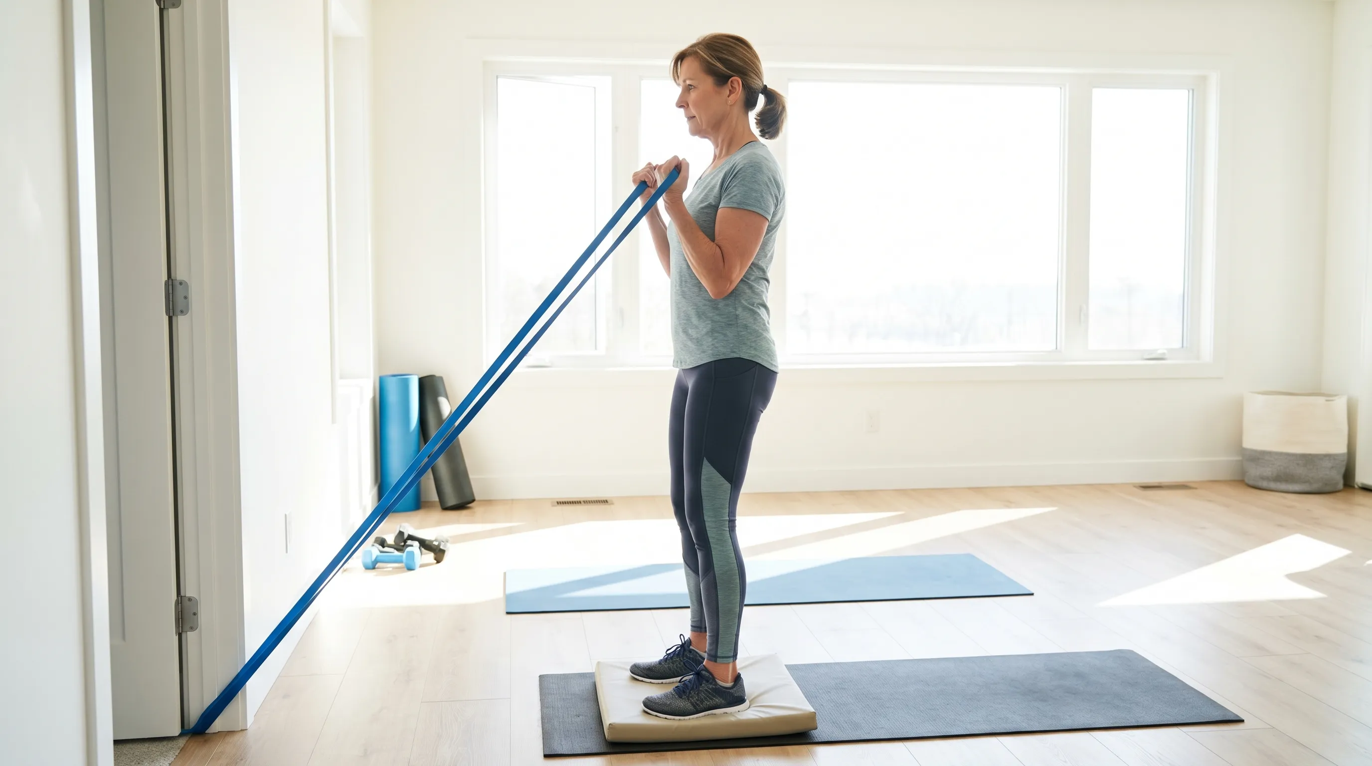
Strengthening the muscles that surround and control the knee dramatically improves joint tracking and reduces abnormal loading. The vastus medialis obliquus (VMO) plays a particularly crucial role in medial patellar stabilization. Isometric quad sets, straight-leg raises, and short-arc quadriceps extensions effectively activate these fibers without generating excessive compressive forces. Hip abductor and external rotator strengthening through clamshells, lateral band walks, and single-leg bridges addresses proximal biomechanical deficits that frequently contribute to distal joint stress. Progression should follow a predictable hierarchy: isometric holds first, followed by controlled eccentric movements, then dynamic functional patterns, and finally sport-specific or high-demand activities.
Begin with seated quad sets: tighten your thigh muscle while pressing the back of your knee flat against the floor or bed. Hold for five seconds, repeat ten times, and perform three sets daily. Transition to mini-squats within a thirty-degree flexion range, ensuring your knees track directly over your second toes. Incorporate single-leg balance work to stimulate proprioceptive receptors that fine-tune neuromuscular control during weight acceptance. Stretching the iliotibial band, hamstrings, and gastrocnemius reduces lateral pull and restores optimal joint alignment. Consistency matters more than intensity; performing moderate-repetition sessions four to five times weekly yields superior long-term outcomes compared to sporadic intense workouts that overwhelm recovering tissues.
Supportive Gear and Lifestyle Modifications
External support strategies complement active rehabilitation by providing temporary mechanical assistance while tissues rebuild strength. Patellar stabilizing braces with lateral buttresses gently guide kneecap tracking during ambulation and exercise. Compression sleeves enhance proprioceptive feedback through cutaneous mechanoreceptor stimulation, helping your nervous system maintain better joint positioning awareness. Selecting footwear with adequate midsole cushioning and arch support alters ground reaction forces before they reach the knee. Individuals with overpronation benefit from stability shoes or custom orthotics that prevent excessive internal tibial rotation. Activity modification does not mean complete withdrawal from movement. Switching from high-impact running to cycling, swimming, or elliptical training maintains cardiovascular fitness while dramatically reducing joint compression. Pacing strategies, such as alternating high-demand days with active recovery sessions, prevent symptom exacerbation and support sustainable participation in daily routines.
Medical Interventions and Clinical Pathways
When conservative measures fail to resolve persistent symptoms, clinical evaluation and advanced interventions become necessary. Understanding what to expect during professional assessment streamlines your decision-making and aligns expectations with realistic treatment trajectories.
Diagnostic Imaging and Orthopedic Evaluations
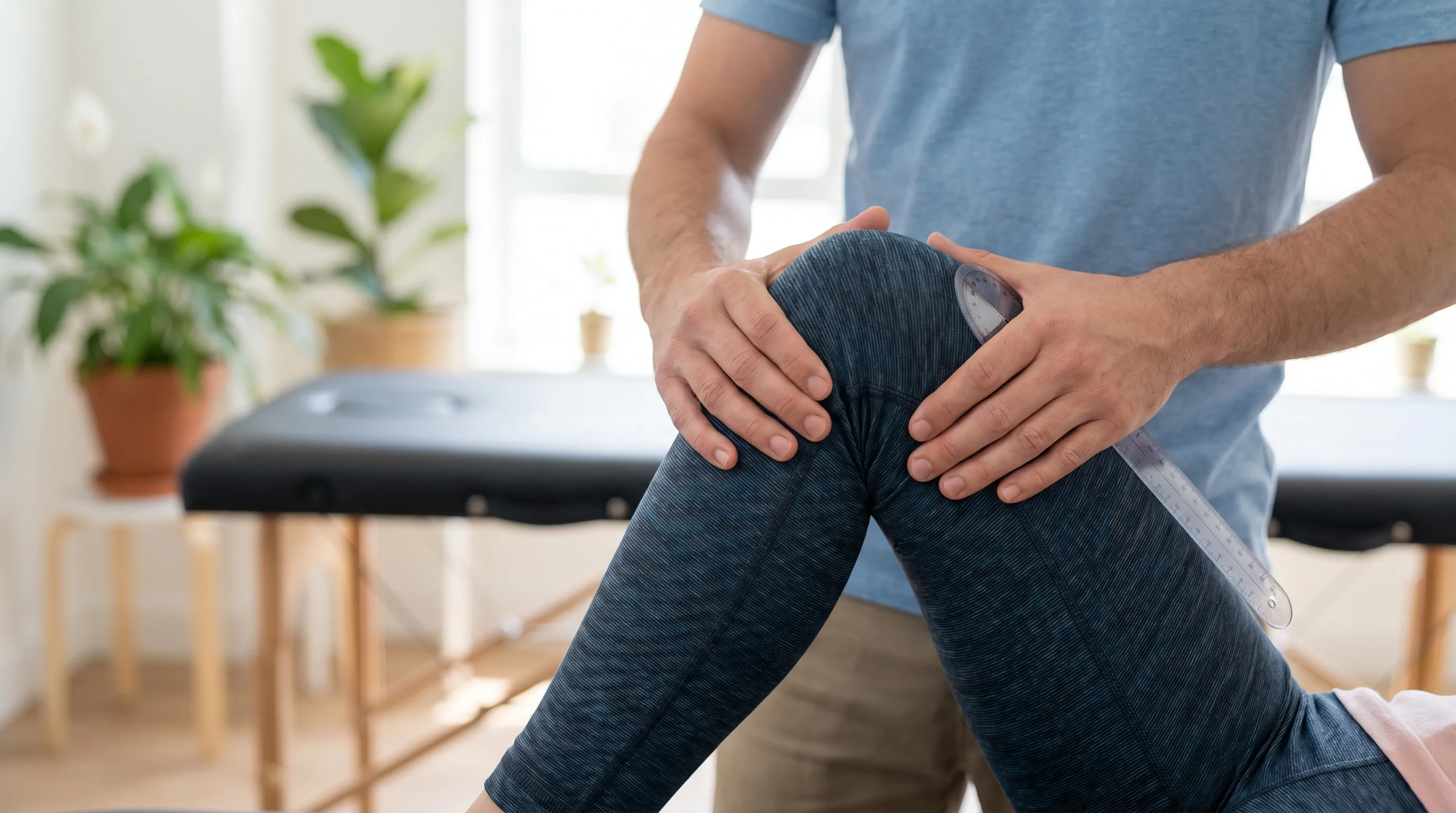
A comprehensive clinical examination remains the cornerstone of accurate knee assessment. Orthopedic specialists evaluate gait patterns, joint effusion, ligamentous laxity, and patellar mobility before advancing to imaging modalities. Weight-bearing X-rays reveal joint space narrowing, subchondral sclerosis, and osteophyte development that plain radiographs frequently miss when performed in non-weight-bearing positions. Magnetic resonance imaging (MRI) excels at visualizing soft tissue integrity, detecting meniscal tears, ligament sprains, tendon degeneration, and early cartilage lesions. Ultrasound provides dynamic assessment of tendons and bursae during movement, capturing abnormalities that static imaging overlooks. Diagnostic accuracy improves significantly when clinical findings correlate directly with imaging results, preventing unnecessary interventions for incidental findings that frequently appear on scans without producing actual symptoms.
Advanced Therapies and Surgical Considerations
When structural damage requires intervention beyond rehabilitation, several procedural options exist depending on the specific pathology. Intra-articular corticosteroid injections rapidly reduce inflammation during severe flare-ups, though repeated use may negatively impact cartilage matrix over time. Hyaluronic acid viscosupplementation aims to restore synovial fluid viscosity, improving lubrication and potentially extending intervals between painful episodes. Platelet-rich plasma (PRP) therapy utilizes concentrated autologous growth factors to stimulate natural tissue repair processes, showing promising results for tendinopathy and early degenerative changes. Arthroscopic surgery addresses mechanical symptoms caused by torn meniscal fragments or loose bodies that physically obstruct joint motion. Realignment osteotomies correct severe maltracking in younger patients with localized cartilage wear, while partial or total knee arthroplasty remains the definitive solution for end-stage osteoarthritis unresponsive to conservative care. Surgical decisions require careful risk-benefit analysis, considering activity demands, overall health status, and realistic recovery expectations.
Prevention and Long-Term Knee Longevity
Sustaining joint health requires proactive habits that address biomechanical efficiency, tissue resilience, and systemic inflammatory balance. Preventive strategies implemented early yield compounding benefits that extend functional independence well into later life stages.
Weight Management and Nutritional Support
Every additional pound of body weight multiplies the mechanical load crossing the knee joint during gait. Biomechanical studies demonstrate that a single step generates compressive forces approximately two to three times your body weight. Losing even modest amounts of excess mass significantly reduces cumulative stress on articular cartilage, ligaments, and meniscal tissue. Nutritional strategies that support joint integrity include adequate omega-3 fatty acid intake to modulate inflammatory pathways, sufficient protein consumption for tissue repair, and antioxidant-rich foods that combat oxidative stress within joint spaces. Vitamin D and calcium remain essential for underlying bone health, while collagen peptides and glucosamine supplements show mixed evidence but remain safe for most individuals seeking additional connective tissue support. Hydration directly impacts synovial fluid viscosity, making consistent water intake a surprisingly practical intervention for joint lubrication and nutrient transport.
Cross-Training and Activity Periodization
Monotonous movement patterns place repetitive stress on identical joint structures, increasing the likelihood of overuse injuries. Cross-training distributes mechanical demands across different tissues while maintaining overall fitness. Combining cycling with swimming, yoga, and strength training creates balanced development that protects vulnerable joints from fatigue-related breakdown. Periodization principles involve systematically varying intensity, volume, and recovery periods to allow tissues adequate time for adaptation and remodeling. Following a three-to-one loading pattern—three weeks of progressive challenge followed by a lighter recovery week—optimizes tissue strengthening while minimizing overtraining syndrome risk. Listening to subtle warning signals like morning stiffness, lingering soreness beyond twenty-four hours, or subtle changes in movement quality enables proactive adjustments before minor irritation evolves into significant injury.
Frequently Asked Questions
Is a clicky sore knee a sign of permanent damage?
Not necessarily. While some clicking indicates cartilage wear or structural changes, many cases result from harmless crepitus or temporary inflammation. Persistent pain, swelling, or mechanical locking warrants professional evaluation to rule out significant tissue damage.
How long does it take for knee pain to improve with exercise?
Most individuals notice meaningful improvement within 4 to 8 weeks of consistent, guided strengthening and mobility work. Tendon and muscle adaptations require progressive overload and adequate recovery time to restore joint stability. Tracking your symptoms weekly helps identify positive trends before complete resolution occurs.
Should I push through knee pain or rest completely?
Complete immobilization often worsens joint stiffness and muscle inhibition. Modern rehabilitation emphasizes relative rest combined with controlled, pain-free movement. Modify activities to avoid sharp pain while maintaining gentle range of motion and low-impact strength work that matches your current tissue tolerance.
Can footwear actually reduce knee clicking and soreness?
Yes. Proper footwear with adequate arch support and cushioning alters lower-limb biomechanics, reducing abnormal rotational forces that reach the knee. Orthotics or stability shoes can significantly decrease patellar tracking stress and joint irritation, particularly for individuals with flat feet or overpronation tendencies.
When should I see a doctor instead of trying home remedies?
Seek medical attention if you experience sudden swelling, joint instability, inability to bear weight, visible deformity, fever, or pain that persists beyond two to three weeks despite conservative self-care and activity modification. Early professional guidance prevents chronic compensation patterns and accelerates targeted recovery.
Conclusion
Managing a clicky sore knee successfully requires shifting from passive symptom suppression to active joint preservation. Understanding the biomechanical roots of your discomfort empowers you to make informed decisions about activity modification, targeted strengthening, and appropriate clinical intervention. Consistent rehabilitation, intelligent load management, and proactive lifestyle habits form the foundation of sustainable joint health. While temporary discomfort remains a normal part of human movement, persistent pain paired with mechanical dysfunction signals that your tissues need structured support to restore balance. By implementing evidence-based strategies, listening carefully to your body’s feedback, and seeking professional guidance when warning signs emerge, you can transform a frustrating joint limitation into an opportunity for long-term resilience and improved functional capacity.
For additional evidence-based guidance on joint health and rehabilitation protocols, visit Arthritis Foundation Exercise Guidelines and OrthoInfo by AAOS. If you prefer visual instruction, this expert-led mobility routine demonstrates safe progression principles:
and . Integrating these resources into your recovery journey supports informed decision-making and sustainable progress toward pain-free mobility.How we work
Researched and drafted with AI assistance, then reviewed by a human editor against the cited sources.
This article is general health information, not medical advice. Consult a qualified healthcare professional about your own situation.