Ab Muscle Tear: Complete Guide to Symptoms, Treatment & Recovery

The human core is engineered for remarkable strength, stability, and dynamic movement, yet it remains surprisingly vulnerable to acute trauma. Whether you are an elite athlete pushing through a high-intensity training session, a weekend warrior experimenting with heavy compound lifts, or someone experiencing a sudden strain during everyday physical labor, an ab muscle tear can instantly disrupt your mobility, compromise your posture, and force an unwelcome pause in your active lifestyle. Understanding how abdominal tissues fail, recognizing the early warning signs, and implementing evidence-based recovery protocols are essential steps toward regaining function and preventing long-term complications. This comprehensive guide explores the biomechanics of abdominal injuries, outlines clinically validated treatment pathways, and provides actionable strategies to support optimal tissue repair while safely guiding you back to peak physical performance.
Abdominal muscle injuries are frequently misunderstood, often dismissed as simple soreness or routine overexertion. However, when the structural integrity of the rectus abdominis, external obliques, or transverse abdominis is compromised, the healing process requires precise physiological support and strategic rehabilitation. Rushing back into training without addressing the underlying tissue damage can lead to chronic instability, compensatory movement patterns, and recurrent injury. By approaching recovery with patience, clinical awareness, and scientifically grounded methodology, you can transform a painful setback into an opportunity for improved neuromuscular control and long-term musculoskeletal resilience.
Understanding Abdominal Muscle Anatomy and Injury Mechanisms
To properly address an ab muscle tear, it is crucial to first understand the structural components involved and the physiological conditions that lead to tissue failure. The anterior abdominal wall consists of multiple overlapping muscle layers designed to protect visceral organs, maintain intra-abdominal pressure, and facilitate spinal flexion, lateral bending, and rotational movements. The most prominent of these is the rectus abdominis, a paired muscle running vertically along the midline of the torso, anchored by the pubic crest below and the costal cartilages of the fifth to seventh ribs above. This muscle is frequently the primary site of acute strain during explosive flexion movements or heavy resistance training.
Free calculatorBMI CalculatorCalculate Body Mass IndexOpen the calculatorFlanking the rectus abdominis are the external and internal obliques, which facilitate lateral flexion and trunk rotation. Deeper still lies the transverse abdominis, a horizontally oriented stabilizing muscle that functions like a natural corset, compressing the abdominal contents and supporting lumbar stability. Each layer works synergistically, distributing mechanical loads across the torso. When one layer experiences excessive tensile stress beyond its physiological tolerance, microtears can rapidly progress into macroscopic fiber disruption, resulting in an ab muscle tear that compromises functional capacity.
What Constitutes an Ab Muscle Tear?
An ab muscle tear occurs when mechanical stress exceeds the elastic limit of muscle fibers, causing partial or complete rupture of the tissue. This typically happens during eccentric loading, where the muscle lengthens while contracting to control movement speed. Examples include lowering a heavy barbell during sit-ups, sudden twisting during athletic competition, or forceful coughing and sneezing in deconditioned individuals. The combination of high force, rapid velocity, and inadequate warm-up creates a perfect storm for structural failure.
The tearing process involves the disruption of sarcomeres, damage to the extracellular matrix, and rupture of microvasculature within the muscle belly. This immediate trauma triggers an acute inflammatory cascade, characterized by localized edema, increased capillary permeability, and recruitment of immune cells to clear cellular debris. Understanding this biological timeline is critical for implementing appropriate interventions at each stage of healing.
Grades of Muscle Strains
Medical professionals classify abdominal muscle injuries using a standardized grading system that correlates with tissue damage, functional impairment, and projected recovery duration. Accurate grading guides treatment selection and sets realistic expectations for rehabilitation milestones.
Grade 1 strains represent mild microtrauma with less than five percent of muscle fibers disrupted. Patients typically experience mild discomfort, minimal strength loss, and full range of motion with slight restriction. Healing generally progresses rapidly, often within two to three weeks with conservative management.
Grade 2 strains involve partial tearing of muscle tissue, affecting between five and fifty percent of fibers. Symptoms include moderate to severe pain, palpable swelling, noticeable weakness during contraction, and significant functional limitation. Bruising may appear within forty-eight hours as hemorrhaged blood migrates toward the skin surface. Recovery typically requires four to eight weeks of structured rehabilitation.
Grade 3 strains indicate complete rupture or avulsion of the muscle from its tendinous attachment. Patients report intense, debilitating pain, a visible or palpable defect in the muscle contour, profound weakness, and often an inability to flex the trunk against gravity. Surgical evaluation is frequently warranted for Grade 3 injuries, followed by a prolonged rehabilitation protocol lasting three to six months.
Recognizing the Signs and Symptoms
Early symptom recognition is paramount for preventing secondary tissue damage and initiating timely intervention. Abdominal muscle injuries often present with a distinct cluster of clinical indicators that differentiate them from visceral pain, gastrointestinal distress, or spinal dysfunction.
Immediate vs. Delayed Pain Signals
The initial presentation of an ab muscle tear typically features a sudden, sharp sensation localized to the affected muscle belly during the inciting movement. Patients often describe feeling or hearing a distinct tearing or popping sensation. Immediate pain usually intensifies with deep breathing, coughing, laughing, or attempting to sit up from a supine position.
Within twenty-four to forty-eight hours, pain often transitions from acute and localized to a dull, aching discomfort accompanied by stiffness and tightness. Delayed onset muscle soreness (DOMS) can coexist with true structural injury, but DOMS resolves within three to five days, whereas symptomatic strain pain persists and worsens with mechanical loading. Differentiating between these conditions prevents premature training resumption that could exacerbate fiber disruption.
When to Seek Emergency Medical Care
While most abdominal strains are musculoskeletal in origin, certain symptoms warrant immediate emergency evaluation to rule out life-threatening conditions. Seek urgent medical attention if you experience sudden, severe abdominal pain accompanied by fever, uncontrolled vomiting, gastrointestinal bleeding, radiating pain to the back or groin, or a rigid, board-like abdominal wall. These signs may indicate appendicitis, bowel obstruction, internal organ injury, or vascular emergencies that require immediate surgical intervention.
Additionally, if an ab muscle tear occurs alongside high-impact trauma, such as a motor vehicle collision or fall from significant height, imaging studies are essential to assess for concomitant rib fractures, organ contusion, or fascial herniation. Prompt clinical evaluation ensures accurate diagnosis and prevents delayed complications that could arise from mismanaged musculoskeletal injuries.
Diagnostic Approaches and Clinical Evaluation
Accurate diagnosis forms the foundation of effective treatment planning. Clinicians utilize a combination of patient history, physical examination maneuvers, and diagnostic imaging to confirm the presence, location, and severity of an abdominal muscle injury.
Physical Examination Techniques
A comprehensive musculoskeletal assessment begins with observation, evaluating for asymmetry, swelling, ecchymosis, or postural guarding. Palpation follows, systematically mapping areas of tenderness, crepitus, or palpable muscle defects. Clinicians often perform resisted abdominal contraction tests, asking patients to lift their head and shoulders while supine, which reproduces pain at the injury site if muscle fibers are compromised.
The cough test and Valsalva maneuver help differentiate muscular pain from fascial or intra-abdominal pathology. If coughing significantly exacerbates localized tenderness without causing diffuse visceral pain, a muscular etiology is more likely. Functional movement screening assesses compensatory patterns, spinal mobility, and pelvic alignment, providing insight into biomechanical contributors to the injury.
Imaging and Advanced Diagnostics
Ultrasound imaging serves as the primary diagnostic modality for soft tissue injuries, offering dynamic, real-time visualization of muscle architecture. Sonography can detect fiber discontinuity, hematoma formation, and fluid accumulation, allowing clinicians to grade tear severity without radiation exposure. Studies published in the Journal of Clinical Ultrasound demonstrate that high-resolution ultrasound achieves over ninety percent accuracy in diagnosing abdominal wall injuries when performed by experienced practitioners.
Magnetic resonance imaging (MRI) provides superior soft tissue contrast and three-dimensional visualization, making it invaluable for evaluating chronic injuries, complex tear patterns, or cases where surgical planning is required. MRI sequences can differentiate between acute edema, chronic fibrotic scarring, and adjacent joint or spinal involvement. Computed tomography (CT) is rarely indicated for isolated muscle tears but may be utilized if underlying bony pathology or intra-abdominal organ injury is suspected.
Evidence-Based Treatment Strategies
Effective management of an ab muscle tear requires a phased approach that aligns interventions with the biological timeline of tissue repair. Rushing aggressive treatments during early phases or delaying rehabilitation during later stages can both compromise healing outcomes.
Acute Phase Management (R.I.C.E. Protocol)
The initial seventy-two to ninety-six hours post-injury represent the inflammatory phase, characterized by vasodilation, edema accumulation, and cellular cleanup. The R.I.C.E. protocol (Rest, Ice, Compression, Elevation) remains a cornerstone of acute management, though modern sports medicine has refined its application for abdominal injuries.
Rest involves avoiding activities that provoke pain, including heavy lifting, twisting, and high-impact movements. Gentle diaphragmatic breathing and walking are encouraged to maintain circulation without stressing healing tissues. Ice therapy, applied for fifteen to twenty minutes every two to three hours, reduces metabolic demand, limits edema, and provides analgesic effects through peripheral nerve desensitization. Direct skin contact should be avoided to prevent cryoinjury.
Compression is challenging in the abdominal region but can be achieved using elastic abdominal binders or kinesiology taping applied with professional guidance. Elevation is less applicable to torso injuries, but maintaining a slightly reclined resting position reduces gravitational pressure on the abdominal wall, decreasing tension on healing fibers.
Pharmacological Interventions
Pain management plays a critical role in facilitating early mobility and preventing compensatory movement dysfunction. Acetaminophen is frequently recommended as a first-line analgesic due to its favorable safety profile and minimal impact on the inflammatory cascade. Nonsteroidal anti-inflammatory drugs (NSAIDs) such as ibuprofen or naproxen may provide symptom relief, though emerging research suggests that prolonged NSAID use during the acute phase can blunt satellite cell activation and delay collagen synthesis.
For severe muscle spasm, clinicians may prescribe short-term muscle relaxants to reduce reflexive contraction and improve tissue compliance. Topical analgesics containing menthol, capsaicin, or diclofenac gel offer localized relief without systemic gastrointestinal risks. Patients with preexisting conditions, medication interactions, or severe pain should consult a healthcare provider before initiating pharmacological therapy, as individualized dosing and contraindication screening are essential for safe medication use.
The Role of Physical Therapy
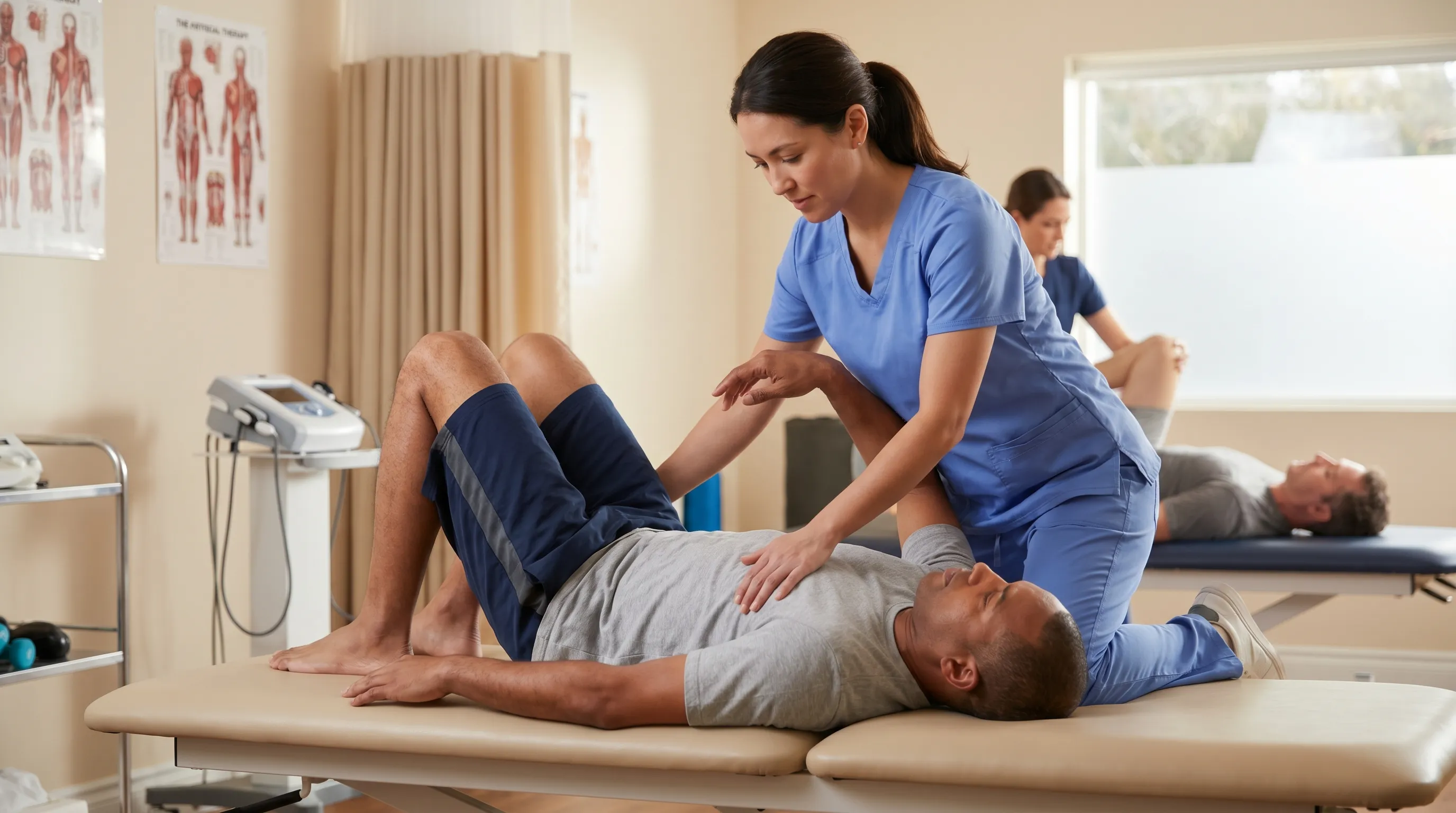
Early physical therapy referral accelerates recovery by introducing graded loading, neuromuscular re-education, and scar tissue mobilization techniques. Manual therapy interventions, including gentle myofascial release, cross-friction massage, and instrument-assisted soft tissue mobilization (IASTM), improve tissue elasticity and prevent adhesions that restrict functional movement. Evidence from the American Physical Therapy Association demonstrates that early manual intervention significantly reduces chronic pain development and improves long-range mobility outcomes.
Therapists also implement therapeutic modalities such as transcutaneous electrical nerve stimulation (TENS) for pain modulation, therapeutic ultrasound for localized heating, and neuromuscular electrical stimulation (NMES) to prevent disuse atrophy during immobilization periods. These adjunctive therapies complement active rehabilitation, creating a comprehensive approach that addresses both symptom management and functional restoration.
Rehabilitation and Progressive Core Recovery
Successful rehabilitation follows a structured progression that prioritizes tissue quality, neuromuscular control, and gradual load tolerance. The healing timeline typically spans inflammatory resolution, proliferative matrix synthesis, and remodeling phases, each requiring specific exercise parameters to optimize outcomes.
Phase One: Gentle Activation and Mobility
Initiated when acute pain subsides and basic daily activities become manageable, this phase focuses on restoring baseline neuromuscular connection without stressing healing fibers. Exercises emphasize low-load isometric contractions that promote blood flow while maintaining structural stability.
Diaphragmatic breathing combined with transverse abdominis engagement serves as the foundational movement, teaching patients to activate deep stabilizers without superficial muscle compensation. Modified pelvic tilts, gentle cat-cow spinal mobility sequences, and supine marching drills improve coordination between abdominal and pelvic floor musculature. Patients are advised to perform movements within a pain-free range, stopping immediately if sharp discomfort emerges.
Phase Two: Strengthening and Stability
Once pain-free range of motion is restored and basic isometric holds become effortless, rehabilitation progresses to dynamic loading that challenges the muscle across multiple planes of movement. This phase introduces controlled eccentric contractions, rotational stability drills, and progressive resistance training.
Exercises include modified planks with knee support, dead bugs with alternating limb extension, bird-dog sequences emphasizing anti-rotation control, and Swiss ball rollouts with limited range. Resistance bands and light dumbbells may be incorporated for functional loading patterns that mimic real-world demands. Research published in the Journal of Sports Rehabilitation indicates that phased eccentric loading significantly improves collagen fiber alignment and tensile strength during the remodeling stage.

Phase Three: Functional Return and Power Development
The final rehabilitation stage prepares the individual for sport-specific demands, heavy lifting, or high-intensity interval training. Exercises incorporate plyometric elements, rotational medicine ball throws, weighted carries, and advanced anti-extension drills that challenge rate of force development.
Return-to-play protocols require objective benchmarking, including pain-free movement through full ranges, symmetrical strength testing, successful completion of sport-specific drills, and physician or physical therapist clearance. Gradual reintroduction of training volume prevents re-injury by allowing the neuromuscular system to adapt to increased mechanical stress. Athletes should monitor fatigue levels closely, as accumulated central nervous system stress can compromise movement quality and increase tissue vulnerability.
Prevention Strategies for Athletes and Everyday Individuals
Injury prevention extends far beyond proper form during exercise sessions. It encompasses lifestyle optimization, periodized training, nutritional support, and proactive mobility management that collectively enhance tissue resilience and mechanical tolerance.
Proper Warm-Up and Mobility Routines
Dynamic warm-ups that elevate core temperature, increase synovial fluid viscosity, and prime neural pathways significantly reduce strain risk. Effective protocols include five to ten minutes of light cardiovascular activity followed by movement-specific drills such as hip circles, thoracic spine rotations, controlled leg swings, and gradual progression from partial to full-range bodyweight movements. Avoiding static stretching prior to high-intensity efforts preserves neuromuscular readiness while preventing temporary force reduction associated with prolonged tissue elongation.
Technique Optimization and Load Management
Biomechanical efficiency directly correlates with injury susceptibility. Maintaining neutral spinal alignment, engaging intra-abdominal pressure before exertion, and avoiding compensatory hyperextension or excessive lumbar flexion reduces shear forces on abdominal attachments. Progressive overload principles should guide training advancement, with volume or intensity increases limited to ten to fifteen percent weekly. Deload weeks, scheduled every four to six weeks, facilitate supercompensation, allowing tissues to adapt without accumulating microtrauma that precedes structural failure.
Nutritional Support for Tissue Repair
Muscle healing demands adequate macronutrient availability, targeted micronutrient intake, and optimal hydration status. Protein consumption of 1.6 to 2.2 grams per kilogram of body weight supports satellite cell proliferation and myofibrillar synthesis. Vitamin C facilitates collagen cross-linking, zinc activates metalloproteinases involved in matrix remodeling, and omega-3 fatty acids modulate inflammatory mediators to support healthy resolution phases.
Hydration maintains extracellular matrix viscosity and cellular transport efficiency. Chronic dehydration reduces tissue elasticity, increases stiffness, and impairs nutrient delivery to healing sites. Incorporating electrolyte-rich fluids, prioritizing sleep quality for growth hormone release, and managing chronic stress to lower cortisol levels collectively create an internal environment conducive to rapid, complete recovery.
| Injury Severity | Fiber Disruption | Primary Symptoms | Estimated Recovery Time | Recommended Intervention |
|---|---|---|---|---|
| Grade 1 Strain | <5% | Mild tenderness, slight weakness, minimal swelling | 2-3 weeks | Active recovery, gentle mobility, progressive loading |
| Grade 2 Strain | 5-50% | Moderate-severe pain, bruising, noticeable weakness, swelling | 4-8 weeks | Physical therapy, controlled rehabilitation, manual therapy |
| Grade 3 Strain | >50% or complete rupture | Severe pain, palpable defect, profound weakness, functional loss | 3-6+ months | Surgical evaluation, structured rehab, gradual return-to-play |
Frequently Asked Questions
How long does it take to fully recover from an ab muscle tear?
Recovery duration correlates directly with injury severity. Grade 1 microtears typically resolve within two to three weeks with appropriate rest and gradual mobility work. Grade 2 partial ruptures require four to eight weeks of structured physical therapy and progressive loading. Complete Grade 3 tears often demand three to six months of rehabilitation, sometimes following surgical repair, to restore full tensile strength and functional capacity.
Can you stretch an ab muscle tear safely?
Stretching should be strictly avoided during the acute inflammatory phase (first seventy-two hours) as elongating compromised fibers disrupts early collagen bridging and increases hemorrhage risk. Gentle, pain-free mobility can begin once swelling subsides and resting pain diminishes. Progressive stretching is reintroduced during the remodeling phase under professional guidance, ensuring controlled tissue elongation that promotes optimal fiber alignment without mechanical overloading.
What does a torn abdominal muscle feel like?
Individuals typically report a sudden, sharp tearing sensation during the inciting movement, followed by localized tenderness that intensifies with trunk flexion, rotation, coughing, or sneezing. Palpable swelling, visible bruising, and a noticeable weakness or gap in the muscle belly may develop within forty-eight hours. Functional impairment ranges from mild discomfort during heavy lifting to complete inability to perform basic abdominal contractions, depending on tear severity.
Do you need surgery for a torn abdominal muscle?
Surgical intervention is rarely necessary for isolated abdominal strains, as the majority heal successfully through conservative management. Surgery becomes indicated for complete Grade 3 ruptures, chronic non-responsive injuries, cases involving avulsion from bony attachments, or concurrent fascial defects resembling athletic pubalgia. A sports medicine specialist or orthopedic surgeon will evaluate imaging findings and functional limitations to determine whether operative repair aligns with your activity goals.
How can you prevent an ab muscle tear during workouts?
Prevention requires a multi-faceted approach emphasizing dynamic warm-ups, progressive load management, and technical proficiency. Incorporating hip and thoracic mobility drills reduces compensatory stress on the abdominal wall during compound movements. Implementing periodized training with scheduled deload weeks prevents accumulated microtrauma. Prioritizing adequate protein intake, maintaining optimal hydration, and ensuring sufficient sleep create physiological conditions that maximize tissue resilience. Finally, avoiding sudden spikes in training volume and seeking coaching for proper exercise mechanics significantly reduce acute strain risk.
Conclusion
An ab muscle tear represents a significant yet highly manageable musculoskeletal injury when approached with clinical awareness, patience, and evidence-based rehabilitation principles. By understanding the anatomical structures involved, recognizing early symptom patterns, and implementing phased recovery protocols, individuals can navigate the healing process efficiently while minimizing the risk of chronic complications. Prioritizing proper warm-ups, technical execution, load management, and nutritional support creates a resilient musculoskeletal foundation that withstands progressive physical demands.
Successful recovery requires listening to your body, respecting biological healing timelines, and seeking professional guidance when pain persists or functional limitations worsen. Whether you are returning to competitive athletics, resuming strength training, or simply seeking pain-free daily movement, a structured approach to rehabilitation ensures long-term core stability and optimal performance. Consistency, informed decision-making, and proactive tissue care remain the cornerstone of sustainable fitness longevity.
For comprehensive rehabilitation guidance and professional assessment of abdominal injuries, consult resources from authoritative organizations such as the American Physical Therapy Association and review clinical protocols published by the National Institutes of Health. Always coordinate with a licensed healthcare provider before initiating new treatment modalities, particularly if symptoms persist beyond expected recovery windows or if underlying medical conditions complicate the healing process. Your core's structural integrity is worth protecting, and with the right approach, you will return stronger, more resilient, and better equipped for the physical challenges ahead.
How we work
Researched and drafted with AI assistance, then reviewed by a human editor against the cited sources.
This article is general health information, not medical advice. Consult a qualified healthcare professional about your own situation.