Understanding Problems After Trigger Finger Surgery: Recovery Guide
Key points
- Rapidly escalating pain unresponsive to prescribed analgesics or elevation
- Fever >100.4°F (38°C), chills, or systemic malaise
- Spreading erythema, warmth, foul-smelling drainage, or wound dehiscence
- Complete loss of sensation or profound, unrelenting numbness in the digit
- Sudden clicking, popping, or recurrent locking suggesting incomplete release or tendon subluxation
- Disproportionate swelling, skin mottling, temperature changes, or extreme touch sensitivity suggestive of CRPS
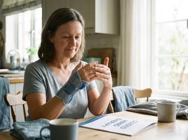
Trigger finger release, medically referred to as A1 pulley release or stenosing tenosynovitis release, is widely recognized as one of the most frequently performed and successful orthopedic procedures worldwide. With documented success rates exceeding 90%, most patients experience immediate and lasting resolution of painful clicking, locking, and inflammation, as noted by clinical data from the Mayo Clinic. However, even with meticulous surgical technique and excellent pre-operative planning, understanding the potential problems after trigger finger surgery is essential for setting realistic expectations and optimizing recovery outcomes. While the vast majority of post-operative concerns are transient and self-limiting, a small percentage of patients may encounter complications that require targeted intervention, extended rehabilitation, or prompt medical evaluation. Navigating the post-surgical landscape requires a balance of physiological awareness, strict adherence to wound care protocols, and proactive hand therapy engagement. This comprehensive guide breaks down the expected recovery trajectory, categorizes common and rare complications, examines patient-specific risk factors, and provides evidence-based strategies to minimize adverse outcomes and restore optimal hand function.
Understanding the Procedure and Expected Recovery
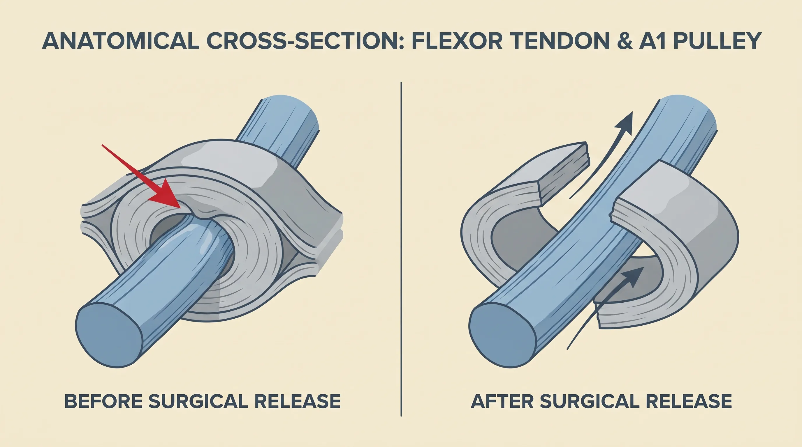
To properly contextualize potential recovery challenges, it is crucial to understand the anatomical and physiological foundation of the procedure. Trigger finger occurs when the flexor tendon becomes entrapped or inflamed beneath the A1 pulley, a fibrous band located at the base of the finger near the palm. This creates friction, swelling, and the characteristic triggering phenomenon. Surgical release involves making a small incision to completely divide the A1 pulley, instantly relieving mechanical constriction and allowing smooth tendon glide. The American Academy of Orthopaedic Surgeons (AAOS) notes that this procedure is highly predictable, with most patients regaining immediate finger motion post-operatively.
Free calculatorA1C CalculatorA1C to average blood glucoseOpen the calculatorThe Physiological Healing Timeline
Post-operative healing follows three distinct biological phases. The inflammatory phase spans the first 3 to 7 days, characterized by localized swelling, erythema, and pain as the body initiates tissue repair. This transitions into the proliferative phase (weeks 1 to 3), where collagen deposition and fibroblast activity strengthen the incision and surrounding tissues. Finally, the remodeling phase extends from month 3 to month 12, during which collagen fibers realign along functional stress lines to restore tensile strength. Understanding this timeline is vital when evaluating problems after trigger finger surgery, as many patients mistakenly interpret normal inflammatory responses as surgical complications. Clinical recovery benchmarks typically allow light daily activities within 1 to 2 weeks, unrestricted normal function by 4 to 6 weeks, and full restoration of grip and pinch strength between 3 to 6 months with appropriate conditioning.
Surgical Approaches: Open vs. Percutaneous Release
The surgical technique selected significantly influences the post-operative course. Open release remains the gold standard, offering direct visualization of neurovascular structures, the flexor tendon, and the pulley system. Percutaneous (needle) release is a minimally invasive alternative performed in-office without an incision. While percutaneous techniques boast faster superficial healing, they carry a marginally higher risk of incomplete release, neurovascular injury, and post-operative stiffness due to lack of direct visualization. Peer-reviewed literature consistently demonstrates that the overall complication rate across both modalities hovers between 1% and 5%, with the open technique providing superior anatomical control during division of the constrictive pulley.
Common, Self-Limiting Problems After Trigger Finger Surgery
The majority of patient-reported concerns following surgery fall into the category of expected, transient physiological responses. These are not true complications but rather predictable healing phenomena that resolve with time and basic conservative management.
Post-Operative Pain and Inflammatory Response
Localized pain and swelling are universal in the first 10 to 14 days. The surgical trauma to soft tissues, combined with post-operative tourniquet deflation, triggers a robust inflammatory cascade. Patients should expect moderate discomfort, particularly during the first 48 to 72 hours. This is effectively managed with strict elevation above heart level, cryotherapy applied in 15-minute intervals, and over-the-counter non-steroidal anti-inflammatory drugs (NSAIDs) or acetaminophen, as tolerated and approved by the surgical team. Persistent, escalating pain beyond two weeks, however, warrants clinical reassessment to rule out infection or complex regional pain syndrome.
Joint Stiffness and the Critical Window for Mobilization
Stiffness represents the most frequently reported early problem after trigger finger surgery. Prolonged immobilization paradoxically increases adhesion formation between the healing tendon and surrounding sheath, severely limiting smooth gliding. The Cleveland Clinic explicitly advises initiating gentle finger mobilization within 24 to 48 hours post-operatively. Controlled range-of-motion exercises prevent capsular tightening and synovial adhesions. Patients are typically instructed to perform full flexion and extension cycles multiple times daily, ensuring that the tendon moves freely through the newly released pulley window. Delayed movement significantly increases the risk of long-term flexion contracture and functional impairment.
Pillar Pain: Pathophysiology and Symptom Management
Pillar pain is a well-documented, highly prevalent phenomenon affecting up to 30% of patients. It manifests as deep, localized tenderness over the thenar and hypothenar eminences immediately adjacent to the surgical incision. The pathophysiology involves disruption of palmar fascial attachments, altered load distribution during grip, and localized soft tissue inflammation during the early healing phase. Crucially, pillar pain does not indicate surgical failure. It typically peaks around weeks 2 to 4 and gradually diminishes over 3 to 6 months. Management focuses on progressive desensitization, scar massage, gentle compressive therapy, and gradual reintroduction of weight-bearing activities. Most patients experience spontaneous resolution without intervention.
Scar Tenderness and Desensitization Protocols
As the incision matures, the superficial nerve endings regenerate and become hypersensitive to tactile stimuli. This manifests as sharp, shooting sensations or intolerance to light touch when washing hands or wearing gloves. Structured desensitization therapy involves progressive tactile stimulation, starting with soft cotton and gradually advancing to textured fabrics, towel rubbing, and gentle tapping. Combined with vitamin E oil or medical-grade silicone gel application, these protocols normalize sensory feedback and improve functional tolerance for daily tasks.
Surgical Complications and Less Common Adverse Events
While rare, recognizing true surgical complications is essential for timely intervention. These adverse events typically occur in less than 3% of cases and often require modified treatment protocols or secondary procedures.
Wound Infection and Delayed Healing
The reported infection rate for open trigger finger release is consistently below 1%, though percutaneous techniques carry a slightly elevated risk due to lack of direct wound irrigation. Superficial infections typically present with localized cellulitis, mild drainage, and warmth, responding rapidly to a course of oral antibiotics targeting Staphylococcus aureus and Streptococcal species. Deep space infections, characterized by severe pain, swelling, systemic fever, and purulent collections beneath the palmar fascia, represent surgical emergencies requiring urgent operative irrigation and debridement. Maintaining a clean, dry dressing for 10 to 14 days, strictly avoiding submersion of the wound, and monitoring glycemic control are foundational infection prevention strategies aligned with CDC surgical site infection guidelines.
Digital Nerve Injury and Neuropathic Sequelae
Digital nerve injury occurs in approximately 0.1% to 0.5% of releases. The radial and ulnar digital nerves run parallel to the flexor tendon sheath, making them vulnerable during pulley division, particularly in patients with anatomical variations or severe pulley hypertrophy. Clinical presentation includes immediate post-operative numbness, paresthesia, or electric shock-like neuropathic pain along the volar aspect of the affected digit. The majority of these injuries represent neuropraxia (temporary conduction block) or axonotmesis (axon disruption with intact sheath), both of which typically recover spontaneously over 6 to 12 months. Complete nerve transection is exceedingly rare and may necessitate microsurgical nerve repair or grafting. Early recognition allows for protective sensory re-education and prevents unrecognized thermal or pressure injuries to the insensate finger.
Tendon Bowstringing and Mechanical Failure
Bowstringing occurs when excessive pulley tissue is released, disrupting the normal biomechanical arc of the flexor tendon. Without adequate pulley constraint, the tendon lifts away from the bone during active flexion, reducing mechanical advantage, grip efficiency, and flexion velocity. This complication is preventable with precise, limited release of the A1 pulley while preserving the proximal cruciate (C1) pulley. If symptomatic bowstringing develops, conservative management with pulley-sparing splinting and targeted strengthening is attempted first. Persistent functional deficits may require secondary pulley reconstruction using autologous tendon grafts or synthetic materials.
Complex Regional Pain Syndrome (CRPS)
CRPS is a rare but devastating complication affecting less than 1% of hand surgery patients. It represents a disproportionate, maladaptive autonomic nervous system response to surgical trauma, characterized by severe burning pain, allodynia, significant edema, temperature asymmetry, and trophic skin changes (thinning, shiny appearance, altered hair/nail growth). Diagnosis follows the Budapest clinical diagnostic criteria, and early recognition is paramount, as delayed treatment drastically worsens prognosis. Management requires a multidisciplinary approach combining aggressive desensitization therapy, progressive mirror box training, graded motor imagery, neuropathic pain medications (gabapentinoids, SNRIs), and occasionally sympathetic nerve blocks. The AAOS emphasizes that maintaining finger motion and avoiding prolonged immobilization are the strongest preventive measures against CRPS development.
High-Risk Populations and Comorbidity Management
Patient-specific physiological factors dramatically influence tissue healing capacity, complication susceptibility, and overall recovery velocity. Identifying high-risk cohorts allows for pre-operative optimization and tailored post-operative protocols.
Diabetes Mellitus and Microvascular Compromise
Diabetes mellitus represents the most significant modifiable risk factor for adverse surgical outcomes. According to NIH and NIDDK research, diabetic patients face a 2 to 3 times higher risk of post-operative stiffness, delayed wound healing, infection, and flexor tendon adhesions compared to non-diabetic counterparts. Chronic hyperglycemia induces microvascular endothelial dysfunction, impairs leukocyte phagocytosis, reduces collagen cross-linking, and accelerates advanced glycation end-product (AGE) accumulation in tendon sheaths. Strict pre-operative glycemic optimization, targeting an HbA1c below 7.5% when possible, is strongly recommended. Post-operatively, continuous glucose monitoring, strict antibiotic compliance, and extended suture retention (14 to 21 days) may be necessary to ensure complete epithelialization.
Rheumatoid Arthritis and Systemic Inflammation
Patients with rheumatoid arthritis (RA) or other systemic connective tissue disorders present unique challenges. Chronic synovitis inherently thickens the flexor tendon sheath, and systemic corticosteroid or biologic immunosuppressants significantly delay tissue repair. Additionally, RA frequently involves multi-tendon involvement, meaning release of a single trigger finger does not prevent triggering in adjacent digits. Surgeons often modify techniques to avoid aggressive soft tissue stripping in RA patients, and hand therapy protocols are extended to address concurrent joint instability and extensor lag. Immunosuppressive medication management requires careful peri-operative coordination with rheumatology to balance surgical wound healing against disease flare prevention.
Tobacco Use and Tendon Vascularization
Nicotine is a potent vasoconstrictor that profoundly impairs cutaneous and tendon microcirculation. The CDC notes that active smoking reduces tissue oxygenation by up to 40%, severely compromising fibroblast proliferation, collagen synthesis, and angiogenesis at the surgical site. Smokers exhibit markedly higher rates of wound dehiscence, infection, tendon adhesion formation, and prolonged stiffness. Complete nicotine cessation at least 4 weeks pre-operatively and 8 weeks post-operatively is non-negotiable for optimal tendon gliding and wound integrity. Even nicotine replacement therapies maintain some vasoactive properties, making comprehensive cessation programs essential for patients undergoing elective hand procedures.
Long-Term Functional Limitations and Rehabilitation
Beyond the initial healing window, some patients experience lingering functional deficits that require structured, progressive rehabilitation to overcome. These problems after trigger finger surgery typically stem from altered biomechanics, inadequate early mobilization, or excessive scar tissue formation.
Early Grip Strength Deficits
Post-operative grip weakness is nearly universal, with objective measurements frequently showing a 15% to 20% reduction compared to the contralateral hand during the first 3 months. This deficit stems from surgical soft tissue trauma, pain inhibition, and temporary disuse atrophy of the intrinsic and extrinsic flexor muscles. Rehabilitation follows a phased progression. Weeks 1 to 3 focus on edema control and passive/active assisted motion. Weeks 3 to 6 introduce isometric strengthening and light resistance putty. Months 2 to 4 incorporate progressive overload with dumbbells, grip trainers, and functional task simulation. Most patients achieve baseline strength parity by month 6, provided therapy compliance remains high and biomechanical loading is gradual to avoid tendon overload.
Adhesion Formation and Restricted Tendon Glide
Adhesion formation represents the most common long-term mechanical complication. During the proliferative healing phase, fibrin deposits can cross-link the healing tendon to the overlying subcutaneous tissue or underlying bone. This restricts smooth excursion, manifesting as stiffness, pain at the end-range of motion, and residual triggering sensations despite successful pulley release. Early mobilization is the primary preventive measure. When adhesions become clinically significant, aggressive hand therapy involving tendon gliding exercises, joint blocking, deep friction massage, and dynamic splinting is initiated. Surgical tenolysis is rarely required but may be considered for patients with persistent, therapy-resistant glide deficits after 6 months of dedicated rehabilitation.
Fixed Flexion Contractures and Joint Integrity
The proximal interphalangeal (PIP) joint is particularly susceptible to fixed flexion contracture if post-operative edema and pain lead to prolonged flexion positioning. The volar plate and collateral ligaments undergo viscoelastic shortening, making passive extension increasingly difficult. Prevention relies on strict extension splinting at night, frequent passive stretching, and avoidance of resting the hand in a curled position. Once established, contractures require prolonged serial casting, static progressive splinting, and in refractory cases, surgical capsular release. Maintaining full joint extension during the critical first 6 weeks is the most effective defense against permanent deformity.
Evidence-Based Strategies to Minimize Complications
Proactive patient participation significantly reduces the incidence and severity of problems after trigger finger surgery. The following evidence-based protocols align with guidelines from the American Society for Surgery of the Hand (ASSH) and peer-reviewed orthopedic literature.
Optimizing Hand Therapy Compliance
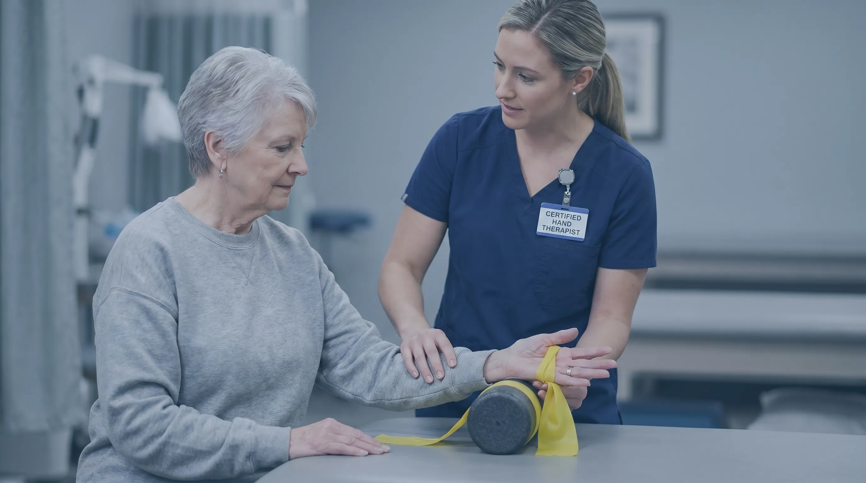
Early and consistent engagement with a certified hand therapist (CHT) is the strongest predictor of successful outcomes. CHTs provide individualized protocols, manual scar mobilization, custom orthotic fabrication, and objective strength/mobility tracking. Patients should initiate therapy within 1 to 2 weeks post-operatively if independent exercise tolerance is limited. Compliance with home exercise programs, typically performed 4 to 6 times daily, prevents capsular contracture and tendon adhesion. Therapists utilize modalities like contrast baths, ultrasound (controversial but occasionally prescribed), and progressive resistive exercise to normalize tissue compliance.
Meticulous Wound Care and Environmental Protection
Infection prevention begins intra-operatively with sterile technique and post-operatively with disciplined dressing management. The incision must remain completely dry and uncovered from water submersion until full epithelialization (usually days 10 to 14). Showering is permitted after 48 hours only if a waterproof barrier dressing is securely applied. Patients should avoid exposure to soil, gardening, or heavy machinery until cleared by their surgeon. Daily inspection for erythema, purulence, or dehiscence enables rapid antibiotic initiation if infection is suspected.
Activity Pacing and Biomechanical Awareness
Premature heavy gripping, repetitive pinching, or forceful vibration exposure during the proliferative healing phase can cause micro-tears in the healing pulley margins, exacerbating inflammation and adhesion formation. Patients must adhere to a strict 3 to 4-week restriction on lifting weights exceeding 5 pounds or performing repetitive forceful activities. Ergonomic modifications, such as utilizing adaptive utensil grips, voice-to-text technology, and two-handed lifting techniques, protect the surgical site while maintaining daily independence. Gradual, symptom-guided return to occupational and recreational activities ensures sustainable tissue adaptation without overloading the healing tendon.
| Factor | Open Release Characteristics | Percutaneous Release Characteristics | Clinical Impact |
|---|---|---|---|
| Incision | 1.5–2.0 cm volar palmar incision | Needle puncture, no open wound | |
| Visualization | Direct anatomical visualization of nerve, tendon, pulley | Blind/ultrasound-guided, higher technical reliance | |
| Infection Rate | <1% | 1–2% (slightly elevated due to skin flora introduction) | |
| Nerve Injury Risk | 0.1–0.3% (direct visualization mitigates risk) | 0.3–0.5% (anatomical variation increases blind risk) | |
| Recurrence Rate | <1% | 1–3% (higher chance of incomplete division) | |
| Pillar Pain Prevalence | 20–30% | 10–20% | |
| Return to Light Duty | 7–10 days | 3–5 days |
Video Resources for Post-Operative Guidance
Visual education significantly improves patient compliance with rehabilitation protocols. The following board-certified hand surgery resources provide clear demonstrations of proper exercise form and recovery expectations.
When to Contact Your Surgeon Immediately
While most recovery challenges are manageable with conservative care, specific red flags demand immediate clinical evaluation. Patients experiencing the following symptoms should not delay medical consultation:
- Rapidly escalating pain unresponsive to prescribed analgesics or elevation
- Fever >100.4°F (38°C), chills, or systemic malaise
- Spreading erythema, warmth, foul-smelling drainage, or wound dehiscence
- Complete loss of sensation or profound, unrelenting numbness in the digit
- Sudden clicking, popping, or recurrent locking suggesting incomplete release or tendon subluxation
- Disproportionate swelling, skin mottling, temperature changes, or extreme touch sensitivity suggestive of CRPS
Early intervention for these warning signs prevents minor complications from progressing into permanent functional deficits or requiring secondary corrective surgery.

Frequently Asked Questions
How long does pillar pain last after trigger finger surgery?
Pillar pain, which manifests as deep tenderness over the fleshy palmar eminences adjacent to the incision, affects approximately 20% to 30% of patients. It typically emerges during the first two weeks as fascial attachments heal and localized inflammation peaks. In most cases, pillar pain gradually subsides between 3 to 6 months post-operatively. Consistent desensitization therapy, gentle massage, and gradual load progression accelerate resolution without indicating surgical failure or long-term damage.
Is it normal to have stiffness after trigger finger release?
Yes, stiffness is a highly predictable physiological response during the first 4 to 6 weeks. Surgical trauma triggers localized fibrosis, and prolonged immobilization encourages collagen cross-linking between the tendon and sheath. Clinical guidelines from the Cleveland Clinic emphasize that initiating gentle finger motion within 24 to 48 hours dramatically reduces adhesion formation. Structured tendon gliding exercises and night extension splinting are the most effective interventions for preventing permanent stiffness.
What is the risk of nerve damage during this procedure?
Digital nerve injury is rare, occurring in 0.1% to 0.5% of cases. The nerves run closely alongside the A1 pulley, making them vulnerable during incision and release, particularly in patients with anatomical variants or severe pulley hypertrophy. Most injuries represent temporary neuropraxia, resolving spontaneously within 6 to 12 months. Complete nerve transection is exceedingly uncommon in open techniques due to direct visualization but requires microsurgical repair if it occurs. Early recognition and sensory protection are critical.
How do I know if my wound is infected after surgery?
Surgical site infections typically present within 5 to 14 days post-procedure. Key indicators include expanding redness beyond the incision line, increasing throbbing pain rather than gradual improvement, purulent or discolored drainage, localized heat, foul odor, and systemic signs like fever or chills. Superficial infections respond well to targeted oral antibiotics. Deep palmar space infections are surgical emergencies requiring prompt irrigation and debridement. Strict wound dryness protocols and early monitoring prevent severe complications.
When should I expect my grip strength to return?
Patients commonly experience a 15% to 20% deficit in grip strength during the first 1 to 3 months due to tissue trauma, pain inhibition, and disuse atrophy. Rehabilitation follows a phased overload model, transitioning from isometric exercises in week 3 to resistance putty and dumbbell strengthening by month 2. Most individuals achieve parity with their unaffected hand between 4 to 6 months, provided hand therapy compliance is high and biomechanical progression is gradual and symptom-guided.
Does trigger finger surgery work for diabetic patients?
Surgery remains highly effective for diabetic patients, though the recovery timeline is extended and complication risks are elevated. Diabetics face a 2 to 3 times higher likelihood of delayed wound healing, infection, and tendon adhesion formation due to microvascular compromise and impaired collagen synthesis. Pre-operative glycemic optimization (target HbA1c <7.5%), extended suture retention, strict infection monitoring, and prolonged hand therapy significantly improve outcomes. Despite the adjusted recovery curve, long-term success rates remain comparable to the general population when comorbidities are actively managed.
Conclusion
Trigger finger release stands as one of the most reliable and high-yield procedures in modern hand surgery. While the prospect of experiencing problems after trigger finger surgery can understandably cause anxiety, the clinical reality is that the vast majority of post-operative concerns are predictable, self-limiting, and effectively managed through disciplined rehabilitation and proper wound care. Temporary pain, swelling, stiffness, and pillar pain represent normal physiological healing responses rather than surgical failures. True complications like infection, digital nerve injury, tendon bowstringing, or CRPS occur in less than 3% of cases and are heavily influenced by patient-specific factors such as diabetes management, tobacco use, and adherence to early mobilization protocols. By understanding the physiological healing timeline, committing to certified hand therapy, maintaining strict wound hygiene, and recognizing red-flag symptoms early, patients can navigate the recovery process confidently and achieve optimal functional restoration. Always consult your board-certified hand surgeon or occupational therapist for personalized medical guidance tailored to your specific surgical technique, comorbidities, and rehabilitation goals. Evidence-based care, proactive patient engagement, and realistic expectation setting form the foundation of a successful, complication-free recovery.
How we work
Researched and drafted with AI assistance, then reviewed by a human editor against the cited sources.
This article is general health information, not medical advice. Consult a qualified healthcare professional about your own situation.