Tooth Extraction Healing Process: Complete Medical Guide
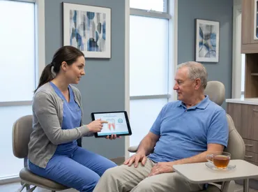
Tooth removal is one of the most common dental procedures performed worldwide, yet the physiological journey that follows is remarkably intricate and often misunderstood. Whether you are preparing for an upcoming procedure or navigating the days immediately following surgery, understanding the underlying biology of recovery can significantly reduce anxiety and improve outcomes. The tooth extraction healing process is not merely about the gum closing over an empty space; it is a highly coordinated cascade of cellular events, vascular responses, and structural adaptations that restore your oral architecture. From the moment the tooth is gently elevated and removed, your body immediately shifts into emergency repair mode, prioritizing clot stabilization, inflammation management, and eventual tissue regeneration. By familiarizing yourself with the precise timelines, recognizing normal versus abnormal symptoms, and adhering to clinically proven aftercare protocols, you can actively support your body’s innate healing capabilities. This comprehensive, evidence-based guide walks you through every biological phase, provides actionable daily management strategies, and addresses common concerns with medical accuracy. Your compliance with post-operative guidelines directly correlates with the speed and quality of your recovery, making patient education an essential component of successful dental surgery outcomes.
Understanding the Biological Phases of the Healing Cascade
The tooth extraction healing process follows the universal principles of secondary intention wound healing, adapted specifically to the dense, vascular environment of the alveolar bone and gingival tissues. Clinically, this progression is divided into four overlapping but distinct physiological phases, each driven by specific cellular mediators, signaling proteins, and structural transformations.
Hemostasis: The Foundation of Clot Formation
Within minutes of tissue disruption, vasoconstriction occurs to minimize blood loss. Platelets rapidly aggregate at the site of vascular injury, adhering to exposed collagen fibers and releasing granules rich in clotting factors. This triggers the coagulation cascade, culminating in the conversion of fibrinogen to fibrin. The resulting meshwork traps red blood cells, leukocytes, and platelets, forming a stable gelatinous clot. This structure is far more than a simple physical barrier; it acts as a provisional extracellular matrix that anchors migrating cells and delivers a concentrated payload of growth factors. Platelet-derived growth factor (PDGF) and transforming growth factor-beta (TGF-β) are immediately secreted, initiating chemotaxis that recruits fibroblasts, endothelial cells, and inflammatory mediators to the wound bed. Protecting this fragile scaffold is the single most critical objective during the first 48 hours.
Inflammatory Phase: Clearing Debris and Initiating Repair
Spanning approximately days one through three, the inflammatory phase is characterized by increased vascular permeability and localized edema. Neutrophils are the first immune responders, arriving to phagocytose bacteria, dead tissue fragments, and foreign debris. Within hours, they are replaced by macrophages, which serve as both cleanup crews and master regulators of the subsequent healing stages. Macrophages secrete a complex array of cytokines, interleukins, and matrix metalloproteinases (MMPs) that dismantle damaged collagen while simultaneously signaling for tissue regeneration. This phase is responsible for the classic post-operative symptoms: localized warmth, moderate swelling, and manageable discomfort. While often uncomfortable, this inflammatory response is entirely beneficial and necessary. Suppressing it prematurely or excessively can actually delay the transition into the next reparative stage.
Proliferative Phase: Tissue Regeneration and Epithelial Migration
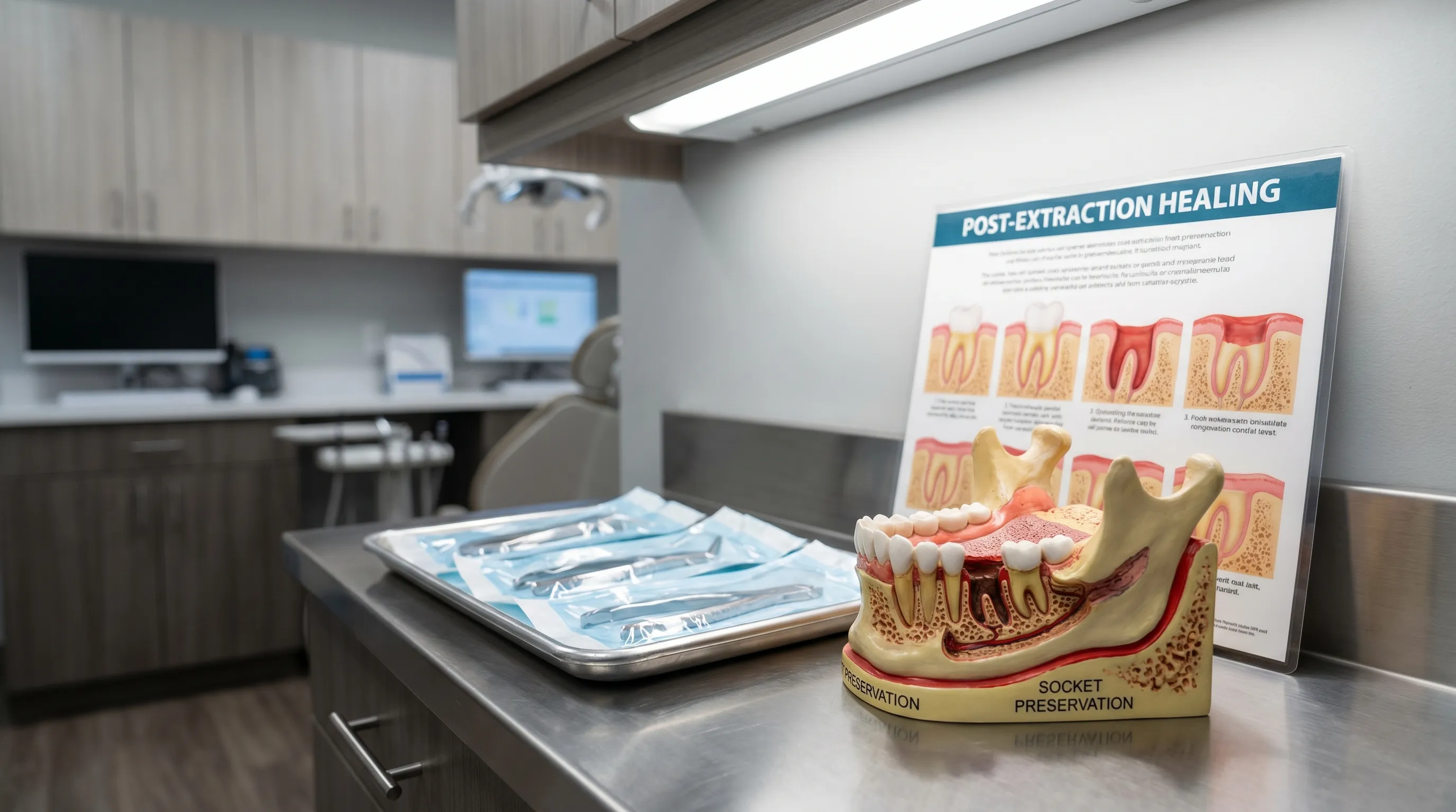
Beginning around day three and continuing through the third week, the proliferative phase shifts the biological priority from defense to reconstruction. Fibroblasts proliferate rapidly, synthesizing type III collagen to form highly vascularized granulation tissue. Simultaneously, angiogenesis occurs as endothelial cells sprout new capillary networks to supply the developing tissue with oxygen and nutrients. Epithelial cells at the wound margins undergo mitosis and begin migrating across the clot’s surface in a continuous, sheet-like fashion. By day ten to fourteen, this migrating epithelium typically establishes a complete seal over the socket entrance. Concurrently, osteoblasts begin laying down immature woven bone along the socket walls, while myofibroblasts exert contractile forces that gradually shrink the wound volume. During this critical window, the socket transitions from a fragile, open cavity to a resilient, biologically active regenerative unit.
Remodeling Phase: Bone Maturation and Structural Integration
The final phase is the most prolonged and determines the long-term structural integrity of the extraction site. Over a period of three to six months, the initially deposited woven bone undergoes systematic replacement by mature lamellar bone. Osteoclasts and osteoblasts work in precise coupling units, resorbing disorganized trabeculae and depositing dense, load-bearing bone matrix in alignment with natural mechanical stress lines. This dynamic process of bone deposition and resorption, known as remodeling, gradually elevates the alveolar ridge height and width. Collagen fibers reorganize into tighter, more parallel bundles, increasing tensile strength. By the conclusion of this phase, the extraction site is radiographically indistinguishable from surrounding healthy bone, completing the tooth extraction healing process at a cellular and architectural level.
The Comprehensive Healing Timeline: What to Expect Day by Day
Translating biological theory into practical recovery milestones requires understanding the chronological progression of tissue repair. Each timeframe presents unique physiological changes, expected symptoms, and specific care requirements that align with your body’s current healing capacity.
First 24 Hours: The Critical Window
The initial twenty-four hours represent the most vulnerable period for the developing blood clot. Immediately following extraction, bleeding should be controlled through firm pressure on sterile gauze for thirty to forty-five minutes. Oozing is normal, but active hemorrhage is not. As the clot organizes, you will experience peak swelling and discomfort within the first twelve to eighteen hours. Applying intermittent cold therapy (twenty minutes on, twenty minutes off) effectively reduces capillary permeability and minimizes tissue edema. Keep your head elevated above heart level, even during sleep, to utilize gravity in limiting fluid accumulation. Avoid physical exertion, hot liquids, and bending forward. Consume only cool or lukewarm liquids during this window.
Days 2 to 3: Transitioning Through Discomfort
As vasoconstriction stabilizes and the inflammatory response begins to modulate, noticeable swelling reduction should occur. Granulation tissue actively fills the socket from the base upward, creating a firm, reddish-pink tissue layer beneath the surface. Pain typically transitions from sharp or throbbing to a dull, manageable ache that responds well to prescribed or over-the-counter analgesics. You may notice a slight salty taste or minimal brownish oozing; this represents normal clot breakdown and lymphatic drainage, not active infection. Begin transitioning to a soft, nutrient-dense diet, maintaining hydration without using straws. Gentle oral hygiene resumes around adjacent teeth, strictly avoiding direct suction or pressure on the surgical site.
Days 7 to 10: Surface Closure and Dietary Shifts
By the end of the first week, epithelial migration is typically complete, and the gingival tissue has closed over the extraction socket. If non-dissolvable sutures were placed, your dentist or oral surgeon will remove them during a brief follow-up appointment. At this stage, granulation tissue is highly vascular and may bleed slightly if accidentally irritated, but structural stability is markedly improved. Most patients can safely return to a normal diet, progressively reintroducing firmer foods on the contralateral side. Mild stiffness in the jaw muscles is common due to prolonged soft-tissue restriction; gentle stretching and warm compresses can restore full mandibular mobility.
Weeks 3 to 4: Soft Tissue Resolution
One month post-extraction, the gingival architecture has fully matured, presenting with healthy pink stippling and firm consistency to palpation. Underneath the surface, osteoblastic activity accelerates, depositing early trabecular bone throughout the socket depth. Swelling and tenderness should be entirely absent. You can resume standard oral hygiene routines, including electric toothbrushes and water flossers, using standard pressure settings. This timeframe is often ideal for evaluating the need for space maintenance appliances or early implant site preparation, depending on your long-term restorative plan.
Months 1 to 6: Complete Bone Remodeling
The final stretch of recovery occurs beneath the gumline. Over months three through six, the immature woven bone undergoes systematic mineralization and lamellar reorganization. Radiographic imaging will reveal progressive radiopacity filling the former radiolucent defect. By month six, the alveolar ridge reaches near-complete density and volume stabilization, marking the definitive conclusion of the tooth extraction healing process. Patients planning dental implants typically wait three to four months for anterior teeth and four to six months for posterior teeth to ensure optimal osseointegration conditions.
Factors That Influence Recovery Speed and Outcomes
Not all extractions heal identically. A wide array of patient-specific variables, procedural complexities, and anatomical considerations directly modulate the velocity and quality of tissue regeneration.
Simple vs. Surgical Extractions
Simple extractions involve teeth with fully erupted crowns and intact, accessible root structures that can be elevated with forceps. Healing is generally straightforward, with minimal tissue trauma and rapid clot stabilization. Surgical extractions, conversely, require mucoperiosteal flap elevation, bone removal (osteotomy), tooth sectioning, and suturing. Impacted wisdom teeth, severely decayed molars, or teeth with hypercementosis or divergent roots fall into this category. The increased surgical field manipulation directly correlates with greater post-operative inflammation, extended soft-tissue healing timelines, and higher complication probabilities.
| Feature | Simple Extraction | Surgical Extraction |
|---|---|---|
| Tissue Trauma | Minimal; forceps delivery only | Moderate to high; flap elevation & bone removal |
| Sutures Required | Rarely | Commonly (resorbable or non-resorbable) |
| Initial Discomfort | Mild to moderate, 24-48 hours | Moderate to severe, 48-72 hours peak |
| Complete Bone Fill | 2-4 months | 4-6+ months |
| Dry Socket Incidence | ~2-5% | ~15-30% for impacted third molars |
Patient-Specific Variables: Age, Health Conditions, and Habits
Systemic health profoundly dictates cellular repair efficiency. Advanced age naturally slows fibroblast proliferation and angiogenesis. Uncontrolled diabetes mellitus impairs neutrophil function, reduces collagen synthesis, and increases susceptibility to secondary infection. Immunocompromised states, whether from autoimmune disorders, chronic corticosteroid use, or oncology treatments, significantly prolong the inflammatory phase and compromise granulation tissue formation. Tobacco use, particularly smoking and vaping, introduces vasoconstrictive chemicals that drastically reduce local blood flow, oxygen delivery, and growth factor bioavailability. Bisphosphonate therapy also carries a rare but serious risk of medication-related osteonecrosis of the jaw (MRONJ).
Location and Anatomical Considerations
Mandibular teeth, especially posterior molars and third molars, are subject to higher masticatory forces and denser bone structure, which can increase surgical difficulty and post-operative discomfort. Maxillary teeth generally possess more trabecular, vascular bone that heals rapidly but carries a slightly higher risk of oroantral communication if sinus proximity exists. Multi-rooted teeth create complex socket topography that requires more extensive clot stabilization across divergent bony partitions.
Recognizing and Managing Common Complications
Vigilance during the first week allows early identification of deviations from the expected recovery trajectory. Understanding complication pathophysiology empowers patients to seek timely intervention and prevents minor issues from escalating into chronic conditions.
Alveolar Osteitis (Dry Socket): Symptoms, Risks, and Treatment
Alveolar osteitis represents the most frequent and painful complication, occurring when the protective blood clot prematurely dislodges, fragments, or undergoes fibrinolysis before tissue maturation. This exposes underlying alveolar bone and nerve endings directly to the oral environment, triggering intense, localized pain that frequently radiates to the ipsilateral ear, temple, or neck. Symptoms typically emerge on day three or four, often described as a deep, unrelenting ache unresponsive to standard analgesics. Halitosis and a visibly empty, greyish socket are hallmark clinical signs. Management involves gentle debridement of the necrotic debris followed by placement of medicated eugenol-based or chlorhexidine dressings. These provide immediate analgesia, promote granulation, and require replacement every 48 to 72 hours until symptoms subside. Risk mitigation includes absolute tobacco cessation, avoiding vigorous rinsing, and strictly following suction-avoidance protocols.
Infection Signs vs. Normal Healing Discomfort
Differentiating between physiological inflammation and pathological infection requires careful symptom assessment. Normal healing presents with manageable pain peaking early, localized swelling that resolves by day five, and low-grade discomfort that improves daily. Signs of secondary bacterial infection include escalating pain beyond day four, fever exceeding 100.4°F (38°C), purulent exudate with foul taste, progressive facial asymmetry, trismus limiting mouth opening beyond two finger-widths, and lymphadenopathy. Infection management requires clinical drainage if abscessed, targeted antibiotic therapy based on culture or broad-spectrum empiric coverage (typically amoxicillin or clindamycin for penicillin allergies), and possible irrigation of the socket.
Prolonged Bleeding and Swelling
Persistent hemorrhage beyond twelve hours usually stems from premature gauze removal, excessive physical activity, or underlying coagulopathy. Management involves biting firmly on a moistened black tea bag for thirty minutes, as the tannic acid acts as a localized astringent promoting vasoconstriction. If swelling fails to resolve after seventy-two hours or worsens after initial improvement, it may indicate hematoma formation, allergic reaction, or developing infection. Consistent cold-to-heat transition therapy, proper head elevation, and adherence to anti-inflammatory medication schedules typically resolve benign edema.
Evidence-Based Aftercare Protocols for Optimal Recovery
Patient compliance with post-operative instructions is the single strongest predictor of uneventful recovery. The following protocols are derived from current dental guidelines and clinical consensus recommendations.
Immediate Post-Operative Care (First 24 Hours)
Upon leaving the clinical setting, maintain continuous gentle pressure on the provided gauze pad. Replace it promptly if bleeding persists. Begin intermittent ice application immediately, never applying heat during this phase, as vasodilation will exacerbate hemorrhage and edema. Rest in a semi-reclined position to minimize venous pressure in the head and neck. Avoid driving, operating machinery, or making important decisions if sedation was administered.
Nutrition, Hydration, and Lifestyle Adjustments
Your dietary intake directly supplies the substrates necessary for cellular proliferation. Prioritize high-protein, calorie-dense soft foods that require minimal mastication. Excellent options include Greek yogurt, blended soups (served lukewarm), mashed sweet potatoes, scrambled eggs, avocado, and nutritional shakes. Maintain aggressive hydration with water and electrolyte solutions to support circulatory volume and tissue perfusion. Absolutely avoid alcohol for at least seventy-two hours, as it interferes with medication metabolism and impairs platelet aggregation. Refrain from smoking, vaping, or using nicotine replacement products for a minimum of one week; studies confirm nicotine reduces local oxygen tension by up to thirty percent, severely compromising angiogenesis.
Oral Hygiene and Wound Protection
Begin gentle saltwater rinses (half teaspoon non-iodized salt in eight ounces warm water) twenty-four hours post-surgery. Perform this rinse after meals and before bed, allowing the solution to passively flow over the site rather than forcefully swishing. Resume brushing all non-surgical teeth using a soft-bristled brush, maintaining a strict one-centimeter buffer zone around the extraction site. Avoid commercial mouthwashes containing alcohol or hydrogen peroxide during the first two weeks, as they delay epithelialization. If prescribed a chlorhexidine gluconate rinse, follow the exact dosing frequency to maximize antimicrobial efficacy without disrupting the clot. For comprehensive visual guidance on post-operative techniques, consider watching this educational overview: . Additional clinical insights can be found here: .

For detailed medical references and patient education materials, consult the Mayo Clinic dental surgery section or review clinical guidelines published by the American Dental Association. The Cleveland Clinic offers excellent recovery timelines, while peer-reviewed studies on wound management are accessible via NIH/PubMed.
Frequently Asked Questions
How long does it take for a tooth extraction socket to completely close?
While the gingival tissue typically establishes a complete epithelial seal within seven to fourteen days, the underlying bone requires significantly longer to regenerate. Complete radiographic filling and structural maturation of the alveolar socket generally takes between three and six months, depending on extraction complexity, patient age, and systemic health status.
Is it normal to experience throbbing pain on day four after tooth removal?
Mild to moderate discomfort usually peaks within the first forty-eight to seventy-two hours and should steadily diminish thereafter. Severe, throbbing, or radiating pain that suddenly emerges or intensifies on day three or four strongly suggests alveolar osteitis (dry socket). This condition requires prompt clinical evaluation and placement of a medicated dressing for rapid relief.
Can I rinse my mouth with water after a tooth extraction?
Vigorous rinsing, spitting, or creating negative intraoral pressure should be strictly avoided for the first twenty-four to forty-eight hours to protect the fragile blood clot. After this critical window, gentle warm saltwater rinses after meals are highly recommended to dislodge food debris and reduce bacterial load without disrupting granulation tissue.
When is it safe to resume brushing near the extraction site?
You may safely resume brushing your non-surgical teeth on the second day using a soft-bristled brush. However, maintain a careful distance from the extraction socket for at least seven days. Once the epithelial layer has matured, typically by the end of the second week, you can gradually reintroduce standard brushing techniques over the healed area.
Do I need antibiotics after every tooth extraction?
No. Contemporary clinical guidelines strongly advise against routine prophylactic antibiotic administration for healthy patients undergoing uncomplicated simple extractions. Antibiotics are exclusively reserved for cases involving pre-existing active infection, immunocompromised status, prosthetic joint considerations, or extensive surgical bone manipulation where infection risk is clinically significant.
Conclusion
Navigating the tooth extraction healing process successfully requires a harmonious blend of biological understanding, disciplined self-care, and timely professional communication. By respecting the fragile early stages of clot formation, recognizing the difference between normal inflammatory responses and pathological deviations, and strictly adhering to evidence-based aftercare protocols, you create the optimal microenvironment for rapid, complication-free recovery. Remember that healing is a dynamic, progressive journey rather than an instantaneous event. Your daily choices regarding nutrition, oral hygiene, rest, and habit modification directly influence the cellular machinery working beneath your gums. Should any symptoms deviate from the expected timeline, never hesitate to contact your dental provider for prompt evaluation. With patience, vigilance, and proper support, your oral tissues will efficiently restore themselves, leaving you ready to embrace long-term dental health and functional comfort.

About the author
Benjamin Carter, MD, is a board-certified otolaryngologist specializing in head and neck surgery, with an expertise in treating throat cancer. He is an associate professor and the residency program director at a medical school in North Carolina.