Posterior Shoulder Dislocation: Complete Guide to Symptoms, Diagnosis, and Recovery
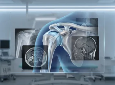
When an unexpected injury occurs, pain often radiates instantly, leaving individuals searching for answers and immediate relief. The shoulder is the most mobile joint in the human body, granting us an extraordinary range of motion for lifting, reaching, and performing everyday tasks. However, this remarkable mobility comes with an inherent trade-off: structural vulnerability. While anterior dislocations dominate orthopedic statistics, a posterior shoulder dislocation represents a rarer yet clinically significant injury that demands specialized attention. Many patients experience profound discomfort, restricted movement, and confusion when standard initial assessments fail to pinpoint the issue. Understanding the mechanics, recognizing the subtle warning signs, and following evidence-based recovery protocols are essential steps toward restoring full function. This comprehensive guide breaks down everything you need to know about diagnosis, treatment, rehabilitation, and long-term joint preservation. By the end, you will have actionable strategies to navigate recovery confidently and reduce the risk of recurrent instability.
Understanding Posterior Shoulder Dislocation
The Anatomy of the Shoulder Joint
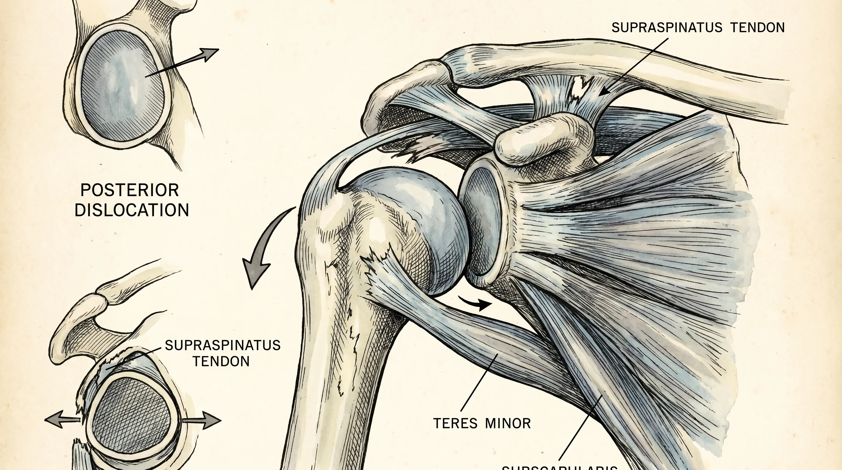
To fully grasp how a posterior shoulder dislocation occurs, it is essential to review the structural composition of the glenohumeral joint. The shoulder operates as a ball-and-socket mechanism, where the spherical humeral head articulates with the shallow glenoid cavity of the scapula. Unlike the hip joint, which benefits from deep osseous congruency, the shoulder relies heavily on soft tissue structures for stability. The glenoid labrum and rotator cuff anatomy act as a fibrocartilaginous rim that deepens the socket by approximately fifty percent, while the joint capsule and four primary rotator cuff tendons (supraspinatus, infraspinatus, teres minor, and subscapularis) dynamically center the humeral head during movement. The posterior capsule and the external rotators, particularly the infraspinatus and teres minor, play a critical role in resisting backward translation of the humeral head. When excessive posterior force overwhelms these restraints, the joint can become displaced, leading to significant capsular stretching, labral tearing, and potential articular cartilage damage.
Free calculatorSleep CalculatorCalculate sleep cyclesOpen the calculatorHow a Posterior Dislocation Differs from Anterior Dislocations
Anterior dislocations account for over ninety percent of all shoulder dislocations and typically involve the humeral head slipping forward and downward, often tearing the anterior inferior labrum in a classic Bankart lesion. In contrast, a posterior shoulder dislocation forces the humeral head backward, frequently impacting it against the anterior rim of the glenoid. This mechanism commonly produces a reverse Hill-Sachs lesion, which is an impaction fracture on the anteromedial aspect of the humeral head. The biomechanical stress patterns differ significantly: anterior injuries usually occur with the arm abducted and externally rotated, whereas posterior injuries typically manifest when the arm is forward-flexed, adducted, and internally rotated at the moment of impact. Clinicians must recognize these distinct vectors of force, as misclassification can lead to inappropriate reduction techniques and delayed rehabilitation protocols. Recognizing the anatomical divergence ensures targeted interventions and optimized healing trajectories.
Causes and Risk Factors
Traumatic Mechanisms
Traumatic events remain the primary drivers of acute posterior shoulder dislocation. High-impact scenarios such as motor vehicle accidents, where an unrestrained driver grips the steering wheel during sudden deceleration, frequently generate massive posteriorly directed forces. Direct blows to the anterior shoulder during contact sports, including football tackles, hockey checks, or martial arts strikes, can forcibly drive the humeral head backward. Additionally, falls onto an outstretched hand with the elbow extended and the arm internally rotated transmit kinetic energy directly along the humeral shaft into the glenohumeral joint. The magnitude of trauma directly correlates with the likelihood of concomitant soft tissue tearing, bony impaction, and neurovascular compromise. Immediate immobilization and rapid transport to an emergency department are critical following high-energy incidents to prevent secondary tissue damage.
Non-Traumatic and Medical Triggers
Not all dislocations result from acute external force. Seizures, particularly during grand mal episodes, produce violent, uncoordinated muscle contractions. The internal rotators of the shoulder, specifically the pectoralis major, latissimus dorsi, and subscapularis, are significantly stronger than their external rotator counterparts. During a seizure, overwhelming internal rotation torque can forcibly displace the humeral head posteriorly. Similarly, electrical shock incidents trigger instantaneous, tetanic muscle spasms that overpower joint stabilizers. Repetitive microtrauma in weightlifters, baseball pitchers, and volleyball players who repeatedly load the shoulder in forward flexion and internal rotation can gradually attenuate the posterior capsule, predisposing them to atraumatic or minimally traumatic displacement. Underlying connective tissue disorders, including Ehlers-Danlos syndrome and generalized joint hypermobility, further compromise capsular integrity and increase baseline susceptibility. Clinical literature on seizure-induced injuries highlights the importance of managing these neurological triggers.
Who Is Most at Risk?
Certain demographics and occupational profiles face elevated risks for posterior shoulder dislocation. Adult males between the ages of twenty and fifty account for the majority of traumatic cases due to higher participation in contact sports, construction work, and high-velocity driving. Patients with a history of uncontrolled epilepsy or chronic alcohol use disorder experience significantly higher rates of atraumatic posterior displacement due to seizure activity or fall-related trauma. Athletes engaged in heavy bench pressing, overhead lifting, or throwing sports must monitor training volume and incorporate dedicated external rotator conditioning to maintain dynamic equilibrium. Elderly individuals with diminished muscle mass, delayed proprioceptive reflexes, and osteoporotic bone structure remain vulnerable to low-energy falls that can precipitate complex joint injuries. Implementing targeted screening and preventive conditioning programs for high-risk populations drastically reduces incidence rates and improves long-term joint health.
Recognizing Symptoms and Clinical Signs
Immediate Physical Symptoms
Following a displacement event, patients typically report sudden, sharp pain localized deep within the shoulder joint, often radiating toward the neck and upper arm. Swelling may develop rapidly as inflammatory mediators flood the injured tissues. A characteristic clinical hallmark is the inability to externally rotate or abduct the arm past thirty degrees. Many individuals instinctively hold the affected limb close to the chest with the forearm crossed over the abdomen, adopting a protective posture that minimizes posterior capsule tension. Visible asymmetry may become apparent when comparing both shoulders, with the affected side potentially displaying a prominent coracoid process or a flattened anterior contour. Palpation over the posterior shoulder often reveals localized tenderness, muscle guarding, and a palpable gap or step-off along the posterior glenohumeral line.
Functional Limitations and Warning Signs
Beyond acute pain, functional impairment severely disrupts daily activities. Patients struggle with basic movements such as reaching for objects on shelves, dressing, washing hair, or sleeping comfortably on the affected side. Attempting to lift lightweight objects often triggers sharp pain and mechanical catching sensations. Weakness in external rotation and forward flexion becomes pronounced as muscle inhibition sets in to protect the compromised joint. Neurovascular warning signs must never be overlooked. Numbness or tingling along the medial arm, particularly in the ulnar distribution, or diminished radial and brachial pulses may indicate brachial plexus stretch injury or axillary artery compromise. Coldness, pallor, or delayed capillary refill in the hand requires immediate emergency intervention. Documenting symptom onset, progression, and neurological status provides clinicians with essential data for urgent management.
Why It Is Frequently Missed on Initial Examination
The notorious diagnostic challenge surrounding posterior shoulder dislocation stems from its subtle radiographic presentation and overlapping symptom profile with more common shoulder pathologies. In emergency settings, trauma protocols often prioritize life-threatening injuries, leaving shoulder assessments cursory or rushed. Standard anteroposterior X-rays may appear surprisingly unremarkable to untrained eyes, lacking the dramatic anterior offset seen in more common dislocations. Subtle indicators like the lightbulb sign, where internal rotation causes the humeral head to lose its normal tuberosity contour and appear rounded, or the widened glenohumeral joint space exceeding six millimeters, are easily overlooked without high suspicion. Clinicians relying solely on physical examination without specialized imaging projections frequently misdiagnose the injury as a simple contusion, rotator cuff strain, or cervical radiculopathy. Maintaining a high index of suspicion, especially after seizures, electrocution, or dashboard impacts, ensures accurate identification and prevents chronic complications.
Diagnostic Evaluation and Imaging
Physical Examination Techniques
A systematic clinical assessment remains the cornerstone of accurate diagnosis. Inspectors first observe for postural abnormalities, muscle atrophy, and asymmetry. Active and passive range of motion testing immediately reveals severe limitations in external rotation and abduction, often halted by mechanical blockage or intense pain. The posterior apprehension test involves applying gentle posteriorly directed pressure while the arm is positioned at ninety degrees of abduction and maximum external rotation, reproducing the sensation of instability. Neurovascular screening evaluates axillary nerve integrity through sensory testing over the lateral deltoid region and motor assessment of deltoid and teres minor contraction. Palpating the posterior glenoid rim may reveal a palpable void or bony irregularity indicative of humeral head displacement. Combining multiple provocative maneuvers with careful neurological mapping provides a comprehensive baseline for treatment planning.
Standard Radiographic Views
Imaging remains indispensable for confirming diagnosis and guiding intervention. While anteroposterior views serve as initial screening tools, they rarely provide definitive visualization of posterior displacement. The axillary lateral projection stands as the gold standard, clearly demonstrating the humeral head positioned posterior to the glenoid cavity. When patient positioning makes true axillary views impractical due to severe pain or immobilization devices, the modified scapular Y-view offers an excellent alternative by superimposing the humeral head over the glenoid rather than anteriorly. The Velpeau view, captured with the arm sling-supported across the chest, further aids in confirming displacement without requiring uncomfortable arm abduction. Radiologists carefully evaluate for reverse Hill-Sachs lesions, glenoid rim fractures, and widened joint intervals. Obtaining high-quality orthogonal projections eliminates diagnostic uncertainty and directs appropriate clinical pathways.
Advanced Imaging Modalities (CT and MRI)
When radiographs reveal complex bony involvement or surgical planning is required, computed tomography provides unparalleled three-dimensional visualization of articular defects. CT arthrography excels at quantifying reverse Hill-Sachs lesion depth and measuring glenoid bone loss, critical parameters for determining operative necessity. Magnetic resonance imaging offers superior soft tissue resolution, delineating posterior labral tears (often termed reverse Bankart lesions), capsular avulsions, and rotator cuff tendinopathy or partial tearing. MR sequences also detect bone marrow edema patterns, helping differentiate acute traumatic injuries from chronic degenerative changes. Advanced imaging protocols should be pursued when conservative management fails to restore stability, when recurrent episodes occur, or when surgical intervention is being considered. Collaborative interpretation by orthopedic surgeons and musculoskeletal radiologists ensures precise anatomical mapping and optimized treatment algorithms.
Treatment Pathways and Medical Management
Initial Reduction Procedures
Prompt reduction minimizes cartilage damage, relieves soft tissue tension, and restores neurovascular integrity. Most acute cases respond well to closed reduction techniques performed under procedural sedation to relax surrounding musculature. The traction-countertraction method remains widely utilized, with steady longitudinal force applied to the affected arm while counterforce stabilizes the scapula. The Stimson technique leverages gravity by suspending the patient prone with the arm hanging off the table edge, allowing gradual muscle fatigue and spontaneous reduction over ten to twenty minutes. For chronic locked dislocations presenting beyond three to six weeks, closed reduction often fails due to capsular contracture and fibrotic tissue ingrowth, necessitating open surgical intervention under general anesthesia. Post-reduction radiographs immediately confirm concentric joint alignment, while repeat neurovascular checks document restored function.
Conservative Management and Immobilization
Following successful reduction, immobilization protocols prioritize tissue healing while preventing stiffening. Patients typically wear a shoulder immobilizer or sling maintaining the arm in slight external rotation or neutral position for two to four weeks, depending on injury severity and capsular integrity. Ice application during the first seventy-two hours reduces inflammation and modulates pain signaling. Early passive pendulum exercises initiated within the first week prevent adhesive capsulitis without stressing healing structures. Anti-inflammatory medications and targeted analgesics manage discomfort during the acute phase. Clinicians closely monitor for signs of recurrent instability or persistent pain, adjusting immobilization duration accordingly. Gradual weaning from the sling coincides with the introduction of active-assisted range of motion exercises, ensuring a smooth transition toward functional recovery.
Surgical Indications and Interventions
Surgical intervention becomes necessary when conservative measures fail to achieve joint stability or when significant structural defects compromise function. Open reduction is indicated for locked dislocations resistant to closed techniques, chronic injuries exceeding six weeks, or cases involving severe neurovascular compromise. Procedures address reverse Hill-Sachs lesions through bone grafting, humeral head reconstruction, or disimpaction techniques. Capsular plication tightens stretched posterior restraints, while posterior labral repairs restore glenohumeral congruency. In extreme cases involving massive humeral head destruction or advanced arthritic changes, hemiarthroplasty or reverse total shoulder arthroplasty may be warranted. Arthroscopic approaches offer minimally invasive advantages for isolated labral and capsular pathologies, reducing postoperative pain and accelerating rehabilitation timelines. Surgeon expertise, patient activity levels, and anatomical considerations dictate the optimal procedural selection.
| Management Pathway | Ideal Candidate Profile | Typical Recovery Duration | Key Advantages | Primary Limitations |
|---|---|---|---|---|
| Conservative Management | First-time dislocation, minimal bone defect (<20% humeral head), intact neurovascular status | 3 to 4 months | Avoids surgical risks, lower cost, preserves native anatomy | Higher recurrence risk if rehab is inadequate, prolonged immobilization may cause stiffness |
| Arthroscopic Stabilization | Recurrent instability, isolated capsulolabral tear, active individuals under 50 | 4 to 6 months | Minimally invasive, faster soft tissue healing, precise tissue repair | Requires surgical expertise, postoperative restrictions, insurance authorization delays |
| Open Reconstruction | Chronic locked dislocation (>6 weeks), large reverse Hill-Sachs lesion (>30%), severe glenoid bone loss | 6 to 9 months | Addresses complex bony defects, reliable stability restoration, comprehensive anatomical correction | Longer hospitalization, higher infection risk, extended rehabilitation period |
Rehabilitation and Recovery Protocol
Phase 1: Protection and Early Motion
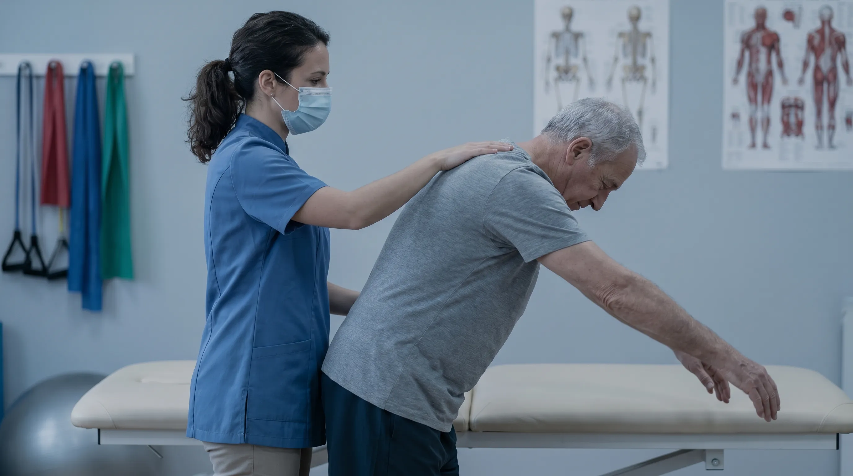
The initial rehabilitation phase spans weeks zero to four, emphasizing tissue protection and edema management. Patients maintain prescribed immobilization while performing hourly finger, wrist, and elbow range of motion exercises to prevent distal stiffness. Scapular retraction drills initiated within days of injury activate stabilizing musculature without stressing the glenohumeral joint. Gentle pendulum exercises utilizing gravity promote synovial fluid distribution and maintain minimal capsular mobility. Isometric contractions targeting the deltoid, rotator cuff, and periscapular muscles rebuild neuromuscular activation patterns without joint shear forces. Ice therapy, compression sleeves, and elevation protocols minimize inflammatory swelling. Patients must avoid active elevation, external rotation beyond neutral, and any resistance training during this critical healing window.
Phase 2: Strengthening and Functional Restoration
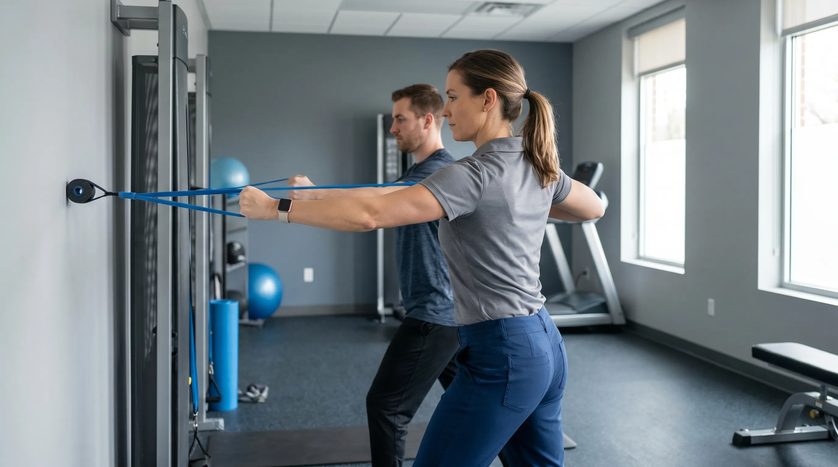
Between weeks four and eight, progressive loading begins as tissue tensile strength improves. Range of motion expands systematically under therapeutic guidance, prioritizing external rotation and forward flexion mobility. Resistance bands introduce controlled tensile stress to rotator cuff and scapular stabilizers, utilizing slow eccentric contractions to enhance tendon resilience. Closed-chain exercises, including wall push-ups and modified plank progressions, improve dynamic joint proprioception. Therapists emphasize scapulohumeral rhythm re-education, ensuring proper kinetic chain sequencing before advancing to isolated strengthening. Pain should remain below four on a ten-point scale during activity; excessive soreness or joint instability indicates premature progression. Consistent adherence to prescribed repetitions and gradual load increments prevents overuse injuries and fosters sustainable tissue adaptation.
Phase 3: Return to Activity and Sports
The final phase, typically spanning months three through six, focuses on sport-specific conditioning and functional integration. Plyometric progressions, medicine ball rotational throws, and dynamic stabilization drills simulate real-world loading patterns while challenging joint control. Isokinetic testing evaluates bilateral strength symmetry, ensuring the affected side achieves at least ninety percent of contralateral torque generation before clearing high-risk activities. Gradual reintroduction to overhead lifting, contact drills, or heavy resistance training occurs under supervised conditions. Athletes incorporate sport-specific agility work and fatigue management strategies to prevent breakdown under competitive stress. Ongoing maintenance programs emphasizing rotator cuff endurance, scapular control, and core stability sustain long-term joint health. Regular follow-up assessments ensure structural integrity and prevent regression.
Complications and Long-Term Outlook
Post-Reduction Complications
Even with optimal initial management, posterior shoulder dislocation carries inherent risks that require vigilant monitoring. Recurrent instability remains the most prevalent complication, particularly in young athletic populations or individuals with underlying capsular laxity. Post-traumatic stiffness frequently develops if immobilization periods extend excessively or if early mobilization protocols are neglected. Axillary nerve neuropraxia, characterized by lateral shoulder numbness and deltoid weakness, often resolves spontaneously within three months but occasionally necessitates electromyographic evaluation. Complex regional pain syndrome manifests in rare cases, presenting with severe burning pain, temperature asymmetry, and abnormal sweating patterns requiring multidisciplinary pain management. Articular cartilage damage sustained during initial displacement accelerates degenerative changes, potentially leading to premature glenohumeral osteoarthritis. Early recognition and targeted intervention mitigate long-term functional impairment.
Chronic Instability and Arthritis Risks
Unaddressed structural deficits predispose the shoulder to chronic mechanical dysfunction. Repetitive micro-subluxations gradually erode labral tissue and stretch capsular restraints, creating a cycle of progressive joint laxity. Patients report persistent clicking sensations, episodic catching, and activity-dependent pain that severely limits occupational and recreational pursuits. Over time, abnormal humeral head tracking concentrates stress on specific articular zones, accelerating cartilage fibrillation and osteophyte formation. Longitudinal studies demonstrate significantly higher rates of glenohumeral arthritis in individuals who sustain traumatic dislocations without adequate rehabilitation or surgical stabilization. Implementing lifelong joint preservation strategies, including targeted strengthening, ergonomic modifications, and activity pacing, substantially delays degenerative progression. Regular imaging surveillance and clinical assessments ensure timely intervention when structural deterioration accelerates.
Prevention and Self-Care Strategies
Ergonomic Adjustments and Fall Prevention
Proactive lifestyle modifications significantly reduce the likelihood of traumatic joint injuries. Workplace environments should prioritize ergonomic workstation setups that minimize repetitive forward shoulder loading and internal rotation postures. Proper lifting techniques emphasizing hip hinge mechanics, neutral spine alignment, and load distribution across bilateral upper extremities prevent sudden joint overload. Fall prevention initiatives prove particularly valuable for older adults, incorporating home safety assessments and environmental modifications, non-slip flooring, adequate lighting, and sturdy handrail installations. Balance training programs enhance proprioceptive awareness and reactive stabilization capacity. Wearing protective gear during high-risk recreational activities, such as padded shoulder guards for cycling or impact-resistant equipment for contact sports, provides an additional mechanical buffer against direct trauma. Environmental hazard elimination remains the most effective primary prevention strategy.
Strengthening and Mobility Exercises
Targeted musculoskeletal conditioning forms the foundation of joint resilience. Rotator cuff strengthening should emphasize balanced development across all four muscle groups, prioritizing eccentric external rotation to counteract dominant internal rotation forces from pectoral and latissimus activation. Scapular stabilizers, including serratus anterior, lower trapezius, and rhomboids, require dedicated attention to maintain optimal glenohumeral positioning during dynamic movements. Thoracic spine mobility drills, including foam roller extensions and seated rotational stretches, reduce compensatory shoulder strain by restoring proper spinal kinematics. Regular dynamic warm-ups incorporating band pull-aparts, wall slides, and arm circles prepare connective tissues for increased load tolerance. Consistency matters more than intensity; performing moderate-volume exercises three to four times weekly yields superior long-term outcomes compared to sporadic high-intensity sessions.
Actionable Daily Habits for Joint Health
Small, sustainable daily practices compound into profound structural benefits over time. Maintaining optimal hydration supports synovial fluid viscosity and nutrient delivery to avascular cartilage structures. Anti-inflammatory dietary patterns rich in omega-3 fatty acids, vitamin C, and zinc accelerate tissue repair and modulate inflammatory cascades. Prioritizing seven to nine hours of quality sleep facilitates hormonal regulation and cellular regeneration essential for musculoskeletal recovery. Stress management techniques, including mindfulness meditation and controlled breathing exercises, reduce muscle tension and prevent protective bracing patterns that strain joint structures. Regular movement breaks every sixty minutes during sedentary work prevent adaptive shortening of anterior shoulder musculature. Listening to early warning signs of joint discomfort, modifying activities before pain becomes severe, and seeking professional guidance promptly preserve long-term function and quality of life.
Frequently Asked Questions
How long does it take to recover from a posterior shoulder dislocation?
Recovery typically spans three to six months, depending on whether the injury was managed conservatively or required surgical stabilization. Initial immobilization lasts about two to four weeks, followed by progressive physical therapy. Full return to heavy lifting or contact sports usually requires six months of structured rehabilitation to ensure joint stability and muscular balance.
Can a posterior shoulder dislocation heal without surgery?
Many cases of posterior shoulder dislocation heal successfully without surgery, particularly when there is minimal bone damage or capsular tearing. Conservative management includes timely closed reduction, temporary sling immobilization, and a structured physical therapy program focused on rotator cuff and periscapular strengthening. Surgery is generally reserved for recurrent instability, large reverse Hill-Sachs lesions exceeding thirty percent, or chronic locked dislocations.
Why is a posterior shoulder dislocation often missed on initial X-rays?
It is frequently overlooked because standard anterior-posterior X-ray views can appear deceptively normal or show only subtle signs, such as the empty glenoid sign or a prominent coracoid process. Without specialized projections like the axillary lateral or modified scapular Y-view, the posterior displacement of the humeral head may not be apparent. Advanced imaging such as CT or MRI is often required to confirm the diagnosis and assess accompanying soft tissue or bony defects.
What are the most common causes of this type of shoulder injury?
The most frequent causes include high-energy trauma such as motor vehicle collisions, falls onto an outstretched arm, direct blows to the front of the shoulder, and forceful muscle contractions from seizures or electrical shocks. Repetitive overhead loading in athletes and underlying connective tissue disorders can also increase susceptibility. Prompt recognition and appropriate immobilization following these mechanisms are crucial for optimal outcomes.
Is it safe to sleep with the arm in a sling after reduction?
Yes, sleeping with the arm properly secured in a prescribed sling or brace is not only safe but highly recommended during the initial two to four weeks post-reduction. Maintaining slight internal rotation and neutral elevation reduces stress on the healing posterior capsule and prevents accidental external rotation movements. Using pillows to support the forearm and keeping the elbow slightly away from the torso can improve comfort and minimize stiffness while sleeping.
Conclusion
Navigating the complexities of a posterior shoulder dislocation requires patience, precise medical guidance, and unwavering commitment to structured rehabilitation. While less common than its anterior counterpart, this injury demands specialized diagnostic approaches and tailored treatment pathways to prevent chronic instability and long-term functional decline. Early recognition through specialized imaging, appropriate immobilization protocols, and progressive therapeutic loading form the cornerstone of successful recovery. Patients who actively engage in their rehabilitation, prioritize tissue healing, and adopt preventative conditioning strategies significantly enhance their prognosis. Whether managed conservatively or through surgical intervention, consistent follow-up care and mindful lifestyle adjustments ensure optimal joint longevity. By understanding the underlying biomechanics and adhering to evidence-based recovery frameworks, individuals can confidently return to daily activities, athletic pursuits, and pain-free movement. Always consult qualified orthopedic specialists to develop a personalized treatment plan aligned with your unique anatomical profile and functional goals.
How we work
Researched and drafted with AI assistance, then reviewed by a human editor against the cited sources.
This article is general health information, not medical advice. Consult a qualified healthcare professional about your own situation.