Jammed Finger vs Broken Finger: Symptoms, Diagnosis & Recovery Guide

You are reaching for a coffee mug, catching a basketball during a weekend pickup game, or simply closing a car door too quickly. In a split second, a sudden force impacts your fingertip, driving it backward against the joint. Your finger immediately swells, throbs, and refuses to bend normally. The next question every patient asks is nearly identical: Is this just a jammed finger vs broken finger? Understanding the distinction is critical because mistaking a fracture for a simple sprain can lead to permanent joint stiffness, malunion, or chronic arthritis, while unnecessarily assuming a break can cause undue anxiety and prolonged immobilization. Finger injuries are among the most common orthopedic complaints, yet they are frequently mismanaged through improper self-diagnosis. This comprehensive guide will walk you through the anatomical differences, clinical signs, diagnostic procedures, treatment pathways, and evidence-based recovery strategies supported by National Institutes of Health (NIH) clinical guidelines. Whether you are an athlete, a manual laborer, or a weekend warrior, knowing exactly how to respond when the difference between a jammed finger vs broken finger becomes apparent can save your hand’s long-term functionality. We will explore the physiological mechanisms behind each injury, compare symptom profiles, outline when to seek urgent care, and provide actionable rehabilitation protocols backed by hand surgery specialists and sports medicine clinicians.
Understanding Finger Anatomy: Why These Injuries Matter
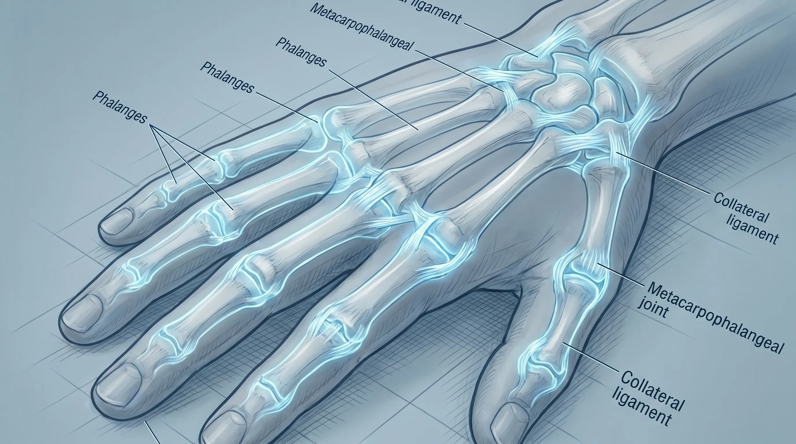
Before diving into clinical differentiation, it is essential to understand the structural complexity of the human finger. Each finger (excluding the thumb) consists of three phalanges: the proximal, middle, and distal bones. The thumb contains two. These bones connect at three primary joints: the metacarpophalangeal (MCP), proximal interphalangeal (PIP), and distal interphalangeal (DIP) joints. Each joint is stabilized by a sophisticated network of collateral ligaments, volar plates, extensor and flexor tendons, and the joint capsule itself. The collateral ligaments prevent side-to-side instability, while the volar plate prevents hyperextension. Tendons, such as the flexor digitorum profundus and extensor digitorum, transmit muscular force to produce grip and extension.
This intricate arrangement makes fingers incredibly mobile but highly vulnerable to traumatic loading. When an axial force strikes the fingertip with the joint partially flexed or extended beyond its physiological range, the ligaments, joint capsule, or bone itself can sustain microscopic or macroscopic damage. The severity of the injury depends entirely on the force magnitude, impact angle, tissue elasticity, and pre-existing joint health. For example, older adults with reduced bone density may fracture from minimal trauma, while younger athletes with stronger ligaments may experience severe sprains instead. Recognizing how these anatomical components interact during trauma provides the foundation for distinguishing a jammed finger vs broken finger with clinical accuracy, as detailed in NIH MedlinePlus anatomical resources.
What Is a Jammed Finger?
A jammed finger, medically referred to as a digital ligament sprain, occurs when the finger is forcefully compressed along its long axis. The impact typically forces the joint capsule and surrounding ligaments to stretch beyond their normal elastic limit. Unlike a fracture, which involves structural bone disruption, a jammed finger primarily damages the soft tissues that stabilize the joint. The injury is graded on a scale similar to other musculoskeletal sprains: Grade I involves mild microscopic tearing with intact joint stability, Grade II features partial ligamentous disruption with mild instability, and Grade III represents a complete ligament rupture that often requires surgical reconstruction.
The Mechanism of Injury
The classic jamming mechanism involves a sudden axial load applied directly to the fingertip while the joint is in a vulnerable position. Sports like basketball, volleyball, baseball, and football frequently produce this injury when a ball strikes an outstretched or improperly positioned finger. The PIP joint is most commonly affected because it bears the greatest mechanical stress during flexion. When the joint is forced past its natural limit, the volar plate can tear, the collateral ligaments can stretch, or the joint capsule can sustain a synovial injury. Blood rushes to the area, causing rapid inflammation, while nerve endings transmit sharp pain signals. The resulting swelling often restricts motion and creates a sensation of tightness or "fullness" within the joint.
Common Causes and Risk Factors
While athletic impacts are the most recognizable cause, jammed fingers frequently occur during everyday activities. Catching a falling object, slamming a finger against a countertop, or even aggressive typing can generate sufficient force to injure vulnerable soft tissues. Risk factors include prior finger injuries that leave residual laxity, poor warm-up routines that reduce tissue elasticity, inadequate protective equipment, and occupations requiring repetitive gripping or impact exposure. Individuals with hypermobility syndrome or connective tissue disorders are also at higher risk for severe sprains due to inherently lax ligaments. Proper taping techniques and technique modification in sports can significantly reduce incidence rates, as outlined by specialists in hand trauma prevention guidelines from the Cleveland Clinic.
What Is a Broken Finger?
A broken finger, clinically termed a phalangeal fracture, occurs when one or more bones in the digit sustain a complete or partial discontinuity. Fractures range from subtle hairline cracks to severe comminuted breaks where the bone shatters into multiple fragments. Unlike soft tissue injuries, fractures disrupt the structural integrity of the skeletal framework, often requiring precise realignment and extended immobilization to ensure proper ossification. The clinical management of fractures depends heavily on location, displacement, intra-articular involvement, and patient-specific factors such as age and occupational demands.
Types of Fractures
Phalangeal fractures are categorized by their pattern and anatomical involvement. Transverse fractures run straight across the bone shaft and often result from direct impact. Oblique or spiral fractures occur with rotational or bending forces, frequently seen when a finger gets caught in equipment or clothing. Comminuted fractures involve three or more bone fragments and typically indicate high-energy trauma. Avulsion fractures occur when a tendon or ligament pulls a small piece of bone away from the joint surface, commonly seen in jamming injuries that masquerade as simple sprains. Intra-articular fractures extend into the joint space and carry the highest risk of post-traumatic arthritis if not anatomically reduced.
When Fractures Require Immediate Attention
Not all fractures demand emergency intervention, but specific red flags indicate urgent orthopedic consultation. Open fractures where bone pierces the skin require immediate surgical debridement to prevent osteomyelitis. Severe displacement, visible angulation, neurovascular compromise (numbness, pallor, coldness, or absent capillary refill), and multi-joint involvement necessitate prompt reduction and stabilization. Delayed treatment can lead to malunion, chronic pain, tendon adhesions, and permanent loss of fine motor skills. Early radiographic evaluation remains the gold standard for accurate classification and treatment planning, consistent with orthopedic recommendations from the Mayo Clinic.
Jammed Finger vs Broken Finger: Key Clinical Differences
Differentiating a jammed finger vs broken finger requires careful observation of symptom patterns, functional limitations, and injury mechanisms. While both conditions share overlapping features like swelling and pain, their clinical trajectories diverge significantly in the acute and subacute phases.
Symptom Comparison
Jammed fingers typically present with diffuse swelling around the joint, moderate to severe pain that peaks within the first 24 hours, and gradual improvement over several days. The finger may appear slightly crooked but usually returns to normal alignment once acute inflammation subsides. Range of motion is restricted due to pain and fluid accumulation rather than mechanical obstruction. Pain is generally localized to the soft tissues and ligament insertions rather than directly over the bone.
Conversely, broken fingers often exhibit sharp, pinpoint bone tenderness that does not diminish with initial rest. Deformity such as shortening, rotational malalignment, or obvious angulation is common. Patients frequently describe a grinding or crackling sensation (crepitus) during movement, which indicates bone fragments rubbing together. Swelling may be more localized and ecchymosis (bruising) often appears rapidly and extends down the finger. Functional impairment is typically more profound, with complete inability to actively flex or extend the digit in severe cases.
Diagnostic Approaches
Clinical examination alone cannot definitively rule out a fracture when evaluating a jammed finger vs broken finger. Physical assessment includes palpation for bony tenderness, assessment of active and passive range of motion, evaluation of rotational alignment by making a fist, and neurovascular screening. Imaging modalities remain indispensable. Standard anteroposterior, lateral, and oblique X-rays provide comprehensive views of fracture patterns. If X-rays appear normal but clinical suspicion remains high, advanced imaging like ultrasound for soft tissue assessment or CT scans for intra-articular fracture delineation may be warranted. Clinical decision rules emphasize that persistent pain, point tenderness, or functional limitation beyond 72 hours warrants radiographic confirmation, per injury management protocols from the Centers for Disease Control and Prevention (CDC).
| Feature | Jammed Finger | Broken Finger |
|---|---|---|
| Primary Tissue Affected | Ligaments, joint capsule, tendons | Phalangeal bone structure |
| Pain Pattern | Diffuse, peaks at 24-48h, gradually improves | Sharp, localized, persists or worsens |
| Visible Deformity | Rare; mild swelling common | Common; angulation, shortening, or rotation |
| Crepitus (Grinding) | Absent | Often present |
| Functional Limitation | Pain-restricted but mechanically intact | Mechanical block; unable to move finger |
| Healing Timeline | 1 to 4 weeks | 4 to 10+ weeks depending on severity |
| Imaging Required | Usually clinical diagnosis; X-ray if severe | Mandatory X-ray for classification |
| Typical Treatment | RICE, buddy taping, splinting | Immobilization, reduction, possible surgery |
How to Tell the Difference: Self-Assessment and Clinical Evaluation
While professional diagnosis remains essential, patients can perform structured self-assessment to gauge injury severity while waiting for medical evaluation. The goal is not to replace clinical examination but to make informed decisions about urgency and initial care.
Self-Assessment Guidelines
Begin with a visual inspection in good lighting. Compare the injured finger with the corresponding finger on the opposite hand. Look for asymmetry, abnormal angulation, or nail bed discoloration. Gently palpate along the length of the phalanges, avoiding excessive pressure. Note whether tenderness is diffuse across the joint or sharply localized to a specific bony prominence. Attempt a slow, controlled flexion and extension. If the finger moves through its full range despite pain, soft tissue injury is more likely. If movement stops abruptly, catches, or triggers severe pain at a specific point, structural bone involvement is probable. Check rotational alignment by slowly curling all fingers into a gentle fist. All fingertips should point toward the scaphoid bone in the wrist without crossing over adjacent digits. Malrotation strongly suggests fracture or severe tendon disruption.
When to Seek Professional Medical Care
Immediate evaluation is warranted if you observe obvious deformity, open wounds, numbness or tingling, cold or pale fingertips, or complete loss of motion. Even seemingly minor injuries require clinical assessment if symptoms fail to improve after 48 hours of consistent rest, ice, compression, and elevation. Patients with diabetes, peripheral neuropathy, or vascular disease should never rely on home management alone due to impaired healing responses. Sports medicine clinics, urgent care centers, and orthopedic hand specialists routinely manage these injuries and can provide accurate radiographic interpretation, customized splinting, and structured follow-up care aligned with World Health Organization (WHO) trauma management standards.
Treatment Pathways: From Home Care to Clinical Intervention
Effective management of a jammed finger vs broken injury depends entirely on accurate classification, injury severity, and individual patient goals. Treatment protocols follow a phased approach designed to control inflammation, protect healing tissues, restore mobility, and rebuild strength.
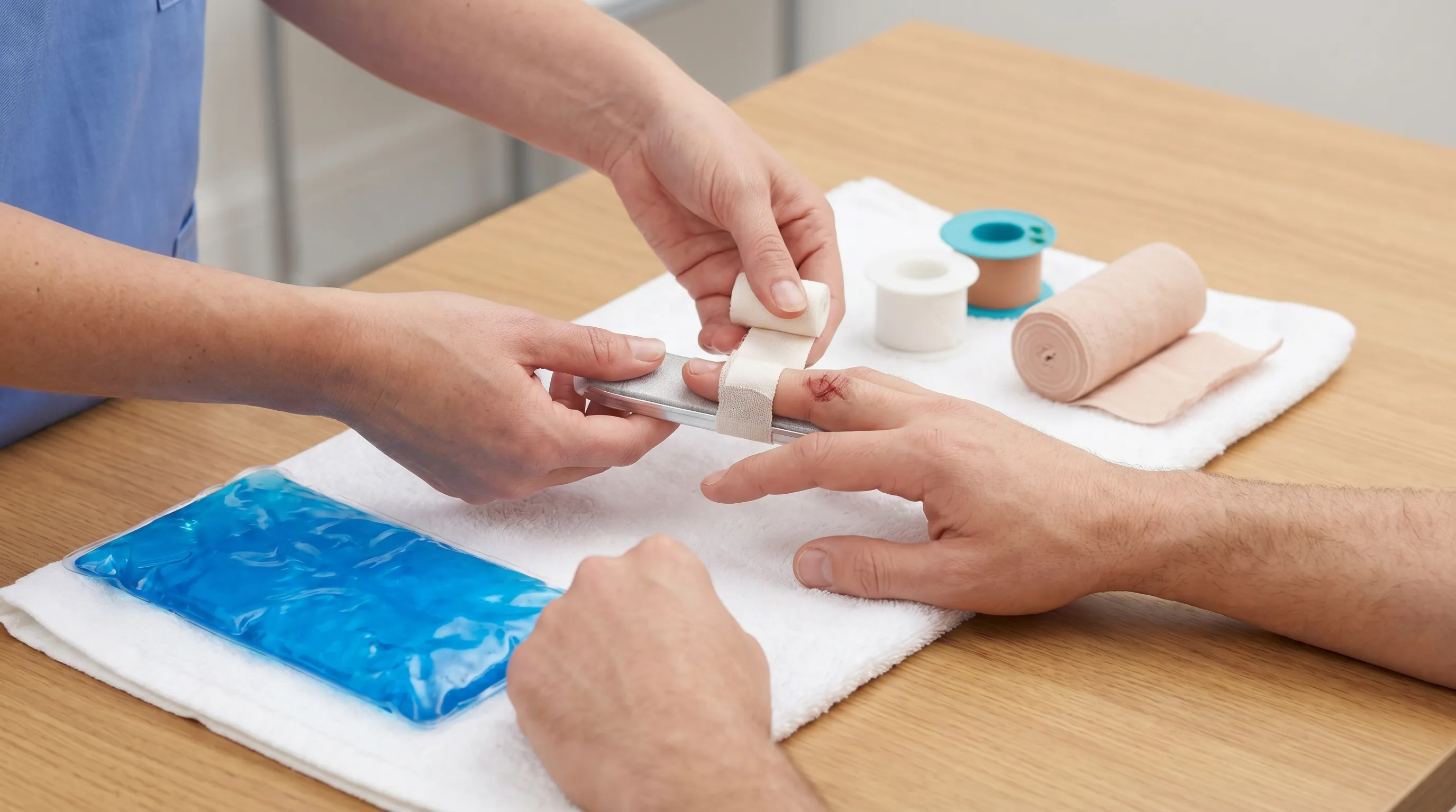
Initial R.I.C.E. Protocol and Splinting
Regardless of whether the diagnosis is a sprain or fracture, the first 72 hours are critical. Rest involves eliminating activities that provoke pain, particularly gripping, catching, or heavy lifting. Ice should be applied in 15-20 minute intervals every 2-3 hours, with a thin cloth barrier to prevent frostbite. Compression using elastic wraps or medical tape reduces edema without restricting arterial flow. Elevation above heart level encourages lymphatic drainage and minimizes throbbing, following standard acute injury protocols detailed by NIH MedlinePlus. For jammed fingers, buddy taping to an adjacent stable digit provides gentle support while maintaining functional alignment. Aluminum foam splints or thermoplastic dorsal splints may be prescribed for moderate sprains to restrict hyperextension while allowing controlled flexion. Patients must avoid removing immobilization prematurely, as early motion can convert a stable injury into a chronic instability syndrome.
Medical Treatments for Fractures
Fracture management follows strict orthopedic principles centered on anatomical reduction and stable fixation. Non-displaced fractures typically heal successfully with custom orthosis or cast immobilization for 3 to 6 weeks. Displaced or unstable fractures require closed reduction, where a specialist manually realigns bone fragments under local anesthesia or sedation, followed by radiographic verification. When closed methods fail, percutaneous pinning (K-wires), external fixation, or open reduction internal fixation (ORIF) with miniature plates and screws becomes necessary. Intra-articular fractures demand millimeter-perfect alignment to prevent post-traumatic osteoarthritis. Post-procedure care includes wound monitoring, neurovascular checks, and staged rehabilitation initiation. Patients must adhere strictly to weight-bearing restrictions and follow-up imaging schedules to track callus formation and alignment maintenance.
Rehabilitation and Recovery Timeline
Recovery from either injury extends far beyond symptom resolution. Proper rehabilitation prevents stiffness, restores proprioception, and rebuilds tendon gliding capacity. The timeline varies significantly between a jammed finger vs broken finger, with fractures requiring more conservative progression.
Exercises to Restore Function
Phase one (weeks 1-2) focuses on edema control and protected range of motion. Gentle tendon gliding exercises, including hook, straight, and composite fist positions, prevent adhesions while respecting healing tissues. Phase two (weeks 3-6) introduces active-assisted stretching and light grip activities. Therapy putty, stress balls, and rice bucket exercises improve intrinsic muscle strength without excessive joint loading. Phase three (weeks 6-12) emphasizes dynamic strengthening, proprioceptive retraining, and sport-specific drills. Contrast baths, paraffin wax therapy, and myofascial release may address residual stiffness. Hand therapists customize progressions based on radiographic healing, pain response, and functional demands. Returning to contact sports or heavy labor prematurely risks re-fracture, ligament attenuation, or tendon rupture.
Nutritional Support for Bone and Ligament Healing
Tissue regeneration requires optimal metabolic support. Protein intake should reach 1.2 to 1.6 grams per kilogram of body weight to supply amino acids necessary for collagen synthesis and muscle preservation. Vitamin D (1000-2000 IU daily) and calcium (1000-1200 mg daily) form the structural foundation of bone mineralization. Vitamin C supports collagen cross-linking and angiogenesis, while magnesium facilitates enzymatic reactions in tissue repair. Omega-3 fatty acids from fish oil modulate inflammatory pathways, preventing chronic synovitis. Hydration maintains disc and joint capsule elasticity, reducing secondary stiffness. Avoiding smoking, excessive alcohol, and high-sodium diets is equally critical, as these factors impair microcirculation and delay ossification, according to bone health guidelines from the Mayo Clinic.
Long-Term Complications of Untreated Injuries
Neglecting proper diagnosis or prematurely returning to activity when evaluating a jammed finger vs broken injury can produce devastating sequelae. Early intervention remains the single most important predictor of long-term hand function.
Arthritis and Joint Stiffness
Post-traumatic osteoarthritis develops when articular cartilage sustains irreversible damage from misaligned fractures or chronic ligament laxity. The inflammatory cascade degrades proteoglycans, leading to joint space narrowing, crepitus, and chronic pain during weather changes or gripping tasks. Adhesive capsulitis and flexion contractures result from prolonged immobilization without supervised therapy. Patients may lose the ability to fully make a fist or grip tools securely, significantly impacting occupational performance.
Malunion and Chronic Deformity
Malunion occurs when fracture fragments heal in suboptimal alignment. Rotational malalignment causes finger scissoring, making it impossible to hold utensils, type efficiently, or wear gloves. Dorsal angulation produces mallet or swan-neck deformities, altering tendon biomechanics and requiring complex reconstructive surgery. Chronic instability from untreated ligament tears forces surrounding muscles to compensate, leading to premature fatigue and secondary tendonitis. Surgical correction becomes more invasive over time, with outcomes declining as degenerative changes progress, as emphasized in rehabilitation literature from the Cleveland Clinic.
Frequently Asked Questions
Can a jammed finger turn into a fracture over time?
No, a sprain cannot biologically transform into a broken bone. However, severe ligament injuries can mask an underlying avulsion fracture or hairline break that goes undetected on initial examination. Persistent swelling or delayed pain often reveals these hidden injuries, which is why radiographic confirmation is standard protocol when symptoms fail to resolve within the expected soft tissue healing window.
How long does it take for a broken finger to heal?
Simple, non-displaced fractures typically require 3 to 6 weeks of immobilization followed by gradual rehabilitation. Complex fractures involving multiple fragments, intra-articular surfaces, or requiring surgical hardware often demand 8 to 12 weeks for complete bony consolidation. Full functional recovery, including grip strength restoration, may take 3 to 6 months depending on patient age, compliance, and rehabilitation intensity.
Is it safe to buddy-tape a fractured finger?
Buddy taping is appropriate only for stable, non-displaced fractures or mild sprains under direct medical supervision. Taping an unstable or angulated fracture can worsen displacement, compress neurovascular structures, or cause tendon entrapment. Always obtain clearance from a hand specialist before initiating any immobilization strategy.
When should I get an X-ray for a finger injury?
Radiographic evaluation is indicated when you observe visible deformity, experience pinpoint bone tenderness, lose active range of motion, or notice rotational misalignment. Additionally, seek imaging if swelling and pain persist beyond 72 hours despite strict adherence to the R.I.C.E. protocol. Early diagnosis significantly improves treatment outcomes and reduces long-term complications.
What exercises help restore mobility after a finger injury?
Begin with passive and active-assisted range-of-motion exercises once acute inflammation subsides. Progress to tendon glides, grip strengthening with therapy putty, finger extension bands, and opposition drills. Incorporate proprioceptive training using textured surfaces and gradual resistance loading. All exercises should remain pain-free and be guided by a certified hand therapist to prevent reinjury or tendon overload.
Conclusion
Distinguishing a jammed finger vs broken finger requires careful attention to symptom patterns, functional testing, and timely radiographic evaluation. While both injuries share overlapping acute features, their underlying pathology, treatment protocols, and recovery trajectories differ significantly. Soft tissue sprains generally respond well to conservative management, early controlled motion, and progressive strengthening. Fractures demand precise anatomical alignment, structured immobilization, and multidisciplinary rehabilitation to restore optimal hand mechanics. Ignoring persistent pain, deformity, or stiffness can lead to permanent joint degeneration, chronic instability, and functional impairment that affects daily life. Prioritize prompt clinical assessment, follow prescribed rehabilitation guidelines, and maintain consistent communication with your healthcare provider throughout the healing process. Your hands perform countless essential tasks, and protecting their structural integrity today ensures pain-free functionality for years to come.
For additional clinical guidance, consult comprehensive finger fracture protocols from the Mayo Clinic, review injury management recommendations from the Cleveland Clinic, and explore evidence-based hand injury resources from the NIH. The CDC and WHO also provide valuable insights on trauma prevention and rehabilitation strategies.
For visual demonstrations of proper splinting techniques and tendon gliding exercises, refer to these expert-led instructional videos:

About the author
Samuel Jones, MD, is a board-certified orthopedic surgeon specializing in joint replacement and orthopedic trauma. He is a team physician for a professional sports team and practices at a renowned orthopedic institute in Georgia.