Eye Hollowing: Causes, Treatments, and Evidence-Based Restoration Strategies

The delicate skin beneath the eyes is among the most reactive and visible indicators of overall facial vitality, yet it is also highly vulnerable to structural changes that can leave the area appearing sunken, shadowed, and fatigued (Cleveland Clinic). Eye hollowing is a common cosmetic and physiological concern that affects millions of individuals across various age groups, ethnicities, and lifestyle backgrounds. Unlike simple wrinkles or fine lines, hollowing involves actual volume depletion within the periorbital region, fundamentally altering facial proportions and creating a tired appearance that often persists despite adequate rest. Understanding the underlying mechanisms of this condition is essential for anyone seeking safe, effective, and long-lasting solutions. Modern dermatology, oculoplastic surgery, and aesthetic medicine have advanced significantly in recent years, offering a comprehensive spectrum of interventions ranging from evidence-based topical regimens to precision surgical techniques. This guide will explore the anatomical foundations, primary triggers, clinical management strategies, and daily preventative practices that can help restore natural under-eye fullness. By combining medical expertise with actionable lifestyle adjustments, individuals can make informed decisions that align with their unique facial structure and wellness goals. Whether you are noticing early signs of volume loss or exploring restorative treatments, the following information provides a clear, research-backed roadmap to achieving healthier, more balanced periorbital aesthetics.
Understanding the Science Behind Eye Hollowing
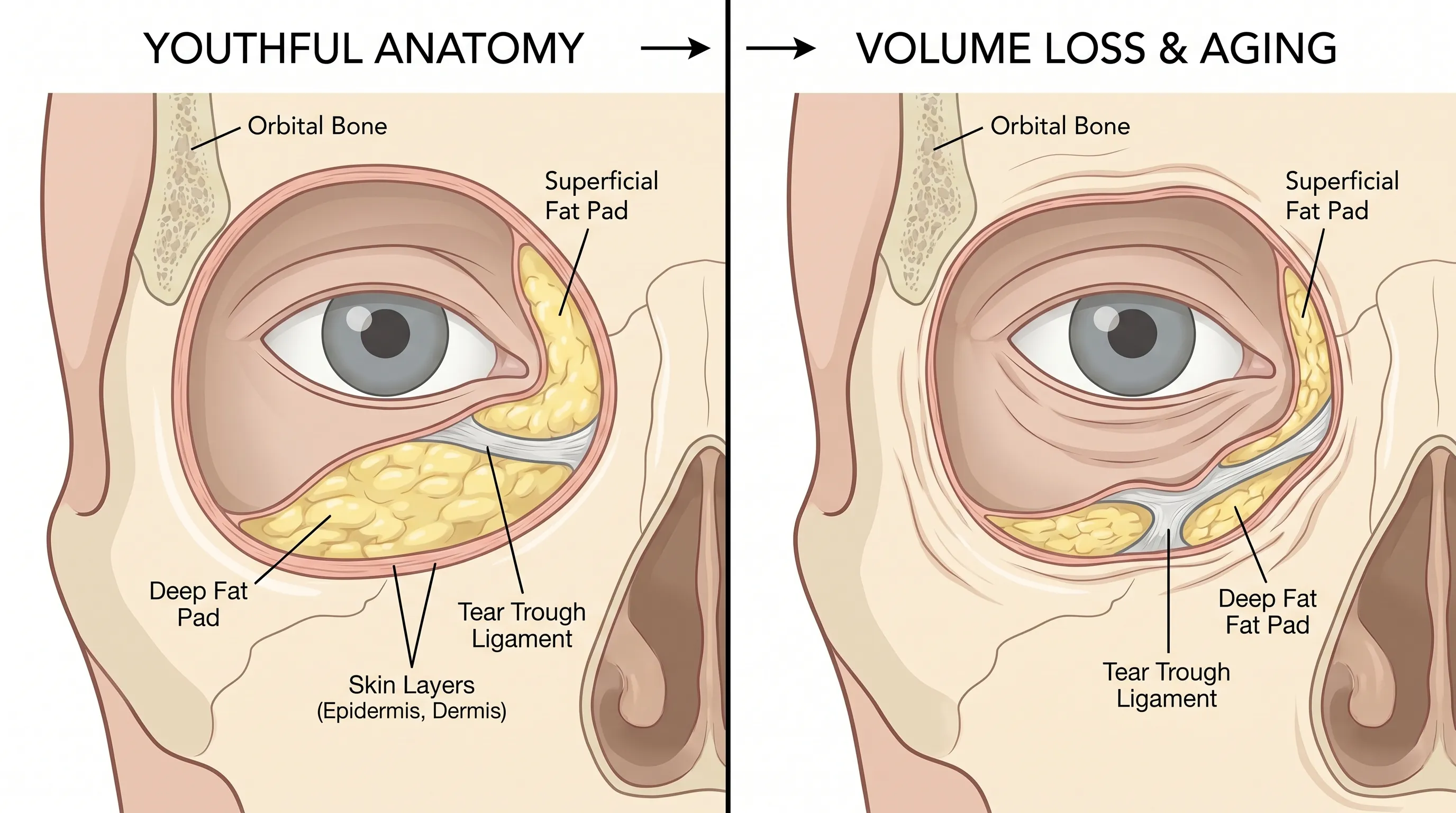
To effectively address under-eye volume depletion, it is crucial to first understand how the periorbital region is constructed and why it undergoes such dramatic transformation over time. The area surrounding the orbit is not merely a flat surface of skin; it is a complex, multi-layered anatomical landscape composed of epidermal, dermal, subcutaneous, and muscular structures, all supported by underlying skeletal framework (NIH). When these layers lose their structural integrity, the characteristic concavity known as eye hollowing becomes visually apparent.
The Complex Anatomy of the Periorbital Region
The lower eyelid and cheek junction is governed by several distinct anatomical compartments. The superficial fat pad, often referred to as the pretarsal fat, sits directly beneath the skin and provides smooth contouring. Beneath this lies the suborbicularis oculi fat (SOOF), which contributes to midface fullness and supports the lower eyelid margin. Deeper still are the deep fat compartments and the malar fat pad, which maintain the transition between the eye and the cheek. These compartments are anchored by fascial ligaments, most notably the orbital retaining ligament and the tear trough ligament, which create a natural depression at the junction of the eyelid and cheek. The thinness of the lower eyelid skin, averaging just 0.5 millimeters in thickness, makes any underlying structural shift immediately visible. Additionally, the presence of the orbicularis oculi muscle provides dynamic movement, but chronic contraction or weakening can alter tension patterns, further influencing the appearance of the area.
How Tissue Volume Shifts Over Time
As the natural aging process unfolds, the periorbital region undergoes a predictable sequence of structural changes. Initially, the skin begins to lose elastin and collagen density, leading to reduced tensile strength and decreased hydration capacity. Simultaneously, adipose tissue within the superficial and deep fat compartments undergoes programmed atrophy and gravitational descent. This dual mechanism of volume loss and tissue migration causes the once-smooth contour of the lower face to develop pronounced depressions. The tear trough ligament remains relatively fixed, while the surrounding tissue sinks away from it, creating a stark contrast between the elevated cheek and the recessed under-eye area. This anatomical separation is what clinically manifests as eye hollowing. The process is further complicated by bone resorption in the maxilla and orbital rim, which reduces foundational support and allows soft tissue to settle into a more concave position. Understanding these biomechanical shifts is the first step toward selecting appropriate therapeutic interventions that address root causes rather than merely masking symptoms.
Primary Causes and Risk Factors for Under-Eye Volume Loss
While chronological aging is the most universally recognized driver of periorbital hollowing, numerous biological, environmental, and behavioral factors accelerate or exacerbate the condition. Identifying your specific risk profile allows for targeted prevention and more effective treatment planning.
Natural Aging and Structural Fat Pad Atrophy
Cellular senescence affects every tissue layer in the face, but the periorbital region is particularly sensitive due to its low sebum production and thin epidermis. Fibroblasts gradually reduce collagen synthesis, while matrix metalloproteinases break down existing extracellular proteins. Adipocytes in the facial fat compartments experience reduced metabolic activity, leading to cellular shrinkage and decreased lipid storage capacity. This progressive volume depletion is compounded by lymphatic drainage inefficiencies, which can cause intermittent fluid retention that alternates with dry, hollowed appearances. Research published in the Journal of Clinical and Aesthetic Dermatology consistently demonstrates that periorbital volume loss accelerates significantly after age 40, though early signs can emerge in the late 20s and 30s depending on genetic predisposition and environmental exposure.
Genetic and Bone Structure Influences
Hereditary factors play a substantial role in determining the baseline architecture and aging trajectory of the lower eyelids. Individuals with naturally prominent infraorbital rims, shallow malar eminences, or tightly bound tear trough ligaments will typically exhibit more pronounced eye hollowing as volume diminishes. Certain ethnic backgrounds demonstrate distinct fat distribution patterns that influence how hollowing presents clinically. Furthermore, genetic variations in collagen synthesis, elastin degradation rates, and inflammatory response thresholds can predispose some individuals to accelerated tissue breakdown. Facial bone morphology is equally critical; a flatter midface provides less foundational support for the overlying soft tissues, making gravitational descent and volume loss more visible. Genetic counseling and family history assessment can sometimes predict the likelihood and timing of periorbital hollowing development.
Lifestyle, Environmental, and Behavioral Factors
Daily habits significantly impact the rate and severity of eye hollowing. Chronic sleep deprivation disrupts cortisol regulation and impairs nocturnal tissue repair, leading to accelerated collagen degradation and impaired microcirculation. Dehydration reduces dermal turgor and exacerbates the sunken appearance, as the skin lacks the internal fluid necessary to maintain structural plumpness. Smoking introduces thousands of pro-inflammatory compounds that directly damage fibroblasts and constrict perifollicular blood vessels, severely compromising nutrient delivery to the delicate under-eye tissues (CDC). Excessive alcohol consumption depletes essential vitamins, disrupts hydration balance, and promotes systemic inflammation that breaks down connective tissue. Environmental stressors such as prolonged exposure to blue light from digital screens, extreme temperature fluctuations, and high particulate pollution generate oxidative stress that degrades the extracellular matrix and accelerates tissue thinning.
Systemic Health Conditions and Nutritional Deficiencies
Certain medical conditions can precipitate or worsen under-eye volume loss. Thyroid dysfunction, particularly hyperthyroidism, can alter fat metabolism and cause periorbital tissue changes. Uncontrolled diabetes impairs microvascular circulation and collagen cross-linking, reducing tissue resilience (Mayo Clinic). Chronic gastrointestinal malabsorption disorders limit the availability of essential amino acids, vitamins, and minerals required for collagen synthesis and cellular repair. Nutritional deficiencies in vitamin C, vitamin K, zinc, and essential fatty acids are strongly correlated with accelerated skin thinning and fat pad atrophy. Prolonged corticosteroid use, whether systemic or topical, can cause localized tissue atrophy that mimics or exacerbates natural volume depletion. Regular metabolic screening and nutritional assessment are valuable components of a comprehensive management strategy for persistent eye hollowing.
Clinical Presentation: Recognizing Eye Hollowing
Accurate identification of periorbital hollowing requires distinguishing it from other under-eye concerns such as pigmentation, puffiness, or vascular prominence. Clinical evaluation typically involves visual inspection, tactile assessment, and patient history review to determine the underlying tissue dynamics at play.
Visual and Tactile Characteristics
Eye hollowing presents as a concave depression along the inferior orbital rim, typically extending from the inner canthus toward the cheek junction. The area may exhibit a bluish, grayish, or brownish hue depending on underlying vascularity, melanin concentration, and shadowing effects. When gently palpated, the skin lacks the underlying firmness and plumpness characteristic of well-supported tissue. In advanced cases, the transition from the eyelid to the cheek becomes sharply defined rather than smoothly blended. Patients often report that the condition appears more pronounced under direct overhead lighting or in photographs taken with flash, due to the accentuated shadows cast by the recessed tissue. Tactile examination by a trained clinician can differentiate between fat atrophy, muscle laxity, and ligamentous prominence.
Impact on Facial Harmony and Perception
The lower eyelid-cheek junction serves as a critical aesthetic landmark that influences overall facial balance. When eye hollowing develops, it disrupts the natural convex curve of the midface, creating an illusion of fatigue, illness, or advanced aging regardless of actual rest or vitality levels. This can significantly impact self-perception, social interactions, and professional confidence. The optical illusion is further compounded by the contrast between the depressed under-eye area and adjacent structures; if the cheeks have descended or the nasolabial folds have deepened, the hollows appear more severe. Restoring periorbital volume is not merely about eliminating a single feature, but about reestablishing proportional harmony that aligns with the individual's natural skeletal framework and soft tissue distribution.
Distinguishing Eye Hollowing from Related Conditions
| Feature | Eye Hollowing (Volume Loss) | Tear Trough Deformity | Under-Eye Puffiness | Dark Circles (Pigmentation) |
|---|---|---|---|---|
| Primary Cause | Fat atrophy & bone resorption | Ligament tethering & fat displacement | Fluid retention & fat herniation | Melanin deposition or vascular prominence |
| Tactile Feel | Soft depression, thin tissue | Sharp ridge, ligament prominence | Soft swelling, fluctuates | Normal tissue thickness, flat |
| Lighting Effect | Worsens in overhead/direct light | Worsens in side lighting | Worsens in frontal lighting | Less affected by lighting angle |
| Age of Onset | Typically 30s onward, progressive | Can appear early (genetic) | Fluctuates, worsens with age/sleep loss | Varies by genetics & lifestyle |
| Primary Treatment | Volume restoration (fillers/fat) | Release & fill combination | Compression, drainage, surgery | Brightening topicals, lasers, lifestyle |
Understanding these distinctions prevents inappropriate treatment selection and ensures that therapeutic interventions target the precise anatomical dysfunction. Many individuals mistakenly treat hollowing as hyperpigmentation or edema, leading to frustration when expected results fail to materialize.
Comprehensive Treatment Modalities and Clinical Approaches
Modern aesthetic medicine offers a tiered approach to addressing periorbital volume loss, ranging from conservative daily regimens to advanced surgical techniques. Selecting the appropriate modality depends on the severity of hollowing, underlying tissue quality, budget considerations, and desired longevity of results.
Topical Skincare and Non-Invasive Strategies
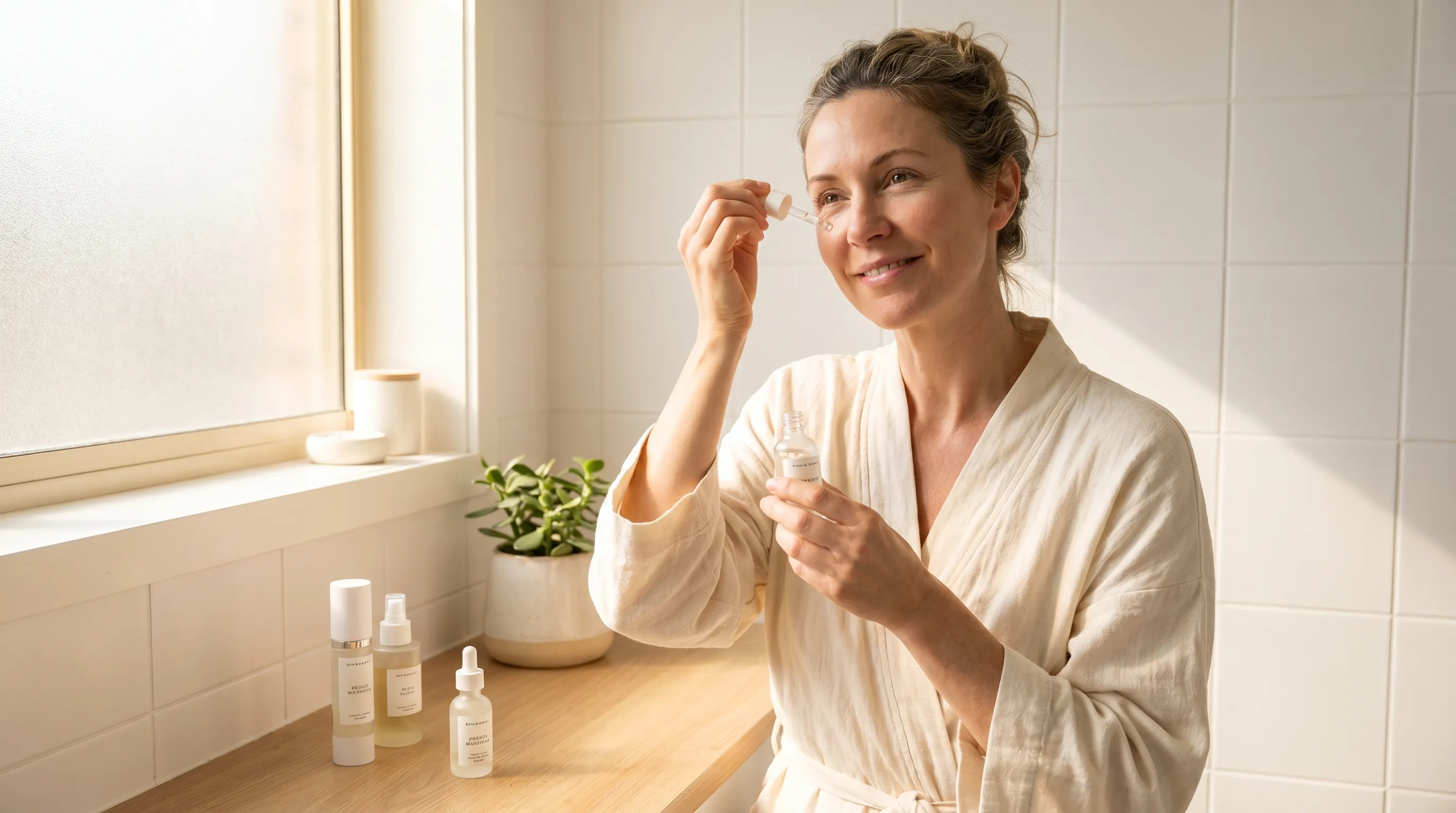
While topical formulations cannot replace lost subcutaneous volume, they play a vital role in optimizing epidermal thickness, enhancing hydration, and stimulating dermal remodeling. High-molecular-weight hyaluronic acid serums provide immediate surface-level plumping by attracting and retaining moisture within the stratum corneum. Peptide complexes such as palmitoyl tripeptide-5 and matrixyl 3000 signal fibroblasts to increase collagen and elastin production over time. Retinoids, particularly low-concentration retinaldehyde or encapsulated retinol, accelerate cellular turnover and thicken the dermis without causing excessive irritation in the sensitive periorbital zone. Vitamin C derivatives stabilize collagen fibers and reduce oxidative damage, while niacinamide strengthens the skin barrier and improves microcirculation. Consistent application of a tailored regimen, combined with gentle massage techniques, can significantly reduce the visual impact of early-stage eye hollowing.
Dermal Fillers and Hyaluronic Acid Restoration
Injectable hyaluronic acid fillers have become the gold standard for non-surgical correction of periorbital hollows. Products specifically formulated for the under-eye area feature high cohesivity, low hydrophilicity, and fine particle sizes to minimize swelling and migration. The cannula-based injection technique is increasingly preferred over traditional needle approaches due to its reduced risk of bruising and vascular compromise. A skilled practitioner typically places micro-deposits of filler along the orbital rim and tear trough ligament to create a smooth transition between the eyelid and cheek. Results are immediately visible, with final settling occurring over 2 to 4 weeks. Maintenance typically ranges from 9 to 18 months depending on metabolic rate, lifestyle factors, and product selection. It is critical to seek treatment from board-certified dermatologists or oculoplastic surgeons, as improper injection depth or volume can lead to lumps, bluish discoloration, or vascular occlusion (Cleveland Clinic).
Autologous Fat Grafting and Surgical Options
For individuals seeking longer-lasting volume restoration, autologous fat transfer utilizes the patient's own adipose tissue harvested from the abdomen, thighs, or flanks. After purification through centrifugation or filtration, the fat is micro-injected into multiple tissue planes beneath the periorbital area. Because the transferred fat establishes its own blood supply, a portion of the graft survives permanently, offering durable structural support. The procedure requires minor surgical intervention and local or general anesthesia, with initial swelling resolving over several weeks. Some volume resorption is expected within the first 3 to 6 months, often necessitating a secondary touch-up session. Blepharoplasty with fat repositioning is another surgical alternative that addresses both hollowing and bulging fat pads simultaneously. By conserving and redistributing native tissue, this approach maintains natural anatomy while restoring youthful contours (Mayo Clinic).
Energy-Based Devices and Adjunctive Therapies
Emerging non-invasive technologies complement traditional volume restoration by improving skin quality, stimulating collagen, and enhancing lymphatic drainage. Radiofrequency microneedling delivers controlled thermal energy to the dermal and subdermal layers, triggering neocollagenesis and tightening lax tissue without surface damage. Low-level laser therapy (LLLT) utilizes specific wavelengths to increase mitochondrial activity in fibroblasts, accelerating tissue repair and microcirculation. Platelet-rich plasma (PRP) injections harness growth factors from the patient's own blood to promote cellular regeneration and enhance the longevity of concurrent treatments. These modalities are rarely standalone solutions for significant hollowing but serve as powerful adjuncts that improve treatment outcomes and extend results. Clinical trials published in dermatology journals continue to validate the synergistic effects of combining volume restoration with tissue-stimulating therapies.
Proactive Prevention and Daily Management
Long-term management of eye hollowing extends far beyond clinical interventions. Implementing sustainable lifestyle practices, nutritional optimization, and environmental protection strategies can dramatically slow tissue degradation and preserve periorbital volume for decades.
Optimized Nutritional Support and Hydration
Adequate protein intake is fundamental for maintaining collagen synthesis and muscle tone. Aim for 0.8 to 1.2 grams of high-quality protein per kilogram of body weight daily, incorporating sources rich in amino acids like glycine, proline, and lysine. Omega-3 fatty acids from fatty fish, flaxseeds, and walnuts reduce systemic inflammation and support cellular membrane integrity. Antioxidant-rich foods, particularly berries, leafy greens, and dark chocolate, neutralize free radicals that degrade dermal structure. Hydration must be consistent and electrolyte-balanced; aim for 2.5 to 3 liters of water daily, supplemented with mineral-rich beverages or broths to maintain osmotic balance in the extracellular matrix. Limiting refined sugars and processed carbohydrates prevents advanced glycation end-product formation, which stiffens collagen and accelerates tissue breakdown.
Sleep Architecture and Circadian Alignment
Quality sleep is when the body performs critical tissue repair, growth hormone release, and cortisol regulation. Establishing a consistent 7 to 9-hour sleep schedule aligned with natural circadian rhythms optimizes nocturnal cellular regeneration. Sleeping on your back with a slightly elevated head reduces fluid pooling that alternately stretches and dehydrates the under-eye area. Use breathable, silk pillowcases to minimize friction-induced microtrauma and collagen disruption. Blue light exposure within 90 minutes of bedtime suppresses melatonin production, delaying restorative sleep cycles and accelerating tissue aging. Implementing a wind-down routine that includes dim lighting, temperature regulation, and relaxation techniques ensures optimal recovery for the delicate periorbital region (CDC).
Advanced Sun Defense and Environmental Protection
Ultraviolet radiation remains the single most destructive external factor for periorbital aging. Daily application of broad-spectrum SPF 30+ sunscreen, formulated with physical blockers like zinc oxide or titanium dioxide, prevents photo-induced collagen degradation and pigmentation changes. Sunglasses with 100% UVA/UVB protection shield the area from direct exposure while reducing squint-induced muscle tension that contributes to tissue laxity. Antioxidant serums containing vitamin E, ferulic acid, and green tea extract provide an additional photoprotective barrier by neutralizing environmental free radicals. Indoor air purification systems and humidifiers maintain optimal moisture levels in dry or climate-controlled environments, preventing chronic transepidermal water loss that exacerbates hollowing. Consistent UV protection is strongly recommended by global health authorities to prevent premature periorbital aging (WHO).
Choosing the Right Treatment Path
Navigating the extensive array of interventions for eye hollowing requires careful consideration of your anatomical profile, medical history, financial investment, and long-term aesthetic goals. A systematic, personalized approach ensures optimal outcomes while minimizing unnecessary risks.
Consulting a Board-Certified Specialist
The periorbital region is one of the most technically demanding areas to treat, requiring intimate knowledge of vascular anatomy, tissue planes, and dynamic facial movement. Always verify that your provider holds board certification in dermatology, plastic surgery, or oculoplastic surgery. During consultation, request a comprehensive facial assessment that evaluates skin thickness, fat distribution, ligament position, and bone structure. Discuss realistic expectations, potential complications, and maintenance requirements before committing to any procedure. Reputable practitioners will prioritize tissue preservation over aggressive volume augmentation, ensuring results that enhance natural harmony rather than creating artificial fullness.
Realistic Expectations and Recovery Timelines
Understanding the physiological timeline of tissue healing is essential for patient satisfaction. Dermal fillers integrate with surrounding tissue over 4 to 6 weeks, during which initial swelling subsides and the product softens. Fat grafting requires 3 to 6 months for neovascularization to complete and final volume to stabilize. Non-invasive devices typically require a series of 3 to 5 sessions spaced 4 weeks apart to achieve cumulative benefits. Avoiding strenuous exercise, excessive heat exposure, and blood-thinning supplements during the immediate recovery period minimizes bruising and optimizes treatment integration. Patience and adherence to post-procedure protocols significantly influence long-term outcomes.
Long-Term Maintenance Strategies
Sustaining periorbital volume requires an integrated approach that combines periodic clinical touch-ups with unwavering daily care. Annual skin assessments allow practitioners to monitor tissue changes and adjust treatment plans proactively. Gradually transitioning from aggressive interventions to maintenance-level therapies preserves tissue integrity and prevents overcorrection. Maintaining consistent hydration, nutritional balance, and sleep quality ensures that treated areas age gracefully alongside natural facial dynamics. Documenting progress through standardized photography helps track changes objectively and guides future decision-making. Remember that healthy periorbital aesthetics are not about reversing time, but about optimizing structural support and tissue vitality at every life stage.
Frequently Asked Questions
What Exactly Causes Eye Hollowing?
Eye hollowing is primarily driven by the natural aging process, which leads to fat pad atrophy, collagen degradation, and bone resorption in the orbital region. Genetic predisposition plays a significant role, determining baseline facial structure and tissue resilience. Lifestyle factors such as chronic dehydration, poor sleep quality, smoking, and prolonged sun exposure accelerate volume loss by impairing cellular repair and increasing oxidative stress. In some cases, underlying medical conditions or nutritional deficiencies can trigger premature or severe hollowing.
Can Dermal Fillers Permanently Fix Under-Eye Hollows?
No, hyaluronic acid dermal fillers provide temporary volume restoration and typically last between 9 and 18 months. The longevity depends on individual metabolism, product characteristics, injection technique, and lifestyle habits. While autologous fat grafting can offer semi-permanent or permanent results, a portion of the transferred fat is naturally reabsorbed by the body during the initial healing phase. Regular maintenance treatments and consistent skincare are necessary to sustain optimal contour and tissue health over time.
Is Eye Hollowing a Sign of Underlying Health Issues?
Gradual hollowing is generally a normal aging process, but sudden or severe volume loss may indicate systemic concerns. Conditions such as thyroid disorders, uncontrolled diabetes, malabsorption syndromes, autoimmune diseases, or chronic inflammatory states can accelerate tissue breakdown. Significant, unexplained weight loss or persistent fatigue accompanied by periorbital changes warrants medical evaluation to rule out metabolic or endocrine dysfunction. Addressing underlying health issues often improves tissue quality and enhances the effectiveness of cosmetic interventions.
What Skincare Ingredients Actually Help Restore Volume?
Topical products cannot replace lost subcutaneous fat, but specific ingredients significantly improve skin thickness, hydration, and dermal density. Hyaluronic acid provides immediate surface plumping by binding water molecules. Peptides signal fibroblasts to produce new collagen and elastin. Low-dose retinoids thicken the dermis over time while accelerating cellular turnover. Vitamin C stabilizes existing collagen structures and combats photoaging. Ceramides and niacinamide strengthen the skin barrier, preventing transepidermal water loss. Consistent, evidence-based application yields measurable improvements in under-eye contour and vitality.
How Long Is the Recovery Time After Eye Hollowing Treatments?
Recovery timelines vary significantly by procedure type. Dermal fillers typically require 3 to 7 days for initial swelling and bruising to resolve, with final results visible at 2 to 4 weeks. Autologous fat grafting involves 1 to 2 weeks of noticeable swelling and mild discomfort, with gradual tissue settling over 3 to 6 months. Energy-based devices generally involve minimal downtime, with temporary redness or mild swelling subsiding within 24 to 72 hours. Strict adherence to post-procedure care, including sun avoidance, gentle cleansing, and hydration, accelerates healing and optimizes outcomes.
Conclusion
Eye hollowing represents a multifaceted physiological transformation rooted in structural volume loss, tissue aging, and environmental exposure. While it cannot be entirely prevented, its progression can be significantly slowed through proactive skincare, nutritional optimization, sleep hygiene, and comprehensive sun protection. Modern aesthetic medicine offers a spectrum of safe, evidence-based interventions tailored to individual anatomy and desired outcomes. Whether you are exploring topical enhancements, injectable volume restoration, or surgical fat redistribution, success depends on partnering with qualified specialists who prioritize tissue preservation and natural harmony. By combining clinical expertise with sustainable daily habits, you can restore youthful under-eye contours, improve facial balance, and maintain long-term skin vitality. Remember that healthy aesthetics are built on consistency, patience, and a deep respect for the body's natural aging processes. Armed with the knowledge outlined in this guide, you are well-equipped to make informed, confident decisions about your periorbital care journey.
About the author
Elena Vance, MD, is a double board-certified dermatologist and pediatric dermatologist. She is an assistant professor of dermatology at a leading medical university in California and is renowned for her research in autoimmune skin disorders.