Can You Dissolve Bone Spurs Naturally? An Evidence-Based Guide

Key points
- Osteoarthritis: The breakdown of cartilage in joints is the most common cause.
- Joint Instability: When ligaments are loose or muscles are weak, the body may grow extra bone to try to stabilize the joint.
- Repetitive Stress: Activities that place repeated stress on joints, like running or dancing, can contribute to their formation.
- Aging: Natural wear and tear on joints over time.
If you're dealing with the nagging pain of a bone spur, you've likely searched for ways to find relief, hoping for a natural method to make it disappear. The idea of "dissolving" a bone spur naturally is appealing, but it's essential to separate popular hope from biological reality.
According to medical experts at institutions like the Cleveland Clinic, it is not possible to dissolve a bone spur through natural remedies. A bone spur is a hardened, bony growth. However, this doesn't mean you're without options. Many natural, evidence-based strategies can effectively manage the pain, reduce inflammation, and improve your quality of life. This guide will walk you through what bone spurs are, why they can't be "dissolved," and the most effective natural approaches for symptom relief.
Living with a bone spur often involves navigating a complex interplay of mechanical stress, localized inflammation, and nerve sensitivity. Bone spurs, or osteophytes, can develop in numerous locations throughout the musculoskeletal system, with the spine, shoulders, hips, fingers, knees, and heels being the most frequently affected. The discomfort associated with these growths rarely stems from the spur itself; rather, it occurs when the extra bone presses against adjacent soft tissues, rubs against neighboring joints, or compresses nearby nerves. This distinction is vital for anyone seeking relief, as treatment strategies must target the secondary symptoms—swelling, muscle guarding, restricted range of motion, and neuropathic pain—rather than attempting the biologically impossible task of melting away calcified bone tissue. Understanding the true nature of your condition is the first step toward building a sustainable, long-term management plan that prioritizes joint preservation and functional mobility.
Understanding Bone Spurs: Why "Dissolving" Isn't an Option
A bone spur, medically known as an osteophyte, is a smooth, hard bump of extra bone that forms on the end of a bone, typically in a joint. They are not jagged or sharp as the name "spur" might suggest. These growths are the body's reaction to persistent stress, pressure, or inflammation in a specific area.
Common causes include:
- Osteoarthritis: The breakdown of cartilage in joints is the most common cause.
- Joint Instability: When ligaments are loose or muscles are weak, the body may grow extra bone to try to stabilize the joint.
- Repetitive Stress: Activities that place repeated stress on joints, like running or dancing, can contribute to their formation.
- Aging: Natural wear and tear on joints over time.
To fully grasp why osteophytes form and why they resist natural dissolution, it is helpful to understand the underlying physiological principle known as Wolff's Law. This fundamental concept in orthopedics states that bone adapts to the mechanical loads placed upon it. When a joint experiences chronic friction due to cartilage loss, ligament laxity, or poor biomechanics, the body's osteoblasts (bone-forming cells) are stimulated to deposit additional calcium and phosphorus along the edges of the stressed bone. Essentially, the skeleton is attempting to increase the joint's surface area to better distribute weight and stabilize the compromised structure. While well-intentioned from an evolutionary standpoint, this adaptive response often backfires in modern humans. Instead of providing relief, the extra bone encroaches on limited anatomical space, leading to tissue impingement, synovial irritation, and pain.
The diagnostic process for bone spurs typically involves clinical evaluation paired with imaging studies. Standard X-rays are usually sufficient to identify osteophytes, as the dense, calcified tissue appears distinctly bright against softer structures. In cases where nerve compression or soft tissue involvement is suspected, physicians may order magnetic resonance imaging (MRI) or computed tomography (CT) scans to visualize the precise anatomical relationships and assess the extent of surrounding inflammation. It is also crucial to note that not all bone spurs cause symptoms. Many individuals live with osteophytes completely unaware of their presence, only discovering them incidentally during imaging for unrelated complaints. Symptomatic spurs generally develop when inflammation peaks, when biomechanics change rapidly, or when compensatory movement patterns overload secondary joints.
The crucial point is that a bone spur is made of the same tissue as the rest of your skeleton. There is no known food, supplement, or herb that can selectively target and break down this specific bony growth without harming the rest of your bone. Therefore, the goal of natural care shifts from "dissolving" the spur to managing the symptoms it causes.
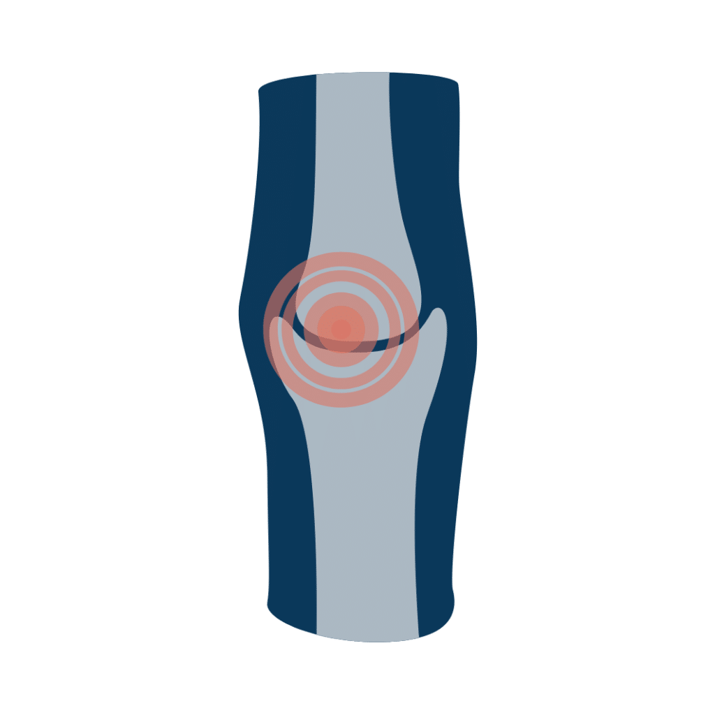 Image Source: Princeton Orthopaedic Associates
Natural Strategies for Managing Bone Spur Symptoms (Palliative Care)
While you can't eliminate the spur itself without medical intervention, you can significantly reduce its impact. These strategies fall under palliative care—they relieve symptoms rather than cure the underlying condition. A comprehensive approach combines systemic anti-inflammatory measures, targeted mechanical unloading, and lifestyle modifications that support long-term musculoskeletal resilience.
Anti-Inflammatory Diet and Nutrition
Inflammation is a primary driver of bone spur pain. Adopting an anti-inflammatory diet can help manage this at a systemic level.
- Omega-3 Fatty Acids: Found in fatty fish (salmon, mackerel), flaxseeds, and walnuts, omega-3s are well-known for their potent anti-inflammatory effects.
- Fruits and Vegetables: Berries, leafy greens (like spinach and kale), and broccoli are packed with antioxidants that fight inflammation.
- Healthy Spices: Turmeric (containing curcumin) and ginger have strong anti-inflammatory properties.
- Maintain a Healthy Weight: Excess weight puts more stress on your joints, particularly in the knees, hips, and feet. Maintaining a healthy weight can significantly reduce this pressure and alleviate pain.
Beyond simply adding beneficial foods, it is equally important to eliminate or drastically reduce dietary triggers that fuel the inflammatory cascade. Highly processed foods, refined carbohydrates, excessive added sugars, and industrial seed oils (high in omega-6 fatty acids) can disrupt the delicate balance between pro-inflammatory and anti-inflammatory mediators in the body. When this balance tips toward chronic inflammation, the synovial fluid surrounding your joints thickens, joint capsules stiffen, and pain signaling intensifies. Shifting toward a Mediterranean-style dietary pattern has consistently demonstrated clinical benefits for joint health, thanks to its high concentration of polyphenols, monounsaturated fats, and fiber-rich whole foods.
Hydration plays an underappreciated yet critical role in joint function and bone spur symptom management. Cartilage is approximately 70% to 80% water, and adequate systemic hydration ensures that synovial fluid remains viscous enough to properly lubricate articular surfaces and absorb mechanical shock. When dehydrated, friction increases within the joint, exacerbating irritation around any existing osteophytes. Additionally, the gut-joint axis is an area of growing research; a diverse, fiber-rich microbiome produces short-chain fatty acids like butyrate that help regulate systemic immune responses. Incorporating fermented foods such as sauerkraut, kimchi, kefir, and yogurt can support intestinal barrier integrity, potentially reducing the translocation of inflammatory endotoxins into the bloodstream that can worsen localized joint pain.
Meal timing and preparation methods also influence inflammation levels. Cooking with high-heat dry methods (like grilling or frying) can produce advanced glycation end products (AGEs), which are known to promote oxidative stress and stiffen connective tissues. Opting for gentler cooking techniques like steaming, poaching, slow-roasting, or braising helps preserve the delicate nutrient profiles of vegetables and proteins while minimizing AGE formation. Combining anti-inflammatory spices like black pepper with turmeric significantly enhances curcumin bioavailability, making natural compounds more effective at the cellular level. Consistency in your dietary approach is paramount; while a single anti-inflammatory meal will not dramatically alter joint pain, sustained nutritional habits create a biochemical environment where tissues heal more efficiently and pain pathways become less sensitized.
The Role of Vitamins and Minerals: Prevention vs. Reversal
There is a great deal of debate about the role of supplements in managing health conditions. For bone spurs, the evidence is clear: no vitamin or mineral can reverse or dissolve an existing spur. However, certain nutrients are vital for maintaining strong bones and healthy cartilage, which can help manage symptoms and prevent further joint degeneration.
- Vitamin D: Essential for absorbing calcium. One study suggested that Vitamin D could help with the resorption of abnormal bone formation, potentially alleviating symptoms. [Source: NIH]
- Vitamin K: Helps maintain cartilage integrity and may help prevent the formation of spurs.
- Calcium: The fundamental building block of bone.
- Magnesium: Important for cartilage function and muscle health. Soaking in an Epsom salt bath is a popular home remedy for delivering magnesium topically and relieving pain.
- Vitamin C: Plays a crucial role in collagen production, which is essential for healthy joints and bones.
Understanding the intricate synergy between these micronutrients is essential for maximizing their therapeutic potential. For instance, Vitamin D3 alone cannot properly direct calcium deposition. Without adequate Vitamin K2, calcium may accumulate in soft tissues, arteries, or around joint margins rather than being efficiently integrated into healthy bone matrix. The D3+K2 partnership ensures proper calcium trafficking, which is particularly relevant for individuals managing osteophyte-related discomfort. Furthermore, magnesium acts as a natural calcium channel blocker, helping to prevent excessive muscle tension and spasms that frequently accompany bone spurs. When muscles remain chronically contracted due to pain or nerve irritation, they pull unevenly on tendon attachments, perpetuating the cycle of mechanical stress that stimulates further osteophyte growth.
The bioavailability and formulation of your supplements matter significantly. Not all magnesium is created equal; magnesium oxide has poor intestinal absorption and is primarily used as a laxative, whereas magnesium glycinate or magnesium malate are highly bioavailable and better suited for musculoskeletal relaxation. Similarly, the form of turmeric or ginger extract, the source of fish oil, and the presence of necessary cofactors can dictate clinical outcomes. Blood testing is highly recommended before embarking on a high-dose supplementation protocol, as both deficiencies and toxicities can exacerbate joint issues. Liver and kidney function should also be evaluated, as these organs are responsible for metabolizing concentrated nutrients.
Beyond core vitamins, other compounds show promise in supporting joint tissue resilience. Collagen peptides, particularly types II and X, provide the amino acid building blocks necessary for cartilage repair and may help cushion joints against friction from bony projections. Hyaluronic acid supplementation supports synovial fluid volume, though oral bioavailability remains debated compared to intra-articular injections. Methylsulfonylmethane (MSM) and boswellia serrata (frankincense) have gained traction in complementary medicine for their ability to downregulate inflammatory cytokines like TNF-alpha and IL-1beta. While none of these will shrink an osteophyte, they can create a more forgiving biomechanical environment, allowing patients to engage in rehabilitative exercises with less discomfort.
Before starting any supplement regimen, it is best to consult with your doctor.
Physical Approaches and Home Remedies
Directly addressing the affected area with physical methods can provide significant and often immediate relief.
- Cold Compress: Applying an ice pack or a bag of frozen vegetables (wrapped in a towel) to the affected area for 10-15 minutes can reduce swelling and numb the pain. This is especially effective after activity. [Source: Medical News Today]
- Exercise and Stretching: Gentle, low-impact exercises like swimming or walking strengthen the muscles around the joint, providing better support and reducing stress. Targeted stretching can improve flexibility and relieve pressure on nerves.
- Supportive Footwear and Orthotics: For bone spurs in the feet (heel spurs), wearing well-fitting, cushioned shoes is critical. Custom or over-the-counter orthotic inserts can also help correct alignment and relieve pressure.
- Topical Applications: Massaging the area with a carrier oil (like coconut or jojoba oil) mixed with essential oils known for their soothing properties, such as lavender or eucalyptus, may provide temporary pain relief.
To maximize the efficacy of physical interventions, it is helpful to employ contrast therapy strategically. Alternating between cryotherapy (cold) and thermotherapy (heat) can dramatically improve local circulation while controlling acute inflammation. Cold therapy constricts blood vessels, limiting edema and numbing overactive pain receptors, making it ideal for post-activity flare-ups or acute exacerbations. Heat therapy, typically applied via a moist heating pad or warm paraffin wax bath, dilates vessels, increases tissue elasticity, and relaxes hypertonic muscles guarding the joint. Applying heat for 15–20 minutes before stretching or gentle movement prepares the connective tissue for elongation, reducing the risk of microtearing and making rehabilitative exercises more comfortable.
Targeted stretching and mobility work must be joint-specific to avoid aggravating the condition. For calcaneal (heel) spurs associated with plantar fasciitis, the classic towel stretch, calf drops on a step, and rolling the arch over a frozen water bottle can decompress the plantar fascia and reduce tension at the calcaneal insertion. Spinal osteophytes often require a different approach, focusing on neural gliding, gentle lumbar rotations, and cat-cow mobilization to maintain segmental motion without compressing the affected nerve roots. Aquatic therapy remains one of the gold standards for multisite bone spur pain; the buoyancy of water offloads up to 90% of body weight, allowing for full-range strengthening movements in a virtually impact-free environment. Resistance band training can further stabilize joints by targeting the smaller, often-neglected stabilizer muscles that work in tandem with larger prime movers.
Proper footwear and ergonomic modifications are non-negotiable components of conservative bone spur management. Shoes with excessive wear, poor arch support, or overly rigid soles alter kinetic chain mechanics, transmitting abnormal forces upward through the ankles, knees, and spine. When selecting footwear, look for a firm heel counter, adequate toe box room, and responsive midsoles that absorb ground reaction forces. For workplace-related joint stress, evaluating desk ergonomics, keyboard placement, and seating support can prevent repetitive microtrauma that fuels chronic inflammation. Additionally, sleep positioning significantly influences overnight tissue recovery. Using strategic pillow placement—such as a knee pillow for side sleepers or a small roll under the knees for back sleepers—maintains spinal neutrality and reduces tension on posterior structures. Incorporating diaphragmatic breathing and progressive muscle relaxation before bed can lower systemic cortisol levels, further dampening inflammatory responses and improving sleep architecture, which is when the majority of musculoskeletal repair occurs.
When Natural Remedies Aren't Enough: Medical Treatments
If pain persists or worsens despite your best efforts with natural remedies, it's time to consult a healthcare professional. A proper diagnosis is key to ensuring you're treating the right condition. A doctor may recommend:
- Over-the-Counter (OTC) Pain Relievers: Nonsteroidal anti-inflammatory drugs (NSAIDs) like ibuprofen (Advil) or naproxen sodium (Aleve) can help manage pain and inflammation.
- Physical Therapy: A physical therapist can design a specific program of exercises and stretches to improve strength and mobility in the affected joint.
- Corticosteroid Injections: For severe inflammation, a doctor can inject a powerful anti-inflammatory medication directly into the affected area for significant, though often temporary, relief.
- Surgery: In rare cases where a bone spur severely limits mobility or causes intense pain that doesn't respond to other treatments, surgery to remove the spur may be considered.
When transitioning to clinical interventions, it is important to understand the mechanisms, timelines, and limitations of each modality. Oral NSAIDs function by inhibiting cyclooxygenase (COX) enzymes, which are responsible for producing prostaglandins—lipid compounds that trigger pain, fever, and swelling. While effective for short-term symptom control, prolonged daily use requires gastrointestinal, renal, and cardiovascular monitoring due to known side effect profiles. A healthcare provider may recommend topical NSAID gels as a safer alternative, delivering targeted relief with minimal systemic absorption.
Physical therapy offers a structured, evidence-based pathway to functional restoration. A licensed therapist will conduct a comprehensive gait and biomechanical analysis to identify faulty movement patterns contributing to joint overload. Treatment typically progresses through phases: pain modulation, mobility restoration, neuromuscular re-education, and eventual return to sport or high-demand activities. Modalities such as therapeutic ultrasound, electrical nerve stimulation (TENS), and instrument-assisted soft tissue mobilization (Graston technique) may be utilized to break up fascial adhesions and desensitize hyper-reactive nerve pathways surrounding the osteophyte.
Injections represent a middle ground between conservative care and operative management. Corticosteroid injections deliver potent anti-inflammatory agents directly to the joint capsule or periosteal region, often providing weeks to months of relief that can be strategically used to enable aggressive physical therapy participation. For osteoarthritis-related spurs, hyaluronic acid (viscosupplementation) injections may be considered to supplement depleted synovial fluid, improving lubrication and shock absorption. Emerging biologic treatments like platelet-rich plasma (PRP) therapy harness the patient's own growth factors to modulate the inflammatory environment and promote tissue healing, though insurance coverage and clinical consensus on long-term efficacy continue to evolve.
Surgical intervention is universally regarded as a last resort, reserved for cases involving progressive neurological deficits, severe mechanical locking, or debilitating pain that profoundly impairs quality of life. The specific procedure depends on anatomical location: microdiscectomy or laminectomy for spinal nerve compression, arthroscopic debridement for knee or shoulder impingement, and plantar fascia release with concomitant spur excision for chronic heel pathology. Recovery protocols vary widely, but all require strict adherence to postoperative rehabilitation to prevent recurrence and optimize tissue remodeling. Modern surgical techniques, including minimally invasive and robotic-assisted approaches, have significantly reduced tissue trauma, hospital stays, and complication rates. Recognizing the warning signs that necessitate prompt medical evaluation—such as sudden weakness, bowel or bladder dysfunction, unrelenting night pain, or rapid swelling—ensures timely intervention and prevents irreversible structural damage.
The Bottom Line
While you cannot "dissolve" a bone spur naturally, you have significant power to manage its symptoms and live comfortably. By focusing on an anti-inflammatory lifestyle, maintaining joint health through proper nutrition and gentle exercise, and using targeted home remedies, you can effectively reduce pain and inflammation. Always partner with a healthcare provider to create a safe and effective management plan tailored to your specific needs.
Long-term success hinges on adopting a proactive rather than reactive mindset toward musculoskeletal health. Tracking your symptoms, identifying personal triggers, and maintaining consistency with your therapeutic routines will yield compounding benefits over time. Pain management is rarely linear; there will be good days and flare-ups. Understanding that fluctuations are normal allows you to adjust your strategies without abandoning them entirely. Integrating mindfulness practices, maintaining social connections, and focusing on functional goals rather than perfection can improve psychological resilience, which directly correlates with improved pain tolerance and recovery outcomes. Your body is remarkably adaptable, and with a comprehensive, multidisciplinary approach grounded in scientific evidence and compassionate self-care, you can navigate the challenges of bone spurs while preserving an active, fulfilling lifestyle.
References
- Bone spurs: What are some natural treatments to dissolve them? - Medical News Today
- Bone Spur (Osteophyte): Causes, Symptoms & Treatment - Cleveland Clinic
- Understanding Bone Spurs: Symptoms, Causes, and Treatments - Princeton Orthopaedic Associates
- Effects of vitamin D with or without calcium on pathological ossification - National Institutes of Health (NIH)
- How To Dissolve Bone Spurs Naturally - Cellaxys
Frequently Asked Questions
Can apple cider vinegar or other acidic home remedies truly dissolve bone spurs?
No, apple cider vinegar and other acidic substances cannot dissolve bone spurs. While popular in alternative health circles, the human digestive and metabolic systems strictly regulate blood pH levels, keeping them tightly balanced between 7.35 and 7.45. Ingested acids do not circulate systemically to break down calcified tissue, and bone is primarily composed of hydroxyapatite crystals, which are highly resistant to dietary acids. Furthermore, excessive consumption of undiluted acidic liquids can erode tooth enamel and irritate the esophageal and gastric linings. While moderate amounts of apple cider vinegar may offer minor digestive benefits or blood sugar modulation, it should not be relied upon for osteophyte reduction. Focusing on proven anti-inflammatory strategies and mechanical unloading will yield far superior clinical outcomes.
How long does it typically take for natural symptom management strategies to produce noticeable relief?
The timeline for symptom improvement varies significantly based on the size and location of the osteophyte, the severity of surrounding inflammation, patient adherence, and underlying metabolic factors. Generally, most patients begin experiencing modest reductions in stiffness and pain within 4 to 6 weeks of consistently implementing dietary modifications, targeted stretching, and proper biomechanical support. Significant functional improvements and sustained pain relief typically require 3 to 6 months of dedicated effort, as connective tissue turnover, muscle strengthening, and systemic inflammation reduction operate on biological cycles that cannot be safely rushed. Tracking progress through a symptom journal, utilizing standardized pain scales, and monitoring range-of-motion metrics can help you objectively evaluate whether your current protocol is effective or requires clinical adjustment.
Are bone spurs the same as plantar fasciitis, and can stretching fix both?
Bone spurs and plantar fasciitis are distinct but frequently interconnected conditions. Plantar fasciitis is an inflammatory condition affecting the thick band of connective tissue that runs along the bottom of the foot, connecting the heel bone to the toes. Chronic traction from an inflamed, tight plantar fascia repeatedly pulls at its calcaneal insertion point, which can eventually stimulate the formation of a heel spur over months or years. While the spur itself is usually not the primary pain generator, the underlying fascial pathology is. Targeted stretching of the calf complex, intrinsic foot strengthening, arch support, and load management are highly effective for resolving plantar fasciitis symptoms. Addressing the soft tissue dysfunction often alleviates the mechanical stress that perpetuates spur-related discomfort, making stretching a cornerstone of conservative treatment for both conditions.
What specific types of exercise or movement should I avoid if I have symptomatic bone spurs?
High-impact, repetitive, and end-range loading activities should be approached with caution or temporarily modified. Exercises that generate significant joint compression or shear forces—such as heavy squatting with improper form, deep plyometric jumps, long-distance running on hard surfaces, and high-velocity sports involving rapid directional changes—can exacerbate inflammation around the spur. Additionally, aggressive passive stretching that forces a joint beyond its comfortable range of motion may irritate compressed nerves or inflamed bursae. Instead of complete rest, which can lead to joint stiffness and muscle atrophy, opt for controlled, low-velocity resistance training within a pain-free range. Focus on tempo control, eccentric strengthening, and proprioceptive balance work. If a specific movement consistently triggers sharp, shooting, or electric-like pain, discontinue that motion and consult a physical therapist to identify safer biomechanical alternatives that maintain fitness without aggravating the affected structure.
Can untreated bone spurs cause permanent nerve damage or require emergency intervention?
Yes, in specific anatomical locations, progressive osteophyte growth can lead to nerve root compression or vascular compromise that may result in lasting neurological deficits if left unaddressed. Cervical and lumbar spinal spurs that narrow the neural foramina or spinal canal can cause radiculopathy (radiating arm or leg pain), sensory deficits, muscle weakness, and in severe cases, myelopathy characterized by gait instability, loss of fine motor skills, or bowel/bladder dysfunction. Peripheral spurs around joints may occasionally entrap nerves, leading to conditions similar to carpal or tarsal tunnel syndrome. Emergency medical evaluation is warranted if you experience sudden, unexplained weakness, numbness that does not resolve, loss of coordination, severe unrelenting night pain, or signs of compromised blood flow (pallor, coolness, or diminished pulses in the affected limb). Early recognition and timely intervention, whether through specialized physical therapy, injections, or surgical decompression, are critical for preventing irreversible tissue damage and preserving long-term neurological function.

About the author
Samuel Jones, MD, is a board-certified orthopedic surgeon specializing in joint replacement and orthopedic trauma. He is a team physician for a professional sports team and practices at a renowned orthopedic institute in Georgia.