Desiccated Disc: A Comprehensive Guide to Causes, Symptoms, and Treatment

Key points
- Nucleus Pulposus: A soft, gel-like center with high water content that provides cushioning. This core is rich in type II collagen and large proteoglycan molecules called aggrecans, which act like molecular sponges. Through osmotic pressure, these molecules draw and bind water, maintaining disc turgor and resilience against compressive forces throughout the day.
- Annulus Fibrosus: A tough, fibrous outer ring that protects the nucleus. It consists of concentric layers (lamellae) of type I collagen fibers arranged in alternating diagonal directions, creating a highly durable, load-bearing structure that contains the pressurized nucleus.
If you've received a medical report mentioning a "desiccated disc," you might be wondering what it means for your health. A desiccated disc is a common condition where the spongy cushions between your vertebrae lose moisture, leading to a variety of symptoms from mild stiffness to significant pain. In clinical practice, this finding is exceptionally common on MRI scans, particularly in adults over the age of 40. While the terminology can sound alarming, disc desiccation is fundamentally a structural change that reflects the natural wear and tear of the spinal column. However, when combined with poor biomechanics, genetic predisposition, or metabolic stressors, it can accelerate into a symptomatic condition that requires targeted intervention. Understanding the underlying mechanisms, recognizing early warning signs, and implementing evidence-based management strategies can dramatically alter your long-term spinal trajectory.
This guide synthesizes expert knowledge from top medical sources to provide a comprehensive overview of disc desiccation, covering everything from its underlying causes and symptoms to the most effective treatment strategies available today.
What is a Desiccated Disc?
Your spine is composed of 33 vertebrae stacked on top of one another. Between most of these bones are tough, spongy intervertebral discs that act as shock absorbers, allowing for flexibility and preventing the bones from grinding together. These discs are avascular structures, meaning they lack a direct blood supply in adulthood. Instead, they rely on a passive diffusion process called imbibition to receive nutrients and oxygen from surrounding capillary beds in the vertebral endplates. This physiological quirk makes them highly vulnerable to dehydration and nutrient deprivation over time.
Each disc has two main parts:
- Nucleus Pulposus: A soft, gel-like center with high water content that provides cushioning. This core is rich in type II collagen and large proteoglycan molecules called aggrecans, which act like molecular sponges. Through osmotic pressure, these molecules draw and bind water, maintaining disc turgor and resilience against compressive forces throughout the day.
- Annulus Fibrosus: A tough, fibrous outer ring that protects the nucleus. It consists of concentric layers (lamellae) of type I collagen fibers arranged in alternating diagonal directions, creating a highly durable, load-bearing structure that contains the pressurized nucleus.
Disc desiccation is the medical term for the dehydration of these discs. As you age, the nucleus pulposus naturally loses its fluid. This occurs as proteoglycans degrade and are replaced by less efficient fibrocartilage, diminishing the disc's ability to retain water through osmotic gradients. This causes the disc to become flatter, harder, and less effective at absorbing shock. Consequently, load distribution shifts abnormally to the posterior facet joints, accelerating osteoarthritis and altering spinal biomechanics. This process is a key feature of a broader condition known as Degenerative Disc Disease (DDD).
 Image Source: Advanced Spine Center
Clinically, desiccation is not merely a localized phenomenon; it initiates a kinetic chain reaction throughout the spine. As the disc loses height, the intervertebral foramina (the openings where spinal nerves exit) can narrow, increasing the likelihood of nerve root impingement. Furthermore, the altered load mechanics force the facet joints, ligaments, and paraspinal muscles to compensate, often leading to myofascial trigger points, muscle spasms, and chronic tension. Understanding this cascade is essential for both patients and clinicians to appreciate why a seemingly isolated finding on an MRI can manifest as widespread musculoskeletal dysfunction.
Symptoms: How Does a Desiccated Disc Feel?
While many people with disc desiccation experience no symptoms, others may face discomfort that ranges from mild to severe. The symptoms often depend on the location and severity of the condition. Importantly, symptom severity does not always correlate directly with the degree of desiccation visible on imaging. Many individuals have advanced desiccation with minimal pain due to successful neural adaptation and stable spinal alignment, while others experience disproportionate pain from mild degeneration due to heightened central sensitization or concurrent inflammatory conditions.
Common signs of a desiccated disc include:
- Pain and Stiffness: This is the most common symptom, localized to the back or neck. Pain may worsen with certain movements like bending or twisting. Patients often report a deep, aching discomfort that is typically mechanical in nature, meaning it fluctuates with posture, activity levels, and rest cycles. Morning stiffness lasting less than 30 minutes is common, as discs gradually rehydrate during sleep but quickly lose height under gravity upon waking.
- Radiating Pain: If the flattened disc presses on a nerve, pain can travel down the arms or legs (a condition known as sciatica if it affects the sciatic nerve). This radicular pain often follows specific dermatomal patterns. For example, L4-L5 involvement typically radiates down the lateral thigh and anterior shin, while L5-S1 issues affect the posterior calf and lateral foot. Cervical desiccation may cause pain that radiates into the shoulder blades or down the medial arm.
- Weakness: Muscles in the arms or legs may feel weak. Chronic nerve compression can lead to motor deficits, manifesting as foot drop, difficulty rising from a chair, or reduced grip strength. This requires prompt clinical evaluation to prevent permanent neurological compromise.
- Numbness or Tingling: A "pins-and-needles" sensation can occur in the extremities. Paresthesia indicates sensory nerve involvement and may be intermittent or constant. In advanced cases, patients report diminished proprioception, making balance and fine motor coordination challenging.
- Reduced Range of Motion: Difficulty moving or bending your spine as you normally would. The loss of disc height restricts flexion, extension, and rotational capacity. Protective muscle guarding further limits mobility as the neuromuscular system attempts to splint the affected spinal segment.
The location of the desiccated disc determines where symptoms are felt. Cervical (neck) desiccation can cause neck pain and headaches, particularly cervicogenic headaches originating from irritated upper cervical nerves and facet joints. Patients may also experience occipital tenderness or visual disturbances linked to muscle tension. Thoracic disc desiccation is rare due to the stabilizing rib cage but can cause mid-back pain and, in severe cases, band-like pain around the ribcage that mimics visceral pathology. Lumbar (lower back) desiccation is a common source of lower back pain, frequently accompanied by pelvic tilt compensations and altered gait mechanics that strain the sacroiliac joints and gluteal musculature.
The Stages of Disc Degeneration and Desiccation
Disc desiccation doesn't happen overnight. It's a progressive condition that experts often describe in four stages. Understanding these stages helps patients and clinicians track disease progression, anticipate complications, and tailor interventions appropriately. The timeline varies significantly based on individual factors, but the biomechanical cascade generally follows a predictable pattern.
- Stage 1: Dysfunction and Dehydration: The disc begins to lose its ability to retain water. Small microtears may form in the outer annulus fibrosus due to repetitive microtrauma and biochemical breakdown. Inflammatory mediators like prostaglandins and cytokines accumulate, irritating nociceptive nerve endings embedded in the outer third of the annulus. You might experience mild, occasional back pain after prolonged sitting, heavy exertion, or awkward lifting. At this stage, lifestyle interventions and targeted physical therapy are highly effective at halting progression.
- Stage 2: Dehydration and Prolapse: The disc continues to dry out and lose height, potentially causing it to bulge (prolapse). The annulus weakens circumferentially, allowing the nucleus to push outward against the compromised outer layers without fully rupturing. This alters spinal kinematics, leading to facet joint impingement and segmental instability. The reduced disc space can cause mild nerve root irritation, leading to more persistent pain, intermittent stiffness, and early signs of radicular symptoms. Patients often notice pain that worsens toward the end of the day as gravity compresses the already compromised disc.
- Stage 3: Stabilization and Herniation: In this advanced stage, the annulus can tear completely, allowing the nucleus to leak out—a herniated disc. The extruded nuclear material may directly compress neural elements, causing acute radiculopathy. Simultaneously, the body recognizes the segmental instability and attempts to stabilize the area by forming bone spurs (osteophytes) along the vertebral margins. While osteophytes represent an adaptive response, they can further narrow the spinal canal (spinal stenosis) and intervertebral foramina, compounding nerve compression. Muscle guarding becomes chronic, and compensatory postural patterns often lead to secondary myofascial pain syndromes.
- Stage 4: Severe Degeneration and Collapse: The disc loses a significant amount of its height and may collapse entirely. The vertebral endplates become sclerotic, and the intervertebral space narrows dramatically. This can lead to bone-on-bone friction, causing chronic, debilitating pain and severely limited mobility. The facet joints hypertrophy significantly, and the ligamentum flavum thickens and buckles inward. Patients often develop a characteristic antalgic posture, altered center of gravity, and marked reduction in functional capacity. Surgical consultation may be necessary if conservative management fails to restore basic mobility or if neurological deficits progress.
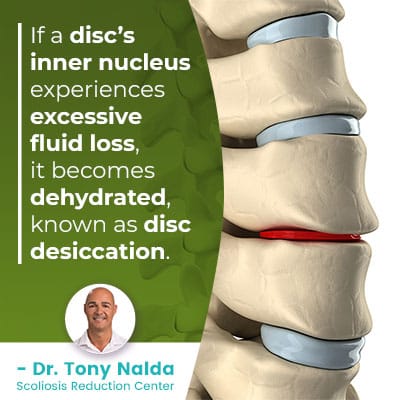 Image Source: Dr. Tony Nalda
It is crucial to recognize that progression through these stages is not inevitable. Many individuals remain in Stage 1 or 2 for decades with proper spinal hygiene, targeted exercise, and metabolic health management. Conversely, aggressive loading, poor nutrition, or untreated mechanical imbalances can accelerate the timeline. Early intervention remains the cornerstone of preserving spinal function and quality of life.
What Causes a Disc to Become Desiccated?
While aging is the primary driver of disc desiccation, several other factors can accelerate the process. The etiology is fundamentally multifactorial, involving a complex interplay between biological aging, mechanical stress, systemic health, and environmental exposures.
- Natural Aging: This is the most common cause. As we get older, our discs naturally lose water content and elasticity. Cellular senescence in the annulus and nucleus reduces proteoglycan synthesis, while matrix metalloproteinases (MMPs) increasingly degrade existing structural proteins. Additionally, capillary networks supplying the vertebral endplates gradually sclerose, impairing nutrient diffusion.
- Repetitive Strain: Jobs or activities involving heavy lifting, frequent bending, or twisting can place excessive wear and tear on the spine. Occupations requiring prolonged static postures (such as truck driving, office work, or assembly line jobs) are particularly detrimental. Static loading reduces intradiscal pressure fluctuations, starving the disc of the pumping mechanism required for nutrient exchange. Dynamic movements under heavy loads increase shear forces that can cause annular microtears.
- Trauma or Injury: A sudden injury from a fall, car accident, or sports can damage a disc and trigger degeneration. Acute trauma disrupts the delicate balance of collagen fibers, initiates localized inflammation, and can compromise endplate integrity. Even if immediate symptoms resolve, the structural vulnerability often leads to accelerated desiccation in subsequent years.
- Lifestyle Factors:
- Obesity: Excess weight puts added pressure on the spine, particularly the lumbar region. Visceral adiposity also creates a pro-inflammatory state, releasing adipokines like leptin and resistin that can penetrate disc tissue and accelerate cellular breakdown.
- Smoking: Nicotine restricts blood flow to the discs, impairing their ability to receive nutrients and stay hydrated. Carbon monoxide reduces oxygen delivery, while other toxins in cigarette smoke directly inhibit proteoglycan synthesis and collagen cross-linking. Smokers are significantly more likely to develop premature DDD.
- Poor Posture: Prolonged slouching or poor ergonomics can create uneven pressure on your discs. Forward head posture increases cervical disc pressure by up to 12 pounds for every inch the head translates forward. Similarly, lumbar flexion under load dramatically increases intradiscal pressure compared to neutral spine alignment.
- Dehydration and Malnutrition: Not drinking enough water or having a poor diet can affect overall tissue health, including your discs. Discs require adequate hydration to maintain osmotic gradients. Chronic systemic dehydration reduces the water available for imbibition, while deficiencies in essential micronutrients impair matrix repair.
- Genetics: Some people may be genetically predisposed to developing degenerative disc disease earlier in life. Polymorphisms in genes encoding collagen types (COL9A2, COL11A2), vitamin D receptors, and inflammatory cytokine regulators (IL-1, TNF-alpha) significantly influence disc resilience and repair capacity. Family history is one of the strongest non-modifiable risk factors for early-onset desiccation.
Additionally, metabolic conditions such as type 2 diabetes mellitus profoundly impact disc health. Advanced glycation end-products (AGEs) accumulate in diabetic patients, causing collagen fibers to become brittle and cross-linked. This reduces disc flexibility and makes the annulus more susceptible to tearing under normal physiological loads. Similarly, osteoporosis can lead to vertebral compression fractures that alter spinal alignment, indirectly accelerating adjacent segment desiccation.
Diagnosis: How Doctors Identify a Desiccated Disc
If you're experiencing persistent back or neck pain, a doctor will typically follow a standard process to diagnose the cause:
- Medical History and Physical Exam: Your doctor will ask about your symptoms, past injuries, and lifestyle. They will then perform a physical exam to test your range of motion, muscle strength, reflexes, and sensation to pinpoint the affected area. Clinicians utilize specific provocative tests such as the straight leg raise (SLR) for lumbar radiculopathy, Spurling's test for cervical radiculopathy, and dermatomal sensory mapping. They also assess for red flag symptoms including bowel/bladder dysfunction, saddle anesthesia, unexplained weight loss, fever, or history of malignancy, which would necessitate immediate advanced imaging or specialist referral.
- Imaging Tests: While a physical exam provides clues, imaging tests are needed to confirm disc desiccation. Radiographic findings must always be correlated with clinical symptoms to avoid over-treatment of incidental findings.
- X-ray: Can show a narrowing of the space between vertebrae, indicating a loss of disc height. Dynamic X-rays (flexion/extension views) may reveal segmental instability or abnormal translational movement. Osteophyte formation and vacuum disc phenomenon (nitrogen gas accumulation within a degenerated disc) are also visible.
- CT Scan: Provides a more detailed cross-sectional view of the spine. Excellent for evaluating bony anatomy, facet joint arthrosis, and calcified ligaments. CT myelography may be utilized when MRI is contraindicated or when assessing nerve root compression within the foramina.
- MRI Scan: This is the gold standard for diagnosing disc desiccation. An MRI can directly visualize the disc's water content, revealing the extent of dehydration. Healthy discs appear bright and white on an MRI (T2-weighted sequences), while desiccated discs look darker. Radiologists often use the Pfirrmann grading scale to classify disc degeneration severity from I (normal) to V (severe collapse/black disc). MRI also beautifully demonstrates soft tissue pathology including annular fissures, bulging, herniation, and nerve root compression.
In complex cases where neurological symptoms persist despite normal or ambiguous imaging, electromyography (EMG) and nerve conduction velocity (NCV) studies may be ordered to objectively assess nerve function and differentiate radiculopathy from peripheral neuropathy or radiculoplexus neuropathy. Blood tests may also be indicated if systemic inflammatory or infectious etiologies are suspected. A multidisciplinary diagnostic approach ensures that treatment targets the actual pain generator rather than incidental radiographic findings.
Treatment Options: Managing a Desiccated Disc
A crucial point to understand is that disc desiccation cannot be reversed. Once a disc has lost its fluid, it cannot be rehydrated. Therefore, treatment focuses on managing pain, improving function, and preventing further degeneration. The overarching goal is to establish spinal stability, optimize neuromuscular control, reduce inflammatory load, and empower patients with self-management tools that sustain long-term functional independence.
Conservative Treatments: The First Line of Defense
For most patients, non-surgical treatments are highly effective. A conservative-first approach is universally recommended. Evidence consistently demonstrates that structured rehabilitation outperforms passive modalities and surgical intervention for the vast majority of degenerative spinal conditions.
- Physical Therapy: This is a cornerstone of treatment. A physical therapist can design a program to strengthen your core and back muscles, improve flexibility, and teach you proper posture and body mechanics. Evidence-based protocols emphasize deep core stabilization (transverse abdominis, multifidus activation), hip hinge mechanics, and thoracic mobility work. The McKenzie Method (Mechanical Diagnosis and Therapy) is frequently utilized to centralize radiating pain and restore pain-free ranges of motion. Manual therapy, including joint mobilization and soft tissue release, addresses secondary myofascial restrictions and improves segmental mobility.
- Pain Medication: Over-the-counter nonsteroidal anti-inflammatory drugs (NSAIDs) like ibuprofen (Advil) and naproxen (Aleve) can help reduce pain and inflammation. Topical NSAIDs or diclofenac gels offer localized relief with minimal systemic absorption. In some cases, a doctor may prescribe stronger medication or muscle relaxants. For neuropathic pain components, gabapentinoids (gabapentin, pregabalin) or serotonin-norepinephrine reuptake inhibitors (SNRIs like duloxetine) may be indicated to modulate central pain processing and reduce nerve hypersensitivity.
- Lifestyle Modifications:
- Maintain a Healthy Weight: Reducing excess weight lessens the load on your spine. Even modest weight loss significantly decreases compressive forces on lumbar discs and reduces systemic inflammatory markers.
- Low-Impact Exercise: Activities like walking, swimming, and yoga can strengthen muscles without jarring the spine. Aquatic therapy is particularly beneficial for older adults or those with acute pain, as buoyancy offloads 70-90% of body weight while allowing resistance training.
- Quit Smoking: This improves blood flow and nutrient delivery to the discs. Smoking cessation also enhances bone healing and reduces postoperative complication rates if surgery eventually becomes necessary.
- Stay Hydrated: Drinking plenty of water is essential for overall tissue health. Establishing a consistent hydration routine supports systemic perfusion and optimizes the remaining disc tissue's osmotic capacity.
- Adjunctive Therapies: Heat and cold therapy, massage, and Transcutaneous Electrical Nerve Stimulation (TENS) units can provide additional symptom relief. Ergonomic assessments for workstations and sleeping environments help maintain neutral spinal alignment during prolonged activities. Lumbar supports or cervical collars may be used temporarily during acute flares but should be avoided long-term to prevent muscular atrophy. Cognitive-behavioral strategies and pain neuroscience education are increasingly integrated to address fear-avoidance behaviors, catastrophizing, and chronic pain centralization.
When Conservative Treatments Aren't Enough
If pain persists after several weeks or months of conservative care, your doctor may suggest more advanced options. Interventional pain management bridges the gap between rehabilitation and surgery, offering targeted relief while preserving anatomical integrity.
- Spinal Injections: Corticosteroid injections delivered into the epidural space around the spinal nerves can provide powerful, short-term relief from inflammation and pain. Transforaminal, interlaminar, and caudal approaches allow precise medication delivery to affected levels. Facet joint injections and medial branch blocks address pain originating from degenerated posterior joints. When performed under fluoroscopic guidance, diagnostic injections can definitively identify pain generators and predict surgical outcomes.
- Surgical Interventions: Surgery is considered a last resort, reserved for cases with severe, debilitating pain, progressive neurological symptoms (like worsening weakness), or signs of spinal instability. Common procedures include:
- Spinal Fusion: The surgeon permanently joins two or more vertebrae to eliminate painful motion. Techniques include anterior lumbar interbody fusion (ALIF), transforaminal lumbar interbody fusion (TLIF), and posterior lateral fusion using cages, bone grafts, and instrumentation. Fusion successfully addresses instability but increases stress on adjacent segments over time.
- Artificial Disc Replacement: The damaged disc is replaced with a prosthetic device designed to preserve motion. Primarily indicated for single-level pathology in younger patients with preserved facet joints, ADR maintains physiological kinematics and reduces adjacent segment disease risk compared to fusion.
- Decompression (Laminectomy or Discectomy): Bone or disc material pressing on a nerve is removed to relieve pressure. Microdiscectomy remains highly effective for radicular pain caused by herniated fragments, while minimally invasive laminectomy addresses symptomatic stenosis. Recovery is typically faster with minimally invasive techniques, though rehabilitation remains critical for long-term success.
Emerging regenerative therapies, including platelet-rich plasma (PRP) injections, bone marrow aspirate concentrate (BMAC), and investigational stem cell therapies, are actively studied for their potential to modulate inflammation and stimulate limited tissue repair. While promising, these modalities currently lack robust, large-scale randomized controlled trial evidence and are generally considered experimental for routine clinical use outside specialized research protocols.
The Role of Diet in Managing Disc Health
While diet can't reverse desiccation, an anti-inflammatory eating plan can help manage symptoms associated with degenerative disc disease. Nutrition directly influences systemic inflammation, cellular repair capacity, and connective tissue resilience. Strategic dietary modifications serve as an adjunct to mechanical rehabilitation, optimizing the internal environment for pain reduction and functional preservation.
Foods to Avoid or Limit
- Sugary and Refined Foods: Sodas, pastries, and white bread can trigger inflammation. Rapid blood glucose spikes increase insulin and IGF-1 levels, promoting inflammatory cytokine release and accelerating matrix metalloproteinase activity that degrades disc collagen.
- Processed and Red Meats: Foods like bacon, sausage, and ham are linked to increased inflammatory markers. High omega-6 fatty acid content disrupts the omega-3 to omega-6 ratio, shifting cellular pathways toward pro-inflammatory eicosanoid production.
- Unhealthy Fats: Limit saturated and trans fats found in fried and processed foods. These lipid profiles contribute to endothelial dysfunction, impairing microcirculation to the vertebral endplates essential for disc nourishment.
- Dehydrating Beverages: Excessive alcohol and caffeine can contribute to dehydration. Diuretic effects reduce total body water availability, compromising the osmotic gradients required for disc imbibition. Alcohol also directly inhibits protein synthesis and impairs sleep architecture, crucial for tissue recovery.
Foods to Emphasize
Focus on a diet rich in whole foods like fruits, vegetables, whole grains, and lean proteins to help manage inflammation and support overall health. Prioritize omega-3 rich sources like wild-caught fatty salmon, sardines, flaxseeds, and walnuts to promote anti-inflammatory prostaglandin pathways. Colorful vegetables provide polyphenols and antioxidants that neutralize reactive oxygen species generated during chronic inflammation. Collagen-supporting nutrients are particularly relevant: vitamin C is essential for collagen cross-linking, copper supports elastin synthesis, and zinc facilitates tissue repair enzymes. Bone broth, while not a magic bullet, provides bioavailable amino acids like glycine and proline that serve as building blocks for connective tissue. Hydration strategies should include electrolyte-balanced fluids rather than plain water alone, as proper sodium and potassium balance ensures cellular fluid retention and optimal tissue hydration.
Emerging research highlights the gut-spine axis, suggesting that microbiome health influences systemic inflammation and pain perception. Fermented foods, prebiotic fibers, and probiotic supplementation may indirectly benefit spinal health by modulating immune function and reducing circulating inflammatory markers. Consistency in dietary patterns matters more than isolated superfoods; adopting a Mediterranean-style eating framework has consistently demonstrated superior outcomes in managing chronic musculoskeletal pain.
Desiccated Disc vs. Herniated Disc: What's the Difference?
It's easy to confuse these terms, but they describe different things:
- A desiccated disc is a state—the disc is dehydrated, thin, and brittle. It's a sign of degeneration.
- A herniated disc (or "slipped disc") is an event—the soft inner gel pushes through a tear in the tough outer wall.
Think of it this way: disc desiccation weakens the disc, making it more prone to tearing and herniating. A desiccated disc is a risk factor for a herniated disc. Clinically, desiccation typically produces axial mechanical pain (localized to the spine) and stiffness, whereas herniation often generates acute radicular symptoms when nuclear material extrudes and chemically irritates or mechanically compresses adjacent nerve roots.
Management strategies overlap in their conservative foundations but diverge in urgency and specific interventions. Desiccated discs rarely require emergency intervention unless severe spinal stenosis develops. Herniated discs, however, can cause acute cauda equina syndrome (a surgical emergency characterized by bowel/bladder incontinence, saddle anesthesia, and profound motor weakness) that mandates immediate decompression. Furthermore, herniated discs frequently undergo spontaneous resorption over 6-12 weeks as the immune system recognizes extruded nuclear material as foreign and phagocytizes it. Desiccation does not resolve spontaneously; it represents permanent matrix alteration. Diagnostic imaging clearly differentiates the two: desiccation appears as low T2 signal intensity with height loss, while herniation shows focal extrusion beyond vertebral margins. Understanding this distinction prevents unnecessary anxiety and guides appropriate clinical decision-making.
Living with a desiccated disc can be challenging, but it is a manageable condition. By working with your healthcare provider to develop a comprehensive treatment plan that includes physical therapy, lifestyle changes, and appropriate pain management, you can significantly improve your quality of life and keep your spine as healthy as possible. Proactive engagement, consistent self-care practices, and regular clinical follow-up create a sustainable framework for maintaining spinal resilience throughout your lifespan.
Frequently Asked Questions
Can a desiccated disc heal itself or become rehydrated?
No, once the nucleus pulposus undergoes significant desiccation and the proteoglycan matrix degrades, the process is irreversible with current medical science. The avascular nature of adult intervertebral discs prevents substantial natural regeneration. While the body cannot fully rehydrate a black disc, targeted exercise, posture optimization, and load management can significantly reduce symptoms, improve function, and slow progression in adjacent segments. Some emerging research explores intradiscal biologic therapies, but these remain experimental.
Does disc desiccation always lead to surgery?
Absolutely not. The vast majority of patients with disc desiccation manage their condition successfully without surgery. Conservative treatments, including structured physical therapy, ergonomic modifications, activity pacing, and targeted pain management, effectively control symptoms for most individuals. Surgery is reserved strictly for cases with progressive neurological deficits, intractable pain unresponsive to exhaustive conservative care, or documented spinal instability that severely impairs quality of life.
How long does it take for symptoms to improve with conservative treatment?
Symptom improvement timelines vary based on severity, chronicity, and individual healing capacity. Acute flare-ups typically show meaningful improvement within 4 to 6 weeks of consistent physical therapy and lifestyle modification. Chronic conditions often require 3 to 6 months of dedicated rehabilitation to rebuild core stability, retrain movement patterns, and desensitize neural pathways. Patience and adherence to prescribed protocols are critical, as premature return to high-load activities frequently triggers relapse.
Are there any specific exercises I should avoid with a desiccated disc?
Yes, exercises that place excessive compressive or shear forces on the spine should be modified or avoided, particularly during acute phases. Heavy deadlifts, sit-ups, toe touches with straight legs, high-impact jumping activities, and deep rotational twisting under load can exacerbate symptoms and increase intradiscal pressure. Instead, focus on neutral-spine strengthening, hip-hinging mechanics with light resistance, swimming, stationary cycling, and controlled core stabilization exercises like bird-dogs, planks (modified if necessary), and glute bridges.
Can I still play sports or remain active with disc desiccation?
Absolutely. Physical activity is essential for maintaining spinal health, muscle tone, and joint mobility. Many athletes with desiccated discs continue to compete at high levels by modifying training volume, prioritizing recovery, and strengthening supportive musculature. Contact sports may require additional protective strategies and careful monitoring. Consult a physical therapist to develop a sport-specific conditioning program that maintains your functional goals while respecting spinal load tolerances. Consistent, graded activity is far superior to sedentary behavior, which accelerates stiffness and muscle atrophy.
Conclusion
A desiccated disc represents a common, age-related structural change characterized by the progressive loss of hydration and elastic properties within the intervertebral discs. While the terminology may sound concerning, it is crucial to recognize that disc desiccation is not a diagnosis of inevitable disability, but rather a physiological marker of spinal aging that often responds exceptionally well to conservative management. Understanding the biomechanical cascade—from proteoglycan degradation and height loss to altered facet joint loading and potential nerve compression—empowers patients to take an active role in their spinal health.
Successful long-term management hinges on a multifaceted approach: targeted physical therapy to restore core stability and movement efficiency, lifestyle modifications that reduce inflammatory load and mechanical stress, and strategic pain management during acute flare-ups. Imaging findings should always be interpreted in the context of clinical symptoms, as radiographic desiccation frequently exists asymptomatically. Early intervention, consistent self-care, and evidence-based rehabilitation remain the gold standards for preserving function and preventing progression to surgical candidacy. By adopting proactive spinal hygiene, maintaining a nutrient-dense anti-inflammatory diet, and prioritizing regular low-impact exercise, individuals can effectively manage desiccated discs and maintain an active, high-quality life. Always collaborate with qualified healthcare professionals to develop personalized treatment plans that address your unique anatomical, functional, and lifestyle needs.
References
- Roland, J. (2017). Disc Desiccation: Symptoms, Causes, and Treatment. Healthline. https://www.healthline.com/health/disc-desiccation
- Lanman, T. (2024). Disc Desiccation: Symptoms, Causes, and Treatment. Spine.MD. https://www.spine.md/insights/articles/disc-desiccation-symptoms-causes-treatment/
- Galan, N. (2018). Disc desiccation: Symptoms, causes, and treatments. Medical News Today. https://www.medicalnewstoday.com/articles/322121
- Donnally III, C.J., & Dulebohn, S.C. (2018). Lumbar Degenerative Disk Disease. Medscape. https://emedicine.medscape.com/article/309767-overview

About the author
Samuel Jones, MD, is a board-certified orthopedic surgeon specializing in joint replacement and orthopedic trauma. He is a team physician for a professional sports team and practices at a renowned orthopedic institute in Georgia.