White Spot Back of Throat: Causes, Treatments & When to See a Doctor

Noticing a white spot back of throat can trigger immediate concern, especially when accompanied by discomfort, difficulty swallowing, or persistent bad breath. The human throat is a complex, highly sensitive passageway that filters air, directs food, and serves as a primary defense point against inhaled or ingested pathogens. When white patches or lesions appear in the pharyngeal or tonsillar region, they typically represent an accumulation of cellular debris, mucus, dead tissue, or microbial colonies rather than a standalone disease. While many cases resolve with simple home care or resolve spontaneously, some indicate underlying infections, immune imbalances, or chronic inflammatory conditions that require professional evaluation. Understanding the precise origin of a white spot back of throat is essential for selecting appropriate interventions and preventing complications. Medical professionals consistently emphasize that accurate diagnosis precedes effective treatment, as the visual presentation alone rarely confirms a specific condition (Mayo Clinic: Sore Throat Symptoms & Causes). This comprehensive guide explores the physiological mechanisms behind throat discoloration, outlines evidence-based treatment pathways, and provides actionable self-care strategies to support rapid recovery while safeguarding long-term respiratory and oral health.
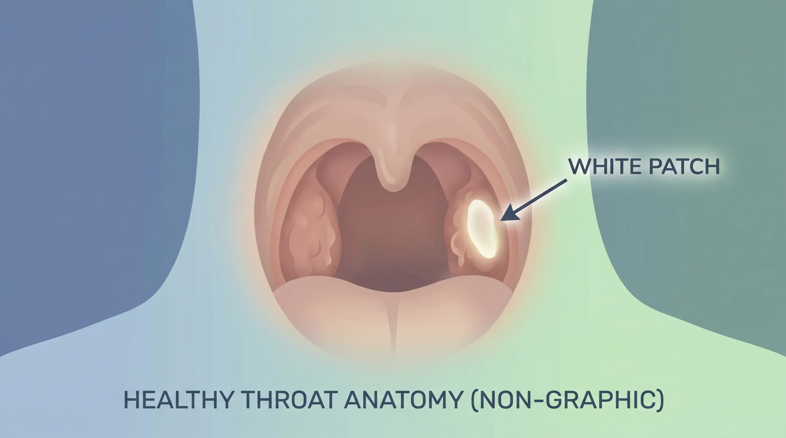
Understanding the Anatomy and Pathophysiology of Throat Discoloration
The back of the throat, medically referred to as the oropharynx, houses the palatine tonsils, lingual tonsils, and lymphoid follicles that collectively form Waldeyer’s ring. These tissues act as immunological sentinels, trapping bacteria, viruses, and environmental particulates before they penetrate deeper respiratory or digestive pathways (Cleveland Clinic: White Patches in the Throat). When immune cells engage with pathogens, they release inflammatory mediators that increase local blood flow, cause tissue swelling, and stimulate mucus production. In many instances, a white spot back of throat emerges when this immune response generates a visible exudate composed of leukocytes, fibrin, epithelial cells, and microbial byproducts. The tonsillar crypts, which are natural crevices designed to maximize surface area for antigen capture, can trap food particles, dead cells, and oral bacteria. Over time, calcified deposits known as tonsilloliths form within these crypts, presenting as pale, firm, or crumbly white nodules that often detach spontaneously or require gentle irrigation.
Beyond localized immune activity, systemic factors heavily influence mucosal integrity. Dehydration, chronic mouth breathing, and reduced salivary flow create an environment where opportunistic fungi like Candida albicans thrive. Saliva naturally contains antimicrobial enzymes and immunoglobulins that regulate oral microbiota; when production declines due to medications, stress, or underlying health conditions, microbial overgrowth can manifest as creamy white patches that may scrape off, leaving erythematous, bleeding tissue beneath (CDC: Thrush (Candidiasis)). Recognizing these foundational mechanisms clarifies why identical visual presentations can stem from vastly different etiologies. A white spot back of throat rarely exists in isolation. Clinical context matters significantly: symptom duration, pain severity, fever presence, and exposure history guide differential diagnosis. Patients who maintain optimal oral hygiene yet still develop recurrent white lesions may require metabolic screening or allergy testing to identify contributing triggers. By appreciating the intricate relationship between local tissue response and systemic wellness, individuals can make informed decisions about when observation suffices and when professional intervention becomes necessary.
Common Medical and Environmental Causes
Identifying the precise origin of a white spot back of throat requires evaluating both infectious and non-infectious pathways. Each cause follows distinct pathophysiological patterns, responds differently to interventions, and carries unique prognostic implications. Physicians routinely categorize these presentations to streamline diagnosis and minimize unnecessary treatments.
Viral and Bacterial Infections
Acute pharyngitis remains the most frequent culprit behind throat discoloration. Group A Streptococcus (GAS) causes streptococcal pharyngitis, characterized by sudden-onset sore throat, fever, swollen anterior cervical lymph nodes, and white or yellow exudate on the tonsils. Unlike common colds, strep throat typically lacks prominent cough or nasal congestion. Rapid antigen detection tests and throat cultures provide definitive diagnosis, guiding targeted antibiotic therapy (CDC: Strep Throat Overview). Viral etiologies, including Epstein-Barr virus (infectious mononucleosis), adenovirus, and influenza, produce similar white patches but often accompany profound fatigue, hepatosplenomegaly, or generalized lymphadenopathy. Infectious mononucleosis frequently presents with extensive tonsillar exudate, palatal petechiae, and prolonged symptom duration. Treatment remains supportive, emphasizing rest, hydration, and analgesia, as antibiotics prove ineffective against viral pathogens and may trigger adverse reactions when misprescribed for EBV-related conditions (Mayo Clinic: Mononucleosis).
Tonsil Stones (Tonsilloliths)
Chronic cryptic tonsils accumulate cellular debris, calcified salts, and anaerobic bacteria, forming tonsilloliths that appear as white or yellowish concretions on the posterior pharyngeal wall or tonsillar surface. These stones rarely cause systemic illness but frequently generate localized halitosis, foreign body sensation, and intermittent throat irritation. Risk factors include chronic tonsillitis, poor oral hygiene, dry mouth, and enlarged tonsillar crypts (Mayo Clinic: Tonsil Stones). While tonsil stones do not require immediate medical intervention unless causing recurrent infection or significant discomfort, conservative management proves highly effective. Regular saline gargling, gentle cotton swab manipulation, or water flossers can dislodge superficial stones. For patients experiencing chronic, debilitating tonsilloliths, tonsillectomy or laser cryptolysis offers definitive resolution.
Oral Thrush (Candidiasis)
Oral candidiasis arises when Candida species overgrow the mucosal lining, producing adherent white plaques that may extend from the posterior pharynx into the buccal cavity. Infants, elderly individuals, immunocompromised patients, and those using inhaled corticosteroids or broad-spectrum antibiotics face elevated risk. A white spot back of throat caused by thrush typically exhibits a cottage-cheese appearance, mild burning, and altered taste perception. Topical nystatin suspensions, clotrimazole troches, or systemic fluconazole resolve most cases within days. Addressing predisposing factors—such as rinsing the mouth after steroid inhaler use or managing uncontrolled diabetes—prevents recurrence.
Autoimmune and Chronic Inflammatory Conditions
Less commonly, persistent white lesions signal underlying autoimmune activity or premalignant changes. Oral lichen planus presents as bilateral, reticular white striae on the buccal mucosa, occasionally extending to the posterior pharynx. Leukoplakia develops as a localized white patch that cannot be scraped away, frequently associated with tobacco use, chronic friction, or HPV exposure (Mayo Clinic: Leukoplakia). Both conditions warrant specialist evaluation and possible biopsy to rule out dysplasia or malignant transformation. Maintaining strict tobacco cessation (WHO: Tobacco Fact Sheet) and scheduling regular oral cancer screenings significantly reduce progression risk for these chronic mucosal disorders.
Recognizing Accompanying Symptoms for Accurate Identification
Symptom clustering dramatically improves diagnostic precision when evaluating a white spot back of throat. Clinicians rely on associated signs to differentiate self-limiting viral illnesses from conditions requiring pharmacological intervention. Pain severity, fever patterns, lymphatic involvement, and systemic fatigue create distinct clinical signatures.
Acute bacterial pharyngitis typically produces sudden, severe odynophagia, high-grade fever exceeding 101°F (38.3°C), tender anterior cervical adenopathy, and palatal erythema without significant cough or rhinorrhea. Patients often report painful swallowing that restricts fluid intake, increasing dehydration risk. In contrast, viral pharyngitis frequently includes cough, nasal discharge, conjunctival injection, and lower-grade fever. Tonsillar exudate may appear patchy and resolve within 3–5 days as the immune system clears the pathogen.
Tonsilloliths generally lack systemic symptoms but cause persistent localized discomfort, halitosis unresponsive to brushing, and occasional mild dysphagia. Patients often discover the lesions accidentally while gargling or using dental floss. The white spot back of throat associated with tonsil stones feels firm rather than inflamed and rarely bleeds when manipulated.
Oral thrush introduces mucosal sensitivity, a metallic or cotton-like taste, and angular cheilitis in severe cases. Scraping the plaque reveals erythematous, occasionally bleeding mucosa beneath, distinguishing it from bacterial exudate that remains intact during gentle examination. Immunocompromised individuals may experience esophageal extension, presenting with substernal pain and progressive dysphagia requiring prompt systemic antifungal therapy.
Red flag symptoms demand immediate medical evaluation. Stridor, respiratory distress, trismus, muffled “hot potato” voice, drooling, neck rigidity, or rapidly expanding unilateral swelling suggest deep space infection, peritonsillar abscess, or epiglottitis. These emergencies bypass routine management and require urgent imaging, needle aspiration, or surgical drainage to secure the airway and prevent sepsis.
Diagnostic Process and Clinical Evaluation
Professional evaluation of a white spot back of throat follows a structured clinical pathway that balances efficiency with diagnostic accuracy. Physicians begin with comprehensive history taking, assessing symptom onset, duration, exposure history, medication use, smoking status, and prior episodes. Physical examination encompasses visualization of the oropharynx, palpation of cervical lymph nodes, inspection of the oral cavity, and assessment of respiratory effort.
When bacterial pharyngitis remains suspected, clinicians utilize validated scoring systems such as the Centor or McIsaac criteria to determine the necessity of rapid strep testing or throat culture (CDC: Clinical Diagnosis & Testing). These criteria evaluate fever, absence of cough, tonsillar exudate or swelling, tender anterior cervical lymphadenopathy, and patient age. A score of 4–5 strongly supports antibiotic therapy, while lower scores favor observation or viral testing.
Laboratory investigations extend beyond rapid antigen detection. Throat cultures remain the gold standard for identifying GAS and antibiotic susceptibility patterns. Complete blood counts may reveal leukocytosis with neutrophilic predominance in bacterial infections or atypical lymphocytosis in EBV. Monospot testing and EBV-specific serology confirm infectious mononucleosis. For persistent or atypical white lesions that fail to resolve within two weeks, referral to an otolaryngologist occurs. Laryngoscopy, mucosal biopsy, and viral PCR panels rule out dysplasia, HPV-related pathology, or atypical infections.
Patients should avoid self-prescribing antibiotics or antifungals prior to evaluation, as these can obscure diagnostic results and foster resistant microbial strains. Maintaining accurate symptom logs, including temperature fluctuations, pain intensity scores, and dietary restrictions tolerated, significantly enhances clinical decision-making during the appointment.
Evidence-Based Treatment and Management Strategies
Therapeutic approaches for a white spot back of throat align directly with confirmed etiology, symptom severity, and patient-specific risk factors. Treatment prioritizes pathogen eradication, symptom mitigation, and tissue healing while minimizing adverse drug effects.
Medical and Prescription Interventions
Bacterial pharyngitis requires targeted antibiotic regimens. First-line therapy includes penicillin V or amoxicillin for ten days, demonstrating superior efficacy in symptom resolution and preventing rheumatic fever or post-streptococcal glomerulonephritis (CDC: Antibiotic Treatment for Strep). Patients with documented beta-lactam allergies receive azithromycin (five days) or clindamycin (ten days). Symptomatic relief runs parallel with antimicrobial therapy, utilizing acetaminophen or ibuprofen for analgesia and antipyresis.
Viral etiologies demand supportive care exclusively. Antiviral medications like oseltamivir prove beneficial only for confirmed influenza within 48 hours of symptom onset. Infectious mononucleosis management emphasizes strict rest, avoiding contact sports for 3–4 weeks to prevent splenic rupture, and utilizing non-sedating antihistamines if concurrent nasal congestion occurs. Corticosteroids are rarely indicated unless severe tonsillar enlargement compromises the airway.
Fungal overgrowth responds rapidly to topical or systemic antifungals. Nystatin oral suspension requires swishing for several minutes before swallowing, ensuring prolonged mucosal contact. Clotrimazole troches dissolve slowly along the buccal surface, maintaining therapeutic concentrations. Recurrent or refractory candidiasis may necessitate oral fluconazole or investigation into underlying immunosuppression, uncontrolled diabetes, or denture-related stomatitis.
Home Remedies and Supportive Care
Complementary self-care significantly accelerates recovery when integrated alongside medical treatment. Warm saline gargles (1/2 teaspoon salt dissolved in 8 ounces warm water) reduce inflammation, loosen exudate, and create an unfavorable pH environment for pathogenic bacteria. Gargling four to six times daily yields measurable symptom improvement within 48 hours.
Hydration remains paramount. Consuming 2–3 liters of water, herbal teas, or clear broths daily thins mucus, soothes irritated mucosa, and prevents dehydration secondary to reduced oral intake. Avoiding acidic, spicy, or heavily seasoned foods minimizes mechanical and chemical irritation. Cold foods like yogurt or ice chips provide temporary numbing effects, particularly beneficial for children and adults experiencing severe odynophagia.
Humidification addresses dry indoor environments that exacerbate throat irritation. Maintaining indoor humidity between 40–60% prevents mucosal desiccation and supports ciliary function. Elevating the head during sleep reduces nocturnal postnasal drip and acid reflux, both of which can trigger reactive white patches in susceptible individuals. Patients utilizing proton pump inhibitors for gastroesophageal reflux should continue therapy as prescribed, as silent reflux frequently manifests as chronic posterior pharyngeal inflammation.
Prevention, Lifestyle Modifications, and Long-Term Throat Health
Sustainable prevention strategies reduce recurrence frequency and enhance overall oropharyngeal resilience. Evidence consistently links consistent oral hygiene, dietary optimization, and behavioral modifications with decreased incidence of throat infections and mucosal abnormalities.
Proper dental care extends beyond cavities and gingivitis. Brushing twice daily with fluoride toothpaste, flossing consistently, and cleaning the tongue surface removes bacterial reservoirs that colonize the posterior pharynx (NIH: Oral Health & Systemic Connections). Replacing toothbrushes every three months or after acute illness prevents reinoculation. Individuals wearing dentures must clean prosthetics nightly and avoid sleeping with them to prevent fungal colonization.
Nutritional status directly influences mucosal immunity. Consuming diets rich in vitamins C, D, zinc, and omega-3 fatty acids supports epithelial barrier integrity and leukocyte function. Fermented foods containing probiotics, including kefir, kimchi, and unsweetened yogurt, promote beneficial microbiota that competitively inhibit pathogenic overgrowth. Limiting refined sugars and alcohol reduces Candida proliferation and prevents mucosal inflammation.
Tobacco cessation stands as the single most impactful intervention for long-term throat health. Smoking damages ciliary clearance, impairs local immune surveillance, and dramatically increases leukoplakia and malignancy risk. Vaping introduces heated chemicals and flavoring agents that trigger chronic pharyngeal irritation and reactive lymphoid hyperplasia. Patients who quit smoking or vaping experience measurable mucosal regeneration within weeks and significantly reduced infection frequency over time (WHO: Health Effects of Tobacco).
Stress management completes the preventive framework. Chronic psychological stress elevates cortisol, suppresses secretory IgA production, and disrupts sleep architecture, collectively weakening pharyngeal defenses. Implementing mindfulness practices, maintaining consistent sleep schedules, and engaging in regular moderate exercise fortify systemic immunity. Scheduling routine dental and medical checkups ensures early detection of chronic conditions before they manifest as visible throat lesions.
| Condition | Primary Symptoms | Contagious | Typical Treatment | Recurrence Risk |
|---|---|---|---|---|
| Strep Throat | Severe sore throat, fever, white exudate, no cough | Yes | 10-day antibiotics | Low with proper hygiene |
| Tonsil Stones | Halitosis, mild throat irritation, visible white nodules | No | Saline gargles, irrigation, tonsillectomy if severe | Moderate without crypt management |
| Oral Thrush | Creamy white patches, altered taste, mucosal sensitivity | Rarely | Antifungal rinses/troches | Moderate if risk factors persist |
| Viral Pharyngitis | Cough, congestion, mild exudate, fatigue | Yes | Supportive care only | Seasonal/epidemic |
| Oral Leukoplakia | Persistent non-scrapable white patch, rough texture | No | Biopsy, tobacco cessation, surgical removal | High without behavioral change |
Frequently Asked Questions
Is a white spot back of throat contagious?
Contagious potential depends entirely on the underlying diagnosis. Viral and bacterial infections like streptococcal pharyngitis, adenovirus, and Epstein-Barr virus spread readily through respiratory droplets, saliva, and contaminated surfaces (CDC: How Strep Throat Spreads). Tonsil stones, oral lichen planus, and leukoplakia are non-infectious and cannot transmit between individuals. Practicing rigorous hand hygiene, avoiding shared utensils, and covering coughs effectively reduce transmission risk during active infections.
How long does it take for a white spot back of throat to heal?
Healing timelines vary by etiology. Viral pharyngitis typically resolves within 5–7 days, with symptom peaking during days 2–4. Bacterial strep throat improves within 24–48 hours of initiating antibiotics but requires completing the full 10-day course. Tonsil stones may persist for weeks or months until dislodged, while oral thrush clears within 3–10 days of antifungal therapy. Persistent white patches lasting beyond two to three weeks warrant clinical evaluation to exclude chronic or premalignent conditions.
Can allergies cause a white spot back of throat?
Allergies rarely produce isolated white patches but frequently contribute to secondary changes. Chronic postnasal drip from allergic rhinitis irritates the posterior pharyngeal mucosa, causing lymphoid follicle hyperplasia that appears as cobblestone-like bumps or mild exudate. Antihistamines, intranasal corticosteroids, and saline nasal irrigation effectively manage underlying allergic inflammation, preventing secondary mucosal changes. If white lesions develop despite allergy control, alternative diagnoses should be pursued.
Should I scrape or pop a white spot back of throat?
Attempting to manually scrape, pick, or rupture throat patches strongly discouraged. The pharyngeal mucosa contains delicate vasculature and lies proximal to major nerves and airway structures. Forceful manipulation can cause hemorrhage, introduce deeper bacterial infection, or trigger severe inflammatory responses. Gentle saline gargling remains the safest method for loosening debris. If a lesion feels embedded or painful to swallow, consult a physician rather than attempting physical removal.
What home tests can identify the cause of throat discoloration?
No validated at-home diagnostic kit definitively identifies throat lesions. Over-the-counter rapid strep tests provide screening but lack culture-level sensitivity and cannot differentiate viral from bacterial exudate. Patients should observe symptom progression, monitor temperature, and document dietary tolerance. Persistent fever, worsening pain, or lesions expanding beyond the tonsillar region require professional throat swabbing, culture, or endoscopic evaluation. Relying solely on home testing delays appropriate therapy and increases complication risk.
Key Takeaways
Discovering a white spot back of throat initiates a diagnostic journey that spans from common, self-limiting infections to chronic inflammatory or premalignent conditions. The human oropharynx responds predictably to microbial invasion, mechanical irritation, and immune dysregulation, producing visible exudates or debris collections that signal underlying physiological shifts. Accurate identification requires evaluating symptom clusters, exposure history, and risk factors rather than relying on visual appearance alone. Medical professionals utilize validated scoring systems, laboratory cultures, and endoscopic visualization to confirm diagnoses and prescribe targeted therapies. Evidence-based treatment prioritizes pathogen eradication when indicated, symptom mitigation, and mucosal restoration through hydration, humidification, and gentle saline irrigation. Preventive strategies encompass rigorous oral hygiene, nutritional optimization, tobacco cessation, stress management, and routine medical screening. By integrating clinical guidance with sustainable lifestyle practices, individuals effectively resolve acute episodes, minimize recurrence frequency, and protect long-term respiratory and oral health. When in doubt, consulting a healthcare provider ensures safe, accurate, and personalized management tailored to individual medical history and symptom presentation.


About the author
Benjamin Carter, MD, is a board-certified otolaryngologist specializing in head and neck surgery, with an expertise in treating throat cancer. He is an associate professor and the residency program director at a medical school in North Carolina.