Piercing Bump vs. Keloid - How to Tell the Difference
Key points
- Irritation and Trauma: Snagging the jewelry on clothing, sleeping on the piercing, or excessive movement can irritate the healing tissue. The mechanical friction disrupts the fragile epithelial lining of the fistula, prompting the body to produce additional protective tissue.
- Improper Aftercare: Over-cleaning, using harsh chemicals like alcohol or hydrogen peroxide, or not cleaning enough can disrupt healing. Alcohol strips the skin of natural moisture and kills healthy fibroblasts, while hydrogen peroxide can cause oxidative damage to new cells. Conversely, neglect allows crust buildup, which harbors bacteria and prolongs inflammation.
- Allergic Reaction: Low-quality jewelry, particularly those containing nickel, can cause contact dermatitis, leading to an inflamed bump. The immune system reacts to metal ions leaching into the tissue, creating a localized hypersensitivity response characterized by redness, itching, and swelling.
- Minor Infection: Bacteria entering the wound can cause a localized infection, sometimes presenting as a pus-filled bump (pustule). Unlike sterile inflammation, an infection involves pathogenic microorganisms like Staphylococcus aureus or Streptococcus species, requiring targeted intervention.
- Pressure and Poor Angle: Jewelry that is inserted at an incorrect angle or is too short can exert uneven pressure on the piercing channel. This compromises blood flow to the area, leading to tissue ischemia and reactive swelling. Additionally, changing jewelry too early before the channel has fully epithelialized can cause micro-tears and subsequent bump formation.
You've got a new piercing, and while you love the look, a mysterious bump has appeared. This common scenario often leads to a pressing question: Is this a normal part of healing, or is it a more serious type of scar known as a keloid?
Distinguishing between a common piercing bump (also known as a hypertrophic scar) and a keloid is crucial, as their causes, characteristics, and treatments are vastly different. This guide, synthesizing information from dermatologists, plastic surgeons, and reputable health sources, will help you identify what's happening with your piercing and determine the best course of action. Understanding the underlying biology, recognizing clinical warning signs, and applying evidence-based aftercare can make the difference between a temporary nuisance and a chronic dermatological issue.
At-a-Glance: Piercing Bump vs. Keloid
While they can look similar at first, several key features set these two conditions apart. This table provides a quick comparison to help you identify the bump on your piercing.
| Feature | Piercing Bump (Hypertrophic Scar) | Keloid |
|---|---|---|
| Onset | Appears relatively quickly, within weeks of piercing. | Delayed onset, typically 3-12 months or even years after the piercing. |
| Location | Stays confined to the immediate area of the piercing wound. | Grows beyond the original piercing boundaries, spreading to surrounding skin. |
| Size & Growth | Stays small and does not grow in size. May shrink over time. | Starts small but continues to grow larger over weeks, months, or years. |
| Appearance | Small, flat or slightly raised lump. Usually pink or reddish. | Raised, often shiny, and irregularly shaped. Can be pink, red, purple, or darker than the surrounding skin. |
| Texture | Generally soft, sometimes tender. May leak clear fluid or pus if irritated. | Firm, dense, and rubbery or hard to the touch. |
| Symptoms | Can be uncomfortable or itchy, but often painless. | Can be itchy, tender, or painful, especially while growing. |
| Permanence | Temporary. Typically resolves with proper care and time. | Permanent. Does not go away without medical treatment. |
| Cause | An inflammatory response to irritation, trauma, or infection at the piercing site. | A genetic predisposition causing an overproduction of collagen and scar tissue. |
A Deeper Look at Piercing Bumps
The vast majority of bumps that appear on new piercings are hypertrophic scars or simple irritation bumps. They are a common part of the body's natural healing process. When the skin is punctured, the immune system immediately initiates a cascade of cellular repair. This process involves inflammation, proliferation, and remodeling. Piercing bumps typically emerge during the proliferation phase, when the body lays down new tissue and blood vessels to close the wound channel. If this phase is prolonged due to external factors, excess granulation tissue can accumulate, forming a visible bump.
What Causes a Piercing Bump?
Your body perceives a new piercing as a wound. A piercing bump is essentially an overenthusiastic healing response, where inflammation leads to a small lump of tissue. Common triggers include:
- Irritation and Trauma: Snagging the jewelry on clothing, sleeping on the piercing, or excessive movement can irritate the healing tissue. The mechanical friction disrupts the fragile epithelial lining of the fistula, prompting the body to produce additional protective tissue.
- Improper Aftercare: Over-cleaning, using harsh chemicals like alcohol or hydrogen peroxide, or not cleaning enough can disrupt healing. Alcohol strips the skin of natural moisture and kills healthy fibroblasts, while hydrogen peroxide can cause oxidative damage to new cells. Conversely, neglect allows crust buildup, which harbors bacteria and prolongs inflammation.
- Allergic Reaction: Low-quality jewelry, particularly those containing nickel, can cause contact dermatitis, leading to an inflamed bump. The immune system reacts to metal ions leaching into the tissue, creating a localized hypersensitivity response characterized by redness, itching, and swelling.
- Minor Infection: Bacteria entering the wound can cause a localized infection, sometimes presenting as a pus-filled bump (pustule). Unlike sterile inflammation, an infection involves pathogenic microorganisms like Staphylococcus aureus or Streptococcus species, requiring targeted intervention.
- Pressure and Poor Angle: Jewelry that is inserted at an incorrect angle or is too short can exert uneven pressure on the piercing channel. This compromises blood flow to the area, leading to tissue ischemia and reactive swelling. Additionally, changing jewelry too early before the channel has fully epithelialized can cause micro-tears and subsequent bump formation.
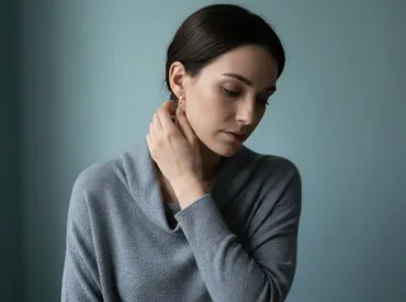
An irritation bump is a common reaction that is usually temporary and localized. Source: Business Insider
It is also important to distinguish between different types of bumps that may present similarly but have distinct etiologies. A granuloma is a specific type of inflammation where the immune system walls off an area it perceives as foreign, often appearing as a moist, red, highly vascularized bump that bleeds easily when touched. While not dangerous, granulomas require gentle handling and consistent saline rinses. In contrast, a true hypertrophic scar represents an excess of collagen deposition that remains within the original wound margins. Both are reactive and reversible, unlike keloidal tissue.
Understanding Keloids: An Overactive Healing Response
A keloid is a fundamentally different and less common condition. It is a type of benign tumor composed of excess scar tissue that forms due to an abnormal healing process. Unlike standard scars that mature and fade over time, keloids represent a dysregulated wound healing pathway that fails to receive proper termination signals.
What Are Keloids and Who Is at Risk?
When skin is injured, fibroblast cells produce collagen to repair the wound. In some individuals, the body doesn't get the signal to stop and produces far too much collagen. This results in a keloid that grows well beyond the initial injury. The underlying mechanism involves an overactive transforming growth factor-beta (TGF-β) signaling pathway, which drives continuous collagen synthesis and impairs normal collagen degradation by matrix metalloproteinases. This creates a dense, disorganized extracellular matrix that expands invasively into adjacent dermal tissue.
According to the American Academy of Dermatology, certain factors significantly increase your risk of developing keloids:
- Genetic Predisposition: Having a personal or family history of keloids is the biggest risk factor. Multiple genes have been implicated, including those regulating immune response and fibroblast activity. If a parent developed keloids, offspring have a significantly higher likelihood of experiencing the same reaction.
- Skin Tone: People with darker skin tones are more prone to keloid formation. Studies indicate that individuals with Fitzpatrick skin types IV through VI have a 15 to 20 times higher incidence rate compared to those with lighter skin tones. The exact reason remains under investigation, but it likely involves variations in melanocyte activity and inflammatory mediator profiles.
- Age: They are most common in people between the ages of 10 and 30. Younger skin tends to have more robust fibroblast activity and faster metabolic turnover, which can paradoxically lead to excessive scar formation when regulatory mechanisms fail. Incidence drops significantly after age 40.
Keloids can develop anywhere on the body, but high-tension areas like the shoulders, chest, jawline, earlobes, and back are particularly vulnerable. Cartilage piercings carry a higher risk than lobe piercings due to the avascular nature of cartilage tissue, which slows initial healing and prolongs the inflammatory window. Psychologically, keloids can cause significant distress, anxiety, and social withdrawal due to their visibility and persistent itching or pain, underscoring the importance of early, medically supervised management.
Navigating the Diagnostic Gray Area in Early Stages
In the first few weeks, it can be difficult to tell a new piercing bump from an emerging keloid. Both may appear as a small, pinkish lump. If you're concerned, watch for these key early differentiators:
- Timing is Key: A bump that appears within the first month is almost always an irritation bump. Keloids take time to develop their characteristic overgrowth of tissue. The delayed presentation of keloids aligns with the prolonged collagen accumulation phase.
- Watch for Growth: Monitor the bump closely. An irritation bump will typically stay the same size or get smaller with good care. A developing keloid will show progressive growth, even if it's slow. Use a consistent measurement technique or take weekly photos under identical lighting to track subtle changes.
- Check the Borders: Pay attention to the edges. A piercing bump will remain neatly contained within the piercing's immediate vicinity. A keloid will start to invade the healthy skin around the piercing, often developing a claw-like or spider-web appearance at the periphery.
Beyond self-observation, clinical evaluation provides definitive clarity. Dermatologists often utilize high-frequency ultrasonography or dermoscopy to assess tissue depth and vascularity. Hypertrophic scars typically show uniform echogenicity with defined margins, whereas keloids demonstrate heterogeneous internal structure, prominent blood vessels at the periphery, and invasive tissue architecture. In ambiguous cases, a skin biopsy may be performed to rule out other dermatological conditions, such as dermatofibroma, epidermoid cysts, or rarely, malignant lesions that can mimic benign scarring. However, biopsy is generally reserved for atypical presentations, as creating another wound carries its own keloid risk in susceptible individuals.
Patients should also document their healing trajectory meticulously. Note any changes in texture, color, size, and associated symptoms. Keep a log of aftercare routines, jewelry changes, and potential trauma events. This historical data proves invaluable when consulting healthcare professionals, as it helps correlate environmental triggers with tissue responses.
Treatment and Management: What You Can Do
The right approach to treatment depends entirely on a correct diagnosis. Misdiagnosing a keloid as an irritation bump can lead to ineffective home treatments and delayed medical care, while aggressively treating a simple bump with invasive methods can cause unnecessary tissue damage.
How to Treat a Piercing Bump at Home
Since most piercing bumps are caused by irritation, the goal is to calm the area and allow it to heal properly. Conservative, evidence-based care is the cornerstone of management.
- Identify and Remove the Irritant: Are you sleeping on it? Is your jewelry too tight? The bump won't resolve until the source of the problem is fixed. Consider using a travel pillow with a hole in the center to keep pressure off ear piercings, or adjust your hairstyle to prevent hair products from migrating to the site.
- Check Your Jewelry: Switch to high-quality, hypoallergenic jewelry made of implant-grade titanium, surgical steel, or solid 14k+ gold. This is often the single most effective solution. Ensure the jewelry is internally threaded or threadless to minimize tissue trauma during insertion and removal. The gauge should match the initial piercing precisely; downsizing too early can cause compression, while keeping a long post can invite snagging.
- Practice Gentle Aftercare: Clean the piercing once or twice daily with a sterile saline solution. Avoid twisting or turning the jewelry, as this disrupts the delicate epithelial bridge forming inside the fistula. Use a 0.9% isotonic sodium chloride spray labeled as "wound wash" with no additives. Spray directly, let it sit for 30 seconds, then gently pat dry with a clean, disposable paper towel. Do not use cotton swabs or balls, which can leave fibers behind.
- Use Warm Compresses: Soaking a clean cloth in warm water and holding it gently on the bump for 5-10 minutes can help improve blood flow and soothe irritation. This encourages lymphatic drainage and softens crust buildup, making removal gentler. Follow immediately with saline application to flush the area.
- Be Patient: Healing takes time. It can take several weeks or months for an irritation bump to fully disappear. Resist the urge to pop, squeeze, or apply unverified home remedies. The tissue is actively remodeling, and interference can reset the healing clock or introduce pathogens.
Important Home Care Warnings: Avoid tea tree oil, undiluted essential oils, aspirin paste, and homemade salt solutions. Tea tree oil is a potent sensitizer that frequently causes allergic contact dermatitis, exacerbating inflammation. Aspirin paste can cause chemical burns on mucosal or delicate tissue. Homemade salt mixtures rarely achieve sterile, isotonic concentrations, leading to cellular dehydration and delayed healing. If a bump shows signs of infection (spreading redness, fever, foul-smelling discharge), seek medical attention promptly, as oral antibiotics may be required.
Professional Treatments for Keloids
Keloids will not go away on their own and require treatment from a dermatologist or plastic surgeon. At-home remedies are ineffective and can worsen the condition. Attempting to excise, cauterize, or chemically burn a keloid without medical supervision often triggers a massive rebound response, causing the lesion to regrow larger than before.
Standard medical treatments include:
- Corticosteroid Injections: Injections directly into the keloid can help shrink and flatten the scar. This is often the first line of treatment. Intralesional triamcinolone acetonide inhibits fibroblast proliferation, reduces collagen synthesis, and induces localized vasoconstriction. Sessions are typically spaced 3 to 4 weeks apart, with noticeable improvement after 2 to 4 treatments. Potential side effects include skin atrophy, hypopigmentation, and telangiectasia, which is why dosing is carefully calibrated by a specialist.
- Surgical Removal: A surgeon can excise the keloid. However, this creates a new wound that is also at high risk of forming another keloid. Surgery is rarely performed in isolation and is usually paired with adjunctive therapies like immediate postoperative radiation or corticosteroid injections to suppress the anticipated healing response.
- Laser Therapy: Pulsed-dye lasers can help flatten keloids and reduce their reddish color by targeting abnormal blood vessels feeding the scar tissue. Fractional CO2 or Nd:YAG lasers may also be used to remodel collagen architecture and improve scar texture. Multiple sessions are typically required, and post-laser care involves strict sun protection and silicone application.
- Cryotherapy: Freezing the keloid with liquid nitrogen can help reduce its hardness and size. Cryotherapy induces vascular thrombosis and cellular necrosis within the scar. It is particularly effective for smaller keloids or when combined with intralesional steroid injections. Temporary blistering, crusting, and depigmentation may occur.
- Pressure Therapy: Applying pressure with silicone gel sheets or special earrings after treatment can help prevent recurrence. Continuous mechanical pressure (typically 24-32 mmHg) alters collagen alignment, reduces tissue hypoxia, and downregulates fibroblast activity. Consistent daily wear for 12 to 18 months is often recommended for optimal results.
Efficacy and Recurrence of Keloid Treatments
A significant challenge with keloids is their high rate of recurrence. Research shows that combination therapies are far more effective than single treatments. For example, surgical removal followed by corticosteroid injections or radiation therapy dramatically lowers the chance of the keloid returning compared to surgery alone, which can have recurrence rates as high as 80%. Multi-modal approaches combining injections, lasers, and other therapies are often recommended for the best long-term results. Emerging treatments, such as topical imiquimod, 5-fluorouracil (5-FU) injections, and botulinum toxin A, show promise in modulating the wound healing cascade and reducing recurrence. Patient compliance with post-treatment care, particularly silicone application and sun avoidance, remains a critical determinant of success.
Proactive Prevention for High-Risk Individuals
If you know you are prone to keloids due to family history or previous scarring, the safest approach is to avoid elective skin trauma like piercings and tattoos. If you decide to proceed, take these proactive steps:
- Consult a Dermatologist First: Discuss your plans and risk factors. A specialist can perform a patch test, evaluate your skin type, and potentially prescribe prophylactic topical agents or recommend preventive silicone therapy protocols.
- Choose an Expert Piercer: A highly experienced professional will cause less trauma to the tissue. Verify credentials, ensure they use single-use sterile needles (never piercing guns, which cause blunt-force trauma and crush tissue), and work in a clinically clean environment.
- Use Proper Jewelry from Day One: Start with high-quality, hypoallergenic materials. ASTM F136 titanium or niobium are optimal choices. Ensure the jewelry design is appropriate for your anatomy, avoiding rings in initial healing piercings due to excessive movement and rotation.
- Follow Aftercare Meticulously: Be vigilant with your cleaning routine to prevent infection and inflammation. Maintain a balanced diet rich in zinc, vitamin C, and protein to support optimal collagen regulation and immune function. Avoid smoking, as nicotine vasoconstricts blood vessels and significantly delays wound healing, increasing scar formation risk.
- Seek Early Intervention: At the very first sign of a suspicious bump that feels firm or seems to be growing, see a dermatologist immediately. Early treatment can help manage keloid growth. Initiating silicone sheeting or low-dose corticosteroids during the proliferative phase can often halt abnormal progression before it becomes clinically entrenched.
- Consider Piercing Location Carefully: Avoid high-tension zones like the upper ear cartilage, sternum, or nape of the neck if you are at high risk. Earlobes generally have better vascular supply and lower keloid incidence compared to helix or tragus piercings. Understanding anatomical risk factors allows for informed decision-making and safer placement choices.
When to See a Doctor or Professional Piercer
While many piercing bumps are manageable at home, it's important to seek professional help if you notice any of the following:
- The bump is growing larger or spreading.
- The bump is very hard, firm, or rubbery.
- You experience severe pain, throbbing, or the area is hot to the touch.
- You see thick, yellow, or green pus, which indicates a significant infection.
- You have a personal or family history of keloids.
- The bump doesn't improve after several weeks of consistent at-home care.
Understanding when to consult a professional piercer versus a medical provider is essential for timely resolution. A certified piercer (APP-certified or equivalent) is ideal for addressing mechanical issues like jewelry sizing, placement correction, aftercare technique, and basic bump evaluation. They can assess whether the jewelry is causing undue pressure or if the fistula is healing at an improper angle.
Conversely, a dermatologist or primary care physician should evaluate suspected infections, systemic symptoms (fever, chills, swollen lymph nodes), rapidly expanding lesions, or any growth that matches keloid characteristics. During a medical consultation, expect a thorough physical examination, discussion of your medical and family history, and potentially a dermoscopic assessment. If infection is suspected, a swab culture may be ordered to guide targeted antibiotic therapy. For confirmed keloids, a personalized treatment roadmap will be developed, often beginning with conservative modalities before progressing to interventional procedures. Delaying medical evaluation for an actively growing keloid or deep infection can result in unnecessary scarring, cartilage damage, or systemic complications, so proactive assessment remains the gold standard in responsible piercing maintenance.
Frequently Asked Questions (FAQ)
Q: How can you tell the difference between a piercing bump and a keloid? A: You can tell the difference by observing several key factors. A piercing bump (hypertrophic scar) typically appears within weeks, stays small and confined to the piercing site, is often soft, and can resolve with proper care. A keloid, however, usually appears months later, grows beyond the original piercing boundary, feels firm or rubbery, and is permanent without medical treatment.
Q: How long do piercing bumps last? A: The duration depends on the type. Simple irritation bumps can disappear in days to weeks once the source of irritation is removed. Hypertrophic scars may take several months to a year to flatten and fade. True keloids are permanent and may continue to grow if not treated by a dermatologist.
Q: Will a piercing bump go away on its own? A: Most common piercing bumps, which are irritation bumps or hypertrotrophic scars, will often go away on their own if you follow proper aftercare and remove any sources of irritation, such as low-quality jewelry or friction. Keloids, however, will not go away on their own and require professional medical treatment.
Q: Can a piercing bump turn into a keloid? A: No, a piercing bump and a keloid are two distinct skin conditions with different causes. A piercing bump (hypertrophic scar) is a localized inflammatory response. A keloid is a genetic condition involving an overproduction of collagen. While both can occur at a piercing site, a hypertrophic scar does not transform into a keloid.
Q: Does tea tree oil work for piercing bumps or keloids? A: Dermatologists and professional piercing organizations strongly advise against using tea tree oil for piercing bumps or keloids. Tea tree oil is highly concentrated and frequently causes allergic contact dermatitis, chemical burns, and excessive drying, which further disrupts the healing process and can exacerbate inflammation. For keloids, it has no proven efficacy in reducing collagen overproduction and may irritate sensitive scar tissue, triggering additional growth.
Q: Should I remove my jewelry if a bump develops? A: Removing jewelry from an irritated piercing can sometimes be beneficial, but it must be done strategically. If the jewelry is the source of irritation (e.g., poor quality metal, wrong size), having it professionally downsized or swapped to a hypoallergenic stud is ideal. However, removing jewelry entirely while an active infection or fluid-filled bump is present can trap bacteria inside the closing fistula, leading to an abscess. If a keloid is suspected, removing jewelry does not shrink the scar and may complicate future treatment. Always consult a professional piercer or dermatologist before removing jewelry during active inflammation.
Q: How does smoking or nutrition affect piercing healing and scar formation? A: Smoking significantly impairs wound healing by introducing carbon monoxide and nicotine, which constrict blood vessels, reduce oxygen delivery to tissues, and impair fibroblast function. This prolongs the inflammatory phase and increases the risk of both persistent irritation bumps and abnormal scarring. Nutrition also plays a critical role; diets deficient in protein, vitamin C, zinc, and omega-3 fatty acids compromise collagen synthesis regulation and immune response. Maintaining adequate hydration and consuming anti-inflammatory foods can support balanced tissue remodeling and reduce the likelihood of excessive bump formation during the healing process.
Q: Are certain piercing locations more prone to keloids? A: Yes, anatomical location significantly influences keloid risk. High-tension areas where skin stretches frequently—such as the upper ear cartilage, shoulders, chest (sternum), upper back, and jawline—are most susceptible. These regions experience constant mechanical stress, which stimulates fibroblast activity and collagen deposition. Earlobe piercings generally carry a lower keloid risk due to their high vascularity and low tension, but individuals with strong genetic predisposition can still develop keloids anywhere skin is punctured. Choosing low-tension sites and following rigorous aftercare protocols can help mitigate location-specific risks.
Conclusion
Navigating the uncertainty of a post-piercing bump requires a clear understanding of tissue behavior, careful observation, and informed decision-making. While the vast majority of bumps are benign, self-limiting irritation responses or localized hypertrophic scarring, recognizing the distinct clinical and pathological features of keloids is essential for long-term skin health. Key differentiators such as onset timing, growth patterns beyond the wound margin, tissue texture, and genetic predisposition provide reliable diagnostic clues that can guide appropriate management strategies. Conservative, evidence-based home care centered around irritant removal, sterile saline cleansing, proper jewelry selection, and patience remains the foundation for resolving common piercing bumps. Conversely, confirmed keloids demand early medical intervention, typically involving combination therapies like corticosteroid injections, silicone pressure therapy, laser treatment, or surgical excision paired with adjuvant modalities to minimize recurrence. Ultimately, successful outcomes hinge on accurate self-assessment, timely professional consultation when red flags emerge, and strict adherence to scientifically supported aftercare protocols. By respecting the body's natural healing processes, avoiding unverified home remedies, and prioritizing preventive measures—especially for high-risk individuals—you can enjoy body modification safely while minimizing complications and achieving optimal aesthetic results. When in doubt, always defer to the expertise of certified professional piercers for mechanical concerns and board-certified dermatologists for medical scar management, ensuring your piercing journey remains both beautiful and healthy.
References:
- American Academy of Dermatology (AAD). "Keloids: Symptoms and Causes." aad.org
- Medical News Today. "Piercing bump vs. keloid: How to tell the difference." medicalnewstoday.com
- The Keloid Plastic Surgery Center. "Keloid vs Piercing Bump: Know the Key Differences." thekeloidplasticsurgerycenter.com
- Verywell Health. "What's the Difference Between a Piercing Bump and a Keloid?" Mayo Clinic
- NHS. "Keloid scars." nhs.uk

About the author
Elena Vance, MD, is a double board-certified dermatologist and pediatric dermatologist. She is an assistant professor of dermatology at a leading medical university in California and is renowned for her research in autoimmune skin disorders.