Why Toenails Fall Off: Causes, Treatments, and Expert Recovery Guide

Experiencing a sudden detachment of a toenail can be an alarming and deeply unsettling event for anyone. The human body relies on the structural integrity of our nail plates for protection, sensation, and everyday mobility. When toenails fall off, it often disrupts daily routines, causes significant discomfort, and raises immediate concerns about underlying health issues. Whether you are noticing gradual lifting after months of wearing tight footwear, sudden separation following an athletic impact, or slow deterioration linked to persistent discoloration, understanding the root cause is the critical first step toward safe recovery. This comprehensive guide explores the medical, environmental, and lifestyle factors behind nail detachment, provides clinically validated treatment pathways, and outlines actionable protocols to support healthy regrowth. By combining dermatological research, podiatric best practices, and evidence-based self-care strategies, you will learn how to protect the exposed nail bed, manage pain effectively, and prevent future complications. If you have ever wondered why this occurs or how to safely navigate the healing process, you will find clear, medically sound guidance tailored to both acute injuries and chronic conditions.
Understanding Why Toenails Detach from the Nail Bed
The toenail is a highly specialized structure composed primarily of compacted keratin layers that serve as a protective shield for the delicate tissues beneath. Each nail is anchored to the underlying nail bed by the hyponychium and supported laterally by the nail folds. The growth process originates in the nail matrix, a region hidden beneath the proximal nail fold where new keratinocytes multiply and harden into the visible nail plate. When the delicate attachment between these layers is compromised, the nail begins to lift, a condition medically known as onycholysis. In more severe cases, where the nail detaches completely from its base and falls away entirely, clinicians refer to it as onychomadesis or traumatic nail avulsion. Both conditions disrupt the protective barrier, leaving the sensitive nail bed vulnerable to friction, moisture, and microbial invasion.
The Anatomy of a Healthy Toenail
A healthy toenail functions as a biomechanical reinforcement for the distal phalanx of each toe. The dorsal surface of the nail plate is smooth and translucent, allowing visualization of the underlying pink nail bed, which is rich in capillaries and nerve endings. The ventral surface contains longitudinal ridges that interlock with corresponding grooves in the nail bed, creating a stable adhesive interface. This anatomical interlocking is maintained by continuous cellular adhesion and the natural secretion of lipids and moisture from surrounding tissues. When systemic or local factors interfere with this balance, the interlocking mechanism fails. Dehydration, inflammation, or physical shearing forces can break the microscopic bonds, causing the nail to separate from its foundation. Understanding this anatomy is essential because it explains why certain interventions, like keeping the area dry or protecting it from pressure, directly influence whether toenails fall off prematurely or remain securely attached.
Onycholysis vs. Onychomadesis: Key Differences
Onycholysis refers to partial or complete lifting of the nail plate from the nail bed without immediate matrix disruption. It typically begins at the distal edge or lateral margins and progresses proximally. This condition is frequently associated with prolonged moisture exposure, chemical irritants, or early-stage fungal colonization. In contrast, onychomadesis involves complete shedding of the nail starting from the proximal end near the cuticle, often following a temporary arrest of matrix activity. This proximal detachment usually signals a significant physiological stressor, such as high fever, severe illness, chemotherapy, or blunt force trauma. Both conditions can eventually lead to situations where toenails fall off entirely, but the underlying pathophysiology differs significantly. Differentiating between them requires evaluating the detachment pattern, accompanying symptoms, and personal medical history, which directly informs whether topical care, systemic treatment, or protective monitoring is the most appropriate clinical response.
Primary Medical Causes of Toenail Loss
When toenails fall off without an obvious acute injury, it typically indicates an underlying pathological process that has progressively weakened the structural integrity of the nail apparatus. Identifying the precise etiology requires careful assessment of clinical presentation, environmental exposures, and systemic health markers. Several well-documented medical conditions consistently correlate with nail detachment, each requiring a distinct therapeutic approach to restore normal nail architecture and prevent recurrence.
Fungal Infections (Onychomycosis)
Fungal nail infections represent one of the most prevalent causes of nail detachment worldwide. Dermatophytes, particularly Trichophyton rubrum and Trichophyton mentagrophytes, invade the keratinized tissue of the nail plate and bed, secreting enzymes that degrade structural proteins. As the infection advances, the nail thickens, discolors, and becomes brittle, eventually separating from its underlying tissue. The warm, moist environment inside closed shoes creates an ideal breeding ground for these pathogens, as noted in CDC fungal infection guidelines. According to the American Academy of Dermatology, onychomycosis accounts for approximately fifty percent of all nail disorders and disproportionately affects individuals with compromised peripheral circulation or immune function. Early intervention with targeted antifungal agents significantly reduces the likelihood of severe detachment, while delayed treatment often results in progressive crumbling and complete shedding.
Trauma and Repetitive Injury
Acute and chronic mechanical trauma remains a primary catalyst for nail loss. Stubbing a toe against a solid surface, dropping heavy objects on the foot, or experiencing sudden deceleration during athletic activities can cause immediate subungual hematoma and pressure necrosis beneath the nail plate. The accumulated blood lifts the nail away from its vascular bed, disrupting cellular adhesion and ultimately leading to detachment. Repetitive microtrauma, often seen in runners, hikers, or individuals wearing improperly fitted footwear, creates a different but equally destructive pathway. Continuous friction against the distal nail edge gradually erodes the attachment points, resulting in chronic onycholysis that slowly progresses until toenails fall off. Addressing biomechanical imbalances through proper footwear selection, toe caps, or custom orthotics is essential for long-term prevention.
Systemic Health Conditions and Nutritional Deficiencies
The health of your toenails often mirrors systemic physiological balance. Endocrine disorders, particularly poorly controlled diabetes mellitus and thyroid dysfunction, impair peripheral microcirculation and reduce cellular turnover rates, making nails more susceptible to detachment. Nutritional deficiencies in essential micronutrients, including zinc, biotin, iron, and protein, compromise keratin synthesis and weaken nail architecture. Chronic kidney disease and liver dysfunction can similarly alter metabolic pathways responsible for tissue maintenance. When toenails fall off in conjunction with unexplained fatigue, weight fluctuations, or widespread dermatological changes, comprehensive metabolic screening becomes necessary to identify and treat the root systemic imbalance rather than focusing exclusively on local foot care.
Psoriasis and Lichen Planus
Autoimmune and inflammatory dermatological conditions frequently manifest in the nail unit. Nail psoriasis affects approximately fifty percent of individuals with cutaneous psoriasis, characterized by pitting, oil drop discoloration, subungual hyperkeratosis, and progressive onycholysis. The inflammatory cascade disrupts matrix keratinization and accelerates abnormal tissue proliferation, causing the nail to separate from the bed. Lichen planus, though less common, can cause severe nail thinning and longitudinal ridging that eventually leads to irreversible detachment if the matrix sustains permanent scarring. Both conditions require coordinated management involving topical corticosteroids, immunomodulators, or biologic therapies prescribed by dermatologists and rheumatologists. Recognizing the distinct visual markers of autoimmune nail disease prevents misdiagnosis as fungal infection and ensures appropriate medical intervention.
Medications and Chemotherapy Effects
Certain pharmacological agents exert direct toxic effects on rapidly dividing cells, including those in the nail matrix. Chemotherapy drugs, retinoids, tetracyclines, and some antimalarial medications temporarily halt nail production, resulting in Beau's lines or complete onychomadesis. Patients undergoing cancer treatment frequently experience multiple nails falling off simultaneously as the matrix enters a resting phase, a phenomenon well-documented by the National Cancer Institute (NIH). While this effect is generally reversible once therapy concludes, the regrowth process requires meticulous wound care and infection prevention. Clinicians often recommend protective footwear and gentle hygiene protocols during treatment cycles to minimize mechanical stress on fragile nails and support smoother recovery post-therapy.
When to See a Doctor vs. Home Management
Determining whether professional intervention is necessary depends on symptom severity, underlying health status, and progression rate. While mild detachment following minor trauma can often be managed safely at home, certain warning signs demand immediate podiatric evaluation to prevent complications.
Red Flag Symptoms Requiring Immediate Medical Attention
Any detachment accompanied by severe, throbbing pain, purulent discharge, expanding erythema, or fever indicates a secondary bacterial infection that requires prescription antibiotics and possible debridement. Individuals with diabetes, peripheral artery disease, or immunosuppression should never attempt home management of detached nails due to elevated risks of ulceration, osteomyelitis, and delayed healing. Spreading discoloration beyond the nail plate, foul odor resistant to routine cleansing, or detachment affecting multiple toes simultaneously warrants prompt clinical assessment, as outlined in Mayo Clinic wound care guidelines. These symptoms suggest progressive pathology that cannot be resolved with over-the-counter remedies alone.
Routine Monitoring and At-Home Assessment
For healthy individuals experiencing gradual, painless detachment without signs of infection, conservative monitoring is appropriate. Keep the affected area clean and thoroughly dry after bathing. Trim any completely detached edges carefully with sterilized clippers to prevent snagging on socks or bedding. Apply a thin layer of petroleum jelly or antibiotic ointment if the nail bed appears raw, then cover with a breathable gauze pad and medical tape to protect against friction. Change dressings daily and inspect for early signs of inflammation or delayed healing. If the nail remains partially attached but loose, secure it temporarily with medical tape rather than pulling it off prematurely. Document the detachment pattern with weekly photographs to track progression and share with a healthcare provider if symptoms worsen over time.

Medical Treatments and Professional Interventions
Once a clinician determines the underlying cause of nail detachment, targeted therapeutic strategies can accelerate healing and prevent recurrence. Modern podiatric and dermatological practice offers a spectrum of evidence-based interventions tailored to individual patient needs.
Antifungal Medications and Topical Therapies
For confirmed onychomycosis, treatment regimens typically combine topical lacquers with systemic antifungal agents when detachment is extensive. Prescription topical solutions containing ciclopirox or efinaconazole penetrate the nail plate to reach the infection site, while oral terbinafine or itraconazole provides systemic eradication. Treatment courses often extend twelve to sixteen weeks due to slow nail growth rates and require periodic liver function monitoring. Adherence to daily application protocols is critical, as inconsistent dosing frequently leads to treatment failure and recurrence. Patients must maintain strict foot hygiene and disinfect shoes regularly to prevent reinfection from residual spores.
Laser Treatments and Surgical Options
Advanced laser therapy utilizes specific wavelengths of near-infrared light to target and destroy fungal pathogens while preserving surrounding healthy tissue. Clinical studies demonstrate improved clearance rates with fewer systemic side effects compared to oral medications, making this an attractive option for patients with contraindications to traditional pharmacotherapy. In cases of complete avulsion, severe onychomadesis, or recurrent ingrown nails causing repeated detachment, surgical removal of the nail plate and partial matrixectomy may be indicated. This procedure eliminates the problematic nail permanently or temporarily while preserving the nail bed for future regrowth. Post-operative care involves strict elevation, antiseptic soaks, and protective padding to ensure optimal healing.
Managing Underlying Systemic Conditions
When nail detachment stems from autoimmune, endocrine, or nutritional deficiencies, addressing the primary condition remains paramount. Optimizing glycemic control in diabetic patients improves peripheral circulation and reduces infection susceptibility. Thyroid hormone normalization restores metabolic balance and cellular turnover rates. Supplementation with zinc, biotin, iron, and high-quality protein supports keratin synthesis and structural reinforcement. Rheumatological or dermatological specialists may prescribe systemic immunomodulators for psoriasis or lichen planus to halt inflammatory matrix damage. Coordinated multidisciplinary care ensures that nail health reflects overall physiological stability rather than serving as an isolated treatment target.
Preventing Future Toenail Detachment
Proactive foot care and lifestyle modifications significantly reduce the likelihood of recurrent detachment. Implementing consistent protective strategies preserves nail integrity and minimizes exposure to mechanical, environmental, and microbial stressors.
Proper Footwear and Sock Selection
Footwear that restricts toe movement or creates excessive pressure against the distal nail edge remains a leading preventable cause of trauma-induced detachment. Select shoes with adequate toe box width, allowing all digits to splay naturally without compression. Ensure a half-inch gap between the longest toe and the shoe tip during standing and walking. Replace athletic footwear every three hundred to five hundred miles, as midsole cushioning degradation increases impact transmission to the toes. Choose moisture-wicking synthetic or wool blend socks that minimize friction and maintain optimal skin hydration. Avoid cotton socks during prolonged physical activity, as retained sweat softens nail structure and increases susceptibility to separation.
Foot Hygiene and Moisture Control
Maintaining a dry, clean foot environment disrupts the conditions that promote fungal colonization and nail bed maceration. Wash feet daily with mild soap and lukewarm water, ensuring thorough drying between all toes using a separate, clean towel. Apply antifungal powder or talc-free moisture absorbers before sock insertion if excessive sweating is present. Alternate shoes daily to allow complete internal drying, and expose footwear to direct sunlight periodically to reduce microbial load. Never walk barefoot in public locker rooms, showers, or pool decks, as these environments harbor high concentrations of dermatophytes. Regular foot care routines significantly decrease the probability of toenails falling off due to preventable environmental exposures, consistent with WHO public health recommendations for hygiene and mobility preservation.
Safe Nail Grooming Practices
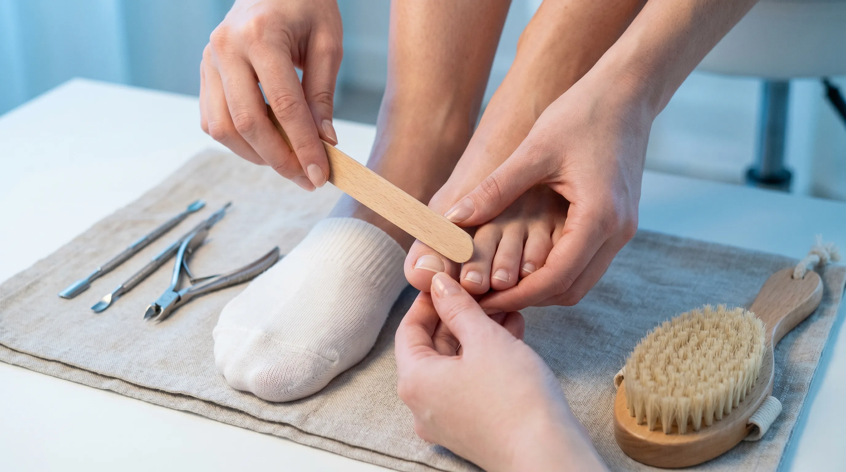
Proper trimming technique directly influences nail attachment stability and structural resilience. Cut toenails straight across using sterilized clippers, avoiding curved edges that encourage lateral ingrowth and pressure-induced lifting. File sharp corners gently with a fine-grit emery board to prevent tearing or catching on fabrics. Never cut nails too short, as excessive exposure of the nail bed removes natural protection and increases vulnerability to trauma. Avoid aggressive cuticle removal, which disrupts the protective seal and introduces pathogens into the matrix region. Schedule professional pedicures only at licensed, hygienic facilities that utilize properly sanitized instruments to prevent cross-contamination and iatrogenic damage.
Living with a Missing or Damaged Toenail
Navigating daily life with a detached or completely missing toenail requires strategic protection, pain management, and realistic expectations regarding the regrowth timeline. The exposed nail bed remains highly sensitive until a new keratin layer forms, making environmental shielding essential.
Protecting the Exposed Nail Bed
The raw tissue beneath a missing nail lacks the protective keratin barrier and remains susceptible to abrasion, infection, and pressure-related pain. Cover the area with a non-adherent silicone dressing or sterile gauze pad secured with hypoallergenic tape. Change protective coverings daily or immediately after they become damp or soiled. Avoid tight shoes, high-impact activities, and prolonged standing during the initial healing phase. When returning to regular footwear, select closed-toe shoes with reinforced toe caps to shield the vulnerable area from accidental impacts. Keep the exposed tissue clean and moisturized with fragrance-free ointments to maintain tissue elasticity and prevent cracking during movement.
Pain Management and Comfort Strategies
Mild to moderate discomfort is common during the first two weeks following detachment. Elevate the affected foot above heart level when resting to reduce localized swelling and pressure. Apply cold packs wrapped in thin cloth for ten to fifteen minutes several times daily to manage inflammation and numb nerve sensitivity. Over-the-counter analgesics, such as ibuprofen or acetaminophen, provide effective relief for routine discomfort. If pain intensifies, becomes constant, or radiates beyond the immediate area, discontinue home management and consult a podiatrist to rule out fracture, deep infection, or nerve involvement. Gradual reintroduction to normal activity should follow tolerance levels without forcing movement that exacerbates symptoms.
Regrowth Timeline and Expectations
Toenails grow significantly slower than fingernails due to reduced peripheral blood flow and constant mechanical stress. Complete regrowth typically requires nine to eighteen months, with an average rate of approximately one millimeter per month, according to NIH research on nail physiology. The new nail emerges from the matrix and gradually extends distally, initially appearing thin and ridged before thickening and smoothing over time. Genetic factors, age, nutritional status, and underlying health conditions influence individual regrowth speed and final nail architecture. Temporary texture variations, mild discoloration, or slight curvature are normal during early stages and typically resolve as the nail matures. Consistent protection and hygiene throughout this extended period maximize the likelihood of healthy, structurally sound replacement.
| Cause of Detachment | Primary Symptoms | Typical Treatment Approach | Recovery Timeline |
|---|---|---|---|
| Fungal Infection | Yellow/brown discoloration, thickening, crumbling, gradual lifting | Oral/topical antifungals, laser therapy, shoe disinfection | 6-12 months with consistent treatment |
| Acute Trauma | Subungual hematoma, immediate pain, sudden separation within days/weeks | Rest, elevation, protective padding, possible surgical drainage | 6-9 months for complete regrowth |
| Repetitive Microtrauma | Lifting from distal/lateral edges, discomfort during activity, gradual progression | Footwear adjustment, toe protectors, biomechanical orthotics | 9-12 months with pressure relief |
| Psoriasis/Autoimmune | Pitting, oil spots, severe lifting, concurrent skin or joint symptoms | Immunomodulators, topical steroids, systemic biologics | Variable; depends on disease control |
| Medication/Chemotherapy | Multiple nails detaching simultaneously, Beau's lines, proximal separation | Supportive care, infection prevention, post-treatment monitoring | Resumes 3-6 months after therapy ends |
Frequently Asked Questions
Can a toenail that has fallen off grow back normally?
Yes, in the vast majority of cases, a completely detached toenail will regenerate fully over six to twelve months. The nail matrix retains the cellular blueprint for healthy production, and as long as it remains undamaged and protected from infection, the new nail will gradually replace the missing plate. Individuals with chronic systemic conditions, repeated trauma, or severe matrix scarring may experience slower regrowth or permanent texture changes. Maintaining strict foot hygiene, wearing properly fitted footwear, and avoiding premature manipulation of the exposed bed significantly improve the quality of replacement nail architecture.
Is it safe to remove a loose toenail at home?
Forced removal of a partially attached toenail is strongly discouraged, as it can tear healthy tissue, damage the underlying matrix, and create an open wound susceptible to bacterial invasion. If a nail is completely detached with minimal resistance and moves freely, you may carefully trim the detached portion using sterilized clippers, sanitize the area with mild antiseptic, and apply a sterile dressing. However, if any portion remains firmly adhered or causes bleeding upon gentle manipulation, leave it intact and consult a healthcare professional. Clinical debridement ensures complete removal without compromising the regenerative structures beneath.
How long does it take for toenails to fall off after an injury?
Following acute blunt force trauma, the nail typically changes color to purple or black within hours as blood accumulates beneath the plate. Over two to six weeks, the body gradually separates the damaged nail from the underlying tissue through natural cellular turnover and fluid reabsorption. The nail becomes increasingly loose and may eventually detach entirely or require minimal assistance during routine washing. During this period, keeping the area clean, avoiding additional pressure, and monitoring for signs of infection ensures a safe transition to the regrowth phase without unnecessary complications.
What are the signs that a fungal infection is causing toenails to fall off?
Fungal-related detachment follows a distinct progressive pattern rather than sudden separation. Initial symptoms include yellow or white streaks along the lateral edges, followed by localized thickening and loss of translucency. The nail gradually becomes brittle, crumbles at the free edge, and develops a noticeable gap between the plate and nail bed. As the infection advances, the nail may emit a mild foul odor and detach entirely without significant trauma. Early dermatological evaluation and mycological testing confirm the diagnosis and enable targeted antifungal intervention before irreversible structural damage occurs.
When should I seek medical attention if my toenails fall off?
Professional evaluation is essential when detachment is accompanied by severe pain, pus, expanding redness, warmth, or fever, as these indicate active infection requiring prescription treatment. Individuals with diabetes, peripheral neuropathy, or vascular disease should never attempt self-management due to impaired healing and elevated complication risks. Sudden detachment across multiple nails, recurrent episodes without clear trauma, or detachment coinciding with systemic symptoms like fatigue or unexplained weight loss warrants comprehensive clinical assessment. Early medical intervention prevents secondary infections, addresses underlying pathology, and accelerates safe recovery.
Conclusion
Experiencing situations where toenails fall off can be deeply concerning, but understanding the underlying mechanisms empowers individuals to respond effectively and safely. Whether the detachment stems from fungal colonization, repetitive footwear pressure, acute trauma, autoimmune activity, or pharmacological effects, each pathway requires targeted intervention and consistent protective care. By recognizing early warning signs, maintaining rigorous foot hygiene, selecting biomechanically appropriate footwear, and seeking professional evaluation when indicated, you can significantly reduce recurrence risks and support optimal nail regrowth. The human body possesses remarkable regenerative capabilities, and with patience, evidence-based treatment, and mindful daily habits, healthy toenail architecture can be fully restored. Prioritizing proactive foot health not only resolves immediate discomfort but also enhances overall mobility, comfort, and long-term wellness. Always consult licensed podiatric or dermatological professionals for personalized guidance, particularly when underlying health conditions complicate the healing process, ensuring that your recovery remains safe, structured, and medically sound.

About the author
Elena Vance, MD, is a double board-certified dermatologist and pediatric dermatologist. She is an assistant professor of dermatology at a leading medical university in California and is renowned for her research in autoimmune skin disorders.