EKG vs EEG: Key Differences, Uses, and What to Expect

Understanding diagnostic testing can be overwhelming, especially when faced with similar-sounding acronyms that evaluate completely different organ systems. If your physician has recommended an electrical assessment, you are likely weighing an ekg vs eeg procedure to determine which test aligns with your symptoms. Both examinations are foundational pillars of modern clinical diagnostics, yet they measure entirely distinct physiological processes using specialized electrode arrays and amplification technologies. The confusion is understandable; after all, both acronyms describe non-invasive, painless procedures that map the body’s inherent electrical signals. However, the clinical pathways, interpretive frameworks, and subsequent treatment plans diverge significantly depending on whether the investigation targets cardiac rhythm stability or neurological cortical activity. In this comprehensive guide, we will dissect the anatomical foundations, procedural nuances, diagnostic applications, and patient preparation protocols for each examination. By understanding the precise differences between an ekg vs eeg, you will be better equipped to engage in informed conversations with your healthcare team, anticipate what to expect during the appointment, and confidently navigate the next steps of your diagnostic journey. Let us explore how these two cornerstone tests function, why they are prescribed, and how they ultimately safeguard your vital health systems.
Understanding the Basics: What Are EKG and EEG Tests?
When medical professionals initiate a diagnostic workup, they frequently rely on electrophysiological recordings to visualize the invisible electrical currents that drive human physiology. Both examinations utilize the same fundamental principle: biological cells generate measurable voltage fluctuations when they depolarize and repolarize during functional activity. Despite this shared technological heritage, the anatomical targets and clinical objectives remain entirely separate.
Free calculatorBMI CalculatorCalculate Body Mass IndexOpen the calculatorElectrocardiogram (EKG/ECG) Explained
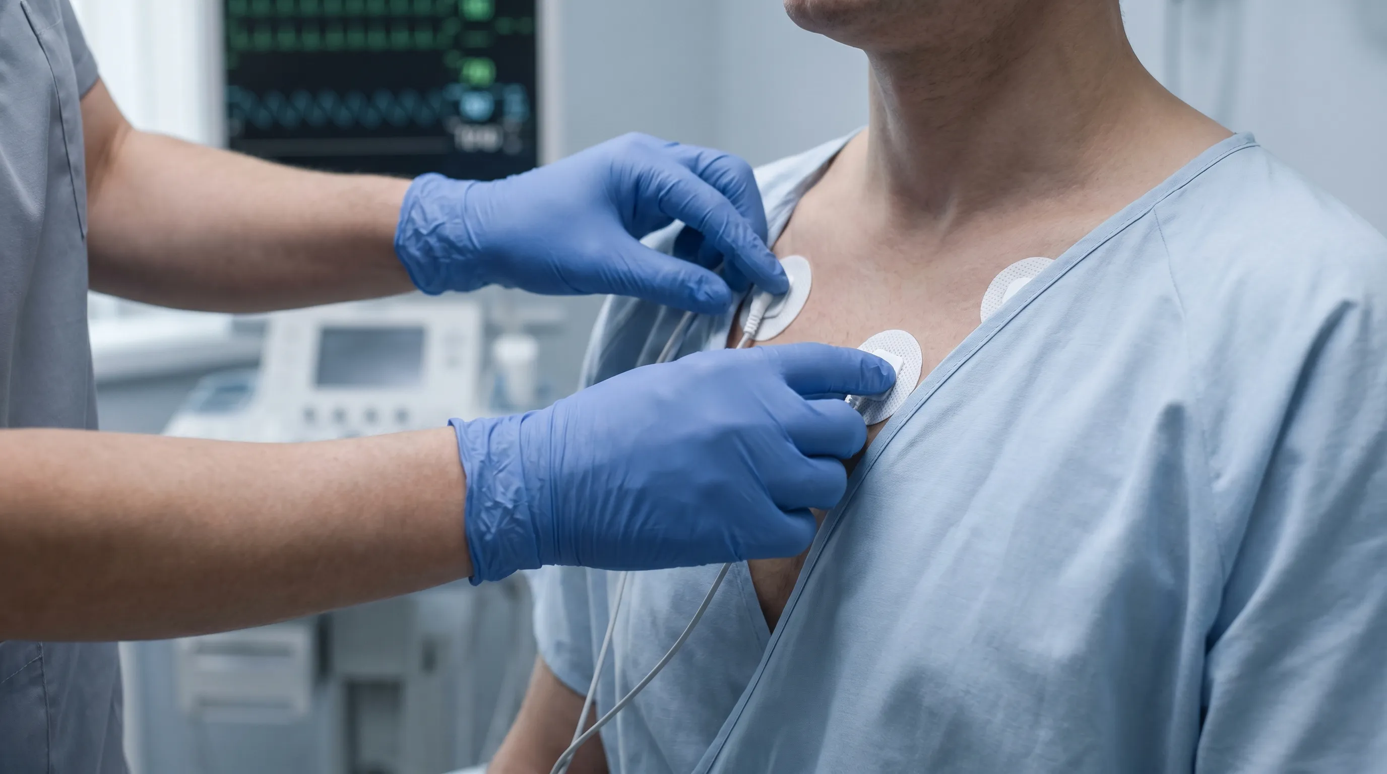
The electrocardiogram, commonly abbreviated as EKG or ECG, is a rapid, non-invasive diagnostic tool designed specifically to capture the heart's electrical conduction sequence. The human heart relies on a highly organized network of specialized pacemaker cells and conducting fibers to coordinate each contraction. This electrical cascade begins in the sinoatrial (SA) node, travels through the atrioventricular (AV) node, descends the bundle of His, and rapidly disperses through the Purkinje fibers to trigger synchronized ventricular contraction. The EKG machine translates these microscopic ionic movements into macroscopic waveforms displayed on a monitor and printed on grid paper. Standard clinical EKGs utilize a 12-lead configuration, placing ten adhesive electrodes on the chest, arms, and legs to create twelve distinct spatial perspectives of the heart's electrical activity. This multi-angle approach allows clinicians to pinpoint ischemic regions, identify conduction blockages, and assess overall myocardial health. The American Heart Association provides extensive clinical guidelines on standardizing these measurements for consistent diagnostic accuracy across different healthcare facilities https://www.heart.org/en/health-topics/consumer-healthcare/what-is-cardiovascular-disease/electrocardiogram.
Electroencephalogram (EEG) Explained
In contrast, the electroencephalogram focuses exclusively on the central nervous system, specifically the cerebral cortex. Billions of interconnected neurons communicate through rapid electrochemical signaling, generating continuous oscillatory electrical fields that can be detected at the scalp surface. An EEG captures these cortical rhythms using a dense array of electrodes strategically positioned according to the international 10-20 system, which standardizes placement relative to anatomical landmarks. These recordings reveal brain wave frequencies categorized into delta (deep sleep, pathological states), theta (drowsiness, meditation), alpha (wakeful relaxation), beta (active concentration, anxiety), and gamma (cognitive processing, perceptual binding). Neurologists analyze these waveforms to assess overall brain health, localize seizure foci, monitor anesthesia depth during surgery, and evaluate sleep architecture. The National Institute of Neurological Disorders and Stroke emphasizes that EEGs remain the gold standard for evaluating seizure disorders due to their exceptional temporal resolution in capturing rapid neuronal firing patterns https://www.ninds.nih.gov/health-information/disorders/epilepsy-seizures.
Primary Functions and Clinical Applications
The decision to order one test over another stems directly from the patient's presenting symptoms, medical history, and suspected underlying pathology. Clinicians follow strict diagnostic algorithms to ensure appropriate test selection, minimizing unnecessary procedures while maximizing diagnostic yield.
When Doctors Order an EKG
Cardiologists and primary care physicians routinely prescribe electrocardiograms to investigate symptoms suggestive of cardiovascular compromise. Common indications include chest pain or pressure, unexplained shortness of breath, palpitations, dizziness, and syncope. Beyond acute symptom evaluation, EKGs serve as essential screening tools during routine physical examinations, preoperative risk assessments, and occupational health clearances for high-stress professions. They are also critical for monitoring patients with established heart disease, hypertension, diabetes, or a family history of sudden cardiac death. Physicians frequently utilize EKGs to evaluate the efficacy and potential cardiotoxicity of prescribed medications, particularly certain antibiotics, antipsychotics, and antiarrhythmic drugs. Stress testing protocols combine physical exertion with continuous EKG monitoring to detect exercise-induced ischemia that may remain silent at rest. For individuals experiencing intermittent symptoms, extended monitoring options like Holter monitors, event recorders, or implantable loop recorders provide weeks to years of continuous cardiac data capture.
When Doctors Order an EEG
Neurologists and sleep medicine specialists primarily utilize electroencephalography to investigate abnormal brain function. The most frequent indication involves diagnosing and classifying seizure types and epilepsy syndromes. When patients present with unexplained loss of consciousness, convulsive movements, staring spells, or unusual sensory experiences, an EEG helps determine whether these events originate from aberrant cortical electrical discharges. Beyond epilepsy, EEGs play a crucial role in evaluating sleep disorders such as narcolepsy, sleep apnea, and parasomnias by tracking the physiological transitions between sleep stages. They are also invaluable for diagnosing encephalopathies, monitoring brain function in comatose patients, and confirming brain death through the demonstration of electrocerebral silence. Functional mapping prior to neurosurgical procedures often incorporates EEG data to preserve eloquent brain regions during tumor resection or epilepsy surgery. Ambulatory EEG devices and polysomnography integrate brain wave monitoring with cardiac, respiratory, and muscular data to provide comprehensive physiological profiling.
Diagnostic Accuracy and Limitations
Both modalities offer exceptional clinical utility, yet neither is infallible. An EKG may appear entirely normal between arrhythmic episodes or in early ischemic stages, necessitating serial testing or advanced imaging. Conversely, an EEG requires active seizure activity or characteristic interictal discharges during the recording window to yield a definitive epilepsy diagnosis; a normal EEG never completely rules out seizure disorders. Sensitivity and specificity improve dramatically when clinicians correlate electrical findings with clinical history, physical examination, laboratory results, and neuroimaging or cardiac imaging studies.
The EKG vs EEG Comparison: Key Differences
Understanding the ekg vs eeg distinction becomes significantly clearer when examining their structural, technical, and procedural variations side-by-side. While both rely on voltage detection, their operational parameters differ substantially.
Anatomical Focus: Heart vs. Brain
The fundamental divergence lies in the target organ. EKG electrodes monitor the myocardium and specialized conduction system, capturing signals traveling through relatively uniform, predictable pathways that generate consistent waveform morphologies. EEG electrodes detect cortical potentials generated by asynchronous neuronal populations firing across complex, three-dimensional networks influenced by neurotransmitters, metabolic states, and external stimuli. This anatomical difference dictates why EKG waveforms appear highly regular and reproducible, whereas EEG patterns exhibit dynamic, fluctuating morphologies that shift with consciousness levels, cognitive tasks, and pathological states.
Technology and Measurement Mechanisms
EKG machines operate with lower frequency filtering (typically 0.05 to 150 Hz) and moderate amplification to capture the heart's slower, higher-voltage electrical currents. Standard paper speed is set at 25 mm/s, and calibration follows a strict 10 mm/mV baseline. EEG systems require significantly higher frequency bandwidths (0.1 to 70 Hz or higher for specialized studies), greater amplification gains, and tighter impedance control because brain signals measure in the microvolt range. Modern digital EEG workstations incorporate advanced artifact rejection algorithms to filter out muscle activity, eye blinks, sweat artifacts, and environmental electromagnetic interference. Both technologies have evolved from analog oscilloscopes to sophisticated cloud-based digital platforms enabling remote specialist interpretation and longitudinal trend analysis.
| Feature | Electrocardiogram (EKG/ECG) | Electroencephalogram (EEG) |
|---|---|---|
| Primary Target Organ | Heart (myocardium & conduction system) | Brain (cerebral cortex & neural networks) |
| Typical Electrode Placement | Chest, arms, legs (10-12 leads) | Scalp, per 10-20 or 10-10 system (19-256+ channels) |
| Signal Amplitude | Millivolts (mV) | Microvolts (μV) |
| Primary Wave Components | P wave, QRS complex, T wave | Delta, Theta, Alpha, Beta, Gamma rhythms |
| Standard Duration | 5-10 minutes (routine); days to months (ambulatory) | 20-90 minutes (routine); 1-7 days (continuous/ambulatory) |
| Common Indications | Arrhythmias, ischemia, infarction, conduction blocks | Epilepsy, sleep disorders, encephalopathy, brain death |
Procedure Duration and Patient Preparation
Routine EKG appointments typically conclude within ten minutes, requiring minimal patient preparation aside from removing upper garments and remaining completely still during the brief recording period. EEG sessions demand significantly longer preparation and recording times. Technicians must meticulously measure the scalp, apply conductive abrasive paste or gel, and secure dozens of electrodes with adhesive collodion or specialized caps. The actual recording lasts twenty minutes to several hours, during which patients must follow activation protocols involving hyperventilation, photic stimulation, or sleep deprivation to provoke diagnostic abnormalities.
Interpreting the Results: What Your Test Shows
Accurate interpretation requires specialized training in electrophysiology, pattern recognition, and clinical correlation. Automated algorithms assist physicians, but human expertise remains indispensable for contextual analysis.
Reading an EKG Report
Clinicians systematically evaluate EKGs by assessing heart rate calculation, rhythm regularity, electrical axis determination, interval measurements (PR, QRS, QTc), ST-segment deviations, and T-wave morphology. Abnormalities like premature ventricular contractions (PVCs), bundle branch blocks, atrial fibrillation, and myocardial ischemia produce highly recognizable waveform distortions. Physicians compare findings against previous recordings to track disease progression or treatment response. The Mayo Clinic outlines standardized interpretation protocols that ensure consistent diagnostic reporting across diverse clinical environments https://www.mayoclinic.org/tests-procedures/electrocardiogram/about/pac-20384988.
Decoding EEG Wave Patterns
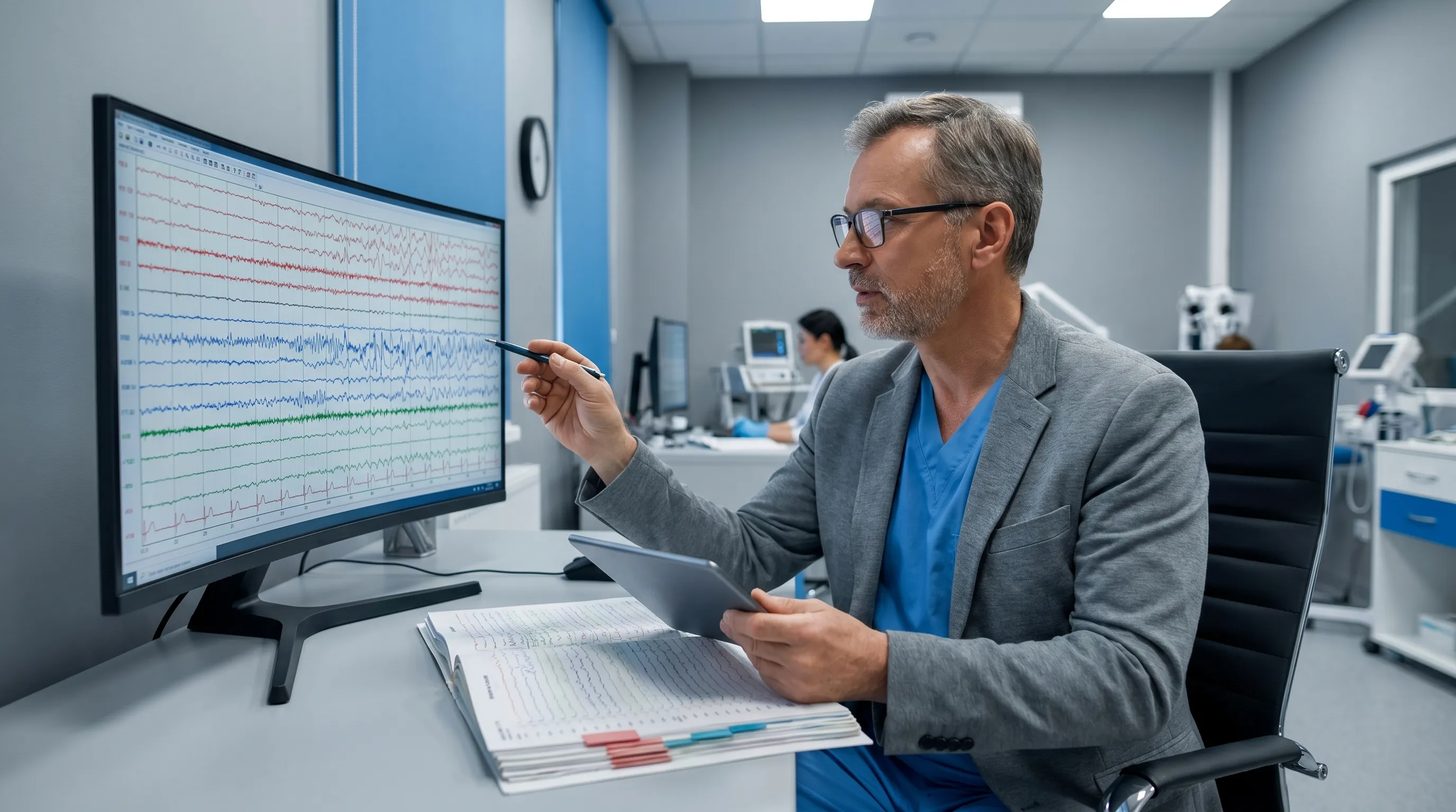
Neurologists analyze EEG recordings by evaluating background rhythm symmetry, frequency distribution, amplitude, reactivity to stimulation, and the presence of epileptiform discharges. Spike waves, sharp waves, and spike-and-wave complexes indicate cortical hyperexcitability and epilepsy risk. Focal slowing suggests localized structural lesions or post-stroke changes, while generalized slowing typically points to metabolic encephalopathy, medication toxicity, or diffuse neurodegenerative processes. Sleep EEGs meticulously track sleep stage transitions, identifying REM sleep behavior disorder, periodic limb movements, or apnea-related arousal fragmentation.
When Results Are Inconclusive
Borderline or non-diagnostic findings are relatively common. In such cases, physicians recommend complementary imaging like echocardiography, cardiac MRI, CT angiography, or brain MRI to visualize structural anatomy. Blood tests evaluating electrolytes, thyroid function, inflammatory markers, and drug levels often clarify underlying metabolic contributors. Patients may undergo repeat testing, prolonged monitoring, or specialized activation studies to capture intermittent phenomena that standard sessions miss.
Safety, Risks, and Patient Considerations
Both procedures rank among the safest diagnostic tools available in modern medicine, carrying virtually no physical risk when performed by trained technicians.
Potential Side Effects and Contraindications
Adverse effects are extremely rare and typically limited to mild skin irritation or temporary redness from electrode adhesives, which resolve quickly after patch removal. Patients with known nickel allergies may react to certain conductive gels or metallic electrode components, prompting technicians to use hypoallergenic alternatives. True contraindications are virtually nonexistent for both tests. However, patients with severe dermatological conditions affecting the chest or scalp may require modified placement strategies or delayed testing to prevent skin breakdown or infection. There is zero radiation exposure, making both examinations safe for pregnant women, infants, and elderly patients.
Insurance Coverage and Cost Implications
Diagnostic EKGs and EEGs are widely covered by major health insurance plans, Medicare, and Medicaid when deemed medically necessary based on documented symptoms or established clinical indications. Routine screening without symptoms may require out-of-pocket payment or prior authorization. Costs vary significantly by region, facility type, and monitoring duration, with routine procedures generally costing substantially less than extended ambulatory or inpatient studies. Patients should verify coverage details with their insurance providers and inquire about cash-pay discounts or hospital financial assistance programs.
Practical Tips for Preparing for Your Test
Proper preparation ensures optimal signal quality, reduces appointment delays, and enhances diagnostic accuracy. Following these evidence-based recommendations will streamline your experience.
Pre-Appointment Guidelines
For EKG appointments, wear a two-piece outfit that allows easy access to your chest, arms, and ankles. Avoid applying lotions, oils, or powders to your torso on the day of testing, as these interfere with electrode adhesion and conductivity. Continue taking prescribed medications unless your physician specifically instructs otherwise. For EEG evaluations, arrive with clean, completely dry hair free of styling products, conditioners, or hair extensions, which severely compromise electrode contact. Your neurologist may request temporary caffeine restriction or sleep deprivation the night before to increase the likelihood of capturing diagnostic abnormalities. Bring a detailed medication list, previous test results, and a trusted companion who can assist with transportation if activation procedures cause temporary lightheadedness.
During the Procedure
Maintain a relaxed posture and breathe normally throughout both examinations. For EKGs, lie completely still with your arms and legs uncrossed to prevent baseline artifact. If instructed to hold your breath briefly, follow the technician's precise timing to avoid motion interference. During EEG recordings, keep your eyes open and closed according to technician prompts. Remain as still as possible, especially during resting-state segments, as jaw clenching, facial grimacing, or limb movements generate electromyographic (EMG) artifact that obscures cortical signals. Immediately report any unusual sensations, chest discomfort, or seizure-like feelings to the monitoring staff so they can annotate the recording and ensure your safety.
Post-Test Care and Next Steps
Once electrodes are removed, gently wash your skin with mild soap and water to eliminate residual adhesive or conductive paste. Apply moisturizer if mild irritation occurs. Resume normal activities immediately after both procedures unless your physician advises otherwise. You may return to work, exercise, and drive safely following standard EKG and EEG sessions. Follow up promptly with your ordering physician to review results, discuss treatment modifications, and establish a monitoring schedule. Keep copies of your reports in your personal health record for future specialist consultations or emergency department visits.
Frequently Asked Questions
What is the main difference between an EKG and an EEG?
The primary distinction lies in the organ system evaluated. An EKG measures the heart's electrical conduction pathways to diagnose arrhythmias, ischemia, and structural heart disease. An EEG records brain wave activity to identify seizure disorders, sleep abnormalities, and neurological impairments. Understanding the ekg vs eeg comparison helps patients and clinicians select the appropriate diagnostic pathway based on presenting symptoms.
Do EKG and EEG tests hurt or require needles?
Neither examination involves needles, incisions, or radiation exposure. Both procedures use painless adhesive electrodes placed on the skin's surface. Patients typically report zero discomfort beyond the mild sensation of cold gel or the slight tug of electrode removal. These non-invasive profiles make them exceptionally well-tolerated across all age groups and medical conditions.
Can a normal EKG or EEG completely rule out disease?
A normal EKG strongly suggests the absence of acute electrical conduction abnormalities or active myocardial injury at the moment of recording. However, intermittent arrhythmias or progressive coronary artery disease may not appear until triggered by exertion or stress. Similarly, a normal EEG captures brain activity only during the recording window. Epileptogenic foci may remain dormant between seizures, requiring prolonged monitoring or sleep-deprived activation to manifest. Negative results provide valuable reassurance but never replace comprehensive clinical evaluation.
How long does it take to receive EKG and EEG results?
Routine EKG tracings are typically reviewed immediately in clinical settings, with formal interpretations available within 24-48 hours. EEG recordings require extensive manual analysis by board-certified neurologists trained in recognizing subtle wave abnormalities, artifact patterns, and sleep staging. Comprehensive EEG reports generally take 3-7 business days to finalize, though urgent inpatient results may be expedited.
Should I adjust my medications before testing?
Never modify prescription dosages without explicit physician authorization. Most patients continue all regular medications for both EKG and EEG procedures. In specific EEG cases, neurologists may temporarily taper anticonvulsants or hypnotics to lower seizure thresholds or capture sleep architecture, but this requires careful clinical supervision to prevent breakthrough events. Always disclose all supplements, over-the-counter drugs, and prescription medications to your testing technician.
Conclusion
Navigating the ekg vs eeg decision becomes straightforward once you recognize their distinct anatomical targets, technological foundations, and diagnostic purposes. The electrocardiogram serves as the cornerstone of cardiovascular assessment, providing instantaneous insights into heart rhythm stability, conduction integrity, and myocardial oxygenation. The electroencephalogram functions as the premier tool for neurological evaluation, mapping complex cortical networks to detect seizure activity, sleep architecture disruptions, and diffuse brain dysfunction. Both procedures deliver critical, life-saving information through safe, non-invasive monitoring that has stood the test of decades of clinical refinement. By understanding their respective indications, preparation requirements, and interpretive frameworks, patients can actively participate in their diagnostic care, ask informed questions, and adhere confidently to prescribed treatment protocols. Whether your healthcare provider recommends cardiac rhythm analysis or cortical wave mapping, modern electrophysiological testing remains an indispensable ally in maintaining long-term health and preventing disease progression. Always consult licensed medical professionals for personalized diagnostic recommendations and treatment planning tailored to your unique physiological profile.
How we work
Researched and drafted with AI assistance, then reviewed by a human editor against the cited sources.
This article is general health information, not medical advice. Consult a qualified healthcare professional about your own situation.