Dilated Pupils and Concussion: What It Means and When to Worry

Key points
- Light: Pupils dilate in dim light and constrict in bright light.
- Stress or excitement: The body’s "fight-or-flight" response can cause pupils to widen due to an adrenaline release.
- Medications or substances: Some eye drops, stimulants, or recreational drugs can cause dilation.
- Injury or illness: Neurological conditions or injuries can affect the nerves controlling the pupil.
When someone suffers a concussion, one of the first things medical professionals check is the person’s pupils. Dilated pupils can be a sign of a head injury, but what does it really mean? How serious is it, and what should you do?
This guide explores the connection between dilated pupils and concussions, explaining what causes pupil changes, why doctors perform pupil exams, and when dilated pupils indicate a medical emergency.
Understanding Pupil Dilation
Your pupil is the black circle in the center of your eye that controls how much light enters. It constricts (gets smaller) in bright light and dilates (gets larger) in the dark.
Pupil size is influenced by several factors:
- Light: Pupils dilate in dim light and constrict in bright light.
- Stress or excitement: The body’s "fight-or-flight" response can cause pupils to widen due to an adrenaline release.
- Medications or substances: Some eye drops, stimulants, or recreational drugs can cause dilation.
- Injury or illness: Neurological conditions or injuries can affect the nerves controlling the pupil.
Normally, pupils are equal in size (about 2–4 mm in bright light, up to 8 mm in the dark) and react symmetrically to light. This reflex is a quick way to gauge if the brain’s autonomic and cranial nerve functions are intact.
The Neurological Pathway of Pupil Response
Understanding how pupil changes occur requires a look at the autonomic nervous system. The pupils are controlled by a delicate balance between two opposing systems: the sympathetic nervous system (which causes dilation) and the parasympathetic nervous system (which causes constriction). Light information travels from the retina through the optic nerve (Cranial Nerve II) to the midbrain. If the light reflex pathway is intact, a signal is sent back through the oculomotor nerve (Cranial Nerve III) to the sphincter pupillae muscle, telling it to constrict.
When a concussion occurs, the brain experiences a complex cascade of neurochemical changes that can temporarily disrupt these pathways. However, because concussions are classified as functional rather than structural injuries, the nerves themselves are rarely severed or permanently damaged. Instead, temporary dysfunction in neural processing or an overwhelming sympathetic surge from trauma can alter pupil behavior. This is why pupil exams remain a cornerstone of the initial neurological assessment: they provide a rapid, non-invasive window into brainstem function and autonomic stability.
What Is a Concussion?
A concussion is a mild traumatic brain injury (TBI) caused by a bump or jolt to the head. This sudden movement can cause the brain to move inside the skull, leading to chemical changes and temporary dysfunction of brain cells.
Key facts about concussions:
- They are serious injuries, even if often called "mild."
- Loss of consciousness is not required for a concussion to occur.
- Common symptoms include headache, confusion, dizziness, nausea, and sensitivity to light or noise.
- Rest is crucial for recovery.
Medical professionals check the pupils after a head injury to perform a quick neurological exam, often using the acronym PERRLA (Pupils Equal, Round, Reactive to Light and Accommodation). Any changes can provide critical clues about the severity of a brain injury.
The Cellular and Metabolic Reality of Concussion
Behind the scenes, a concussion triggers what researchers call the "metabolic crisis." The sudden acceleration-deceleration force stretches and deforms neurons, causing a massive, uncontrolled release of neurotransmitters. This triggers a cascade of potassium flooding out of cells while calcium floods in. To restore the balance, the brain's cellular pumps must work overtime, consuming enormous amounts of glucose (energy).
Simultaneously, blood flow to the brain decreases temporarily, creating an "energy crisis" where demand outstrips supply. This mismatch impairs brain cell function, leading to the cognitive fog, slowed reaction times, and sensory sensitivities characteristic of concussions. The brain also becomes temporarily vulnerable to additional injury, which is why second-impact syndrome—a rare but often fatal condition triggered by a subsequent head trauma during recovery—is taken so seriously. Despite this profound cellular disruption, standard imaging like CT scans or MRIs typically appears normal because the damage is microscopic and functional, not macroscopic or structural.
Dilated Pupils and Concussions: The Connection
So, does a concussion cause dilated pupils? The answer is sometimes, but it's not a definitive sign.
- Mild concussions often do not cause noticeable pupil changes. Many people with a concussion have perfectly normal-looking pupils.
- Stress and adrenaline from the injury can temporarily widen the pupils. However, these pupils will still react to light and return to normal as the person calms down.
- Light sensitivity (photophobia) is a common concussion symptom. This can make a person prefer a dark room, which naturally causes their pupils to dilate. The key is that their pupils will still constrict when light is introduced.
- One pupil being larger than the other is a red flag. This is not a typical sign of a mild concussion and could signal a more severe problem like internal bleeding causing pressure on the brain.
Differentiating Stress-Induced vs. Pathological Dilation
It is crucial for first responders, coaches, and caregivers to distinguish between benign physiological dilation and dangerous neurological changes. Stress-induced dilation is typically bilateral (affecting both eyes) and, most importantly, retains brisk reactivity to light. If you shine a penlight into a stress-dilated pupil, it will promptly constrict, often with a slight overshoot. The person will also typically remain alert, oriented, and able to follow commands, even if they report headache or mild confusion.
Pathological dilation, on the other hand, often presents unilaterally (in one eye) and demonstrates sluggish or absent light reactivity. This type of dilation suggests direct mechanical compression of the third cranial nerve or brainstem dysfunction. In the context of head trauma, recognizing this distinction within the first few minutes can dramatically alter the clinical trajectory, shifting management from sideline monitoring to immediate emergency transport.
Myth vs. Reality
The idea that dilated pupils are a guaranteed sign of a concussion is a myth, often perpetuated by movies. In reality, concussions are diagnosed based on a range of symptoms, not pupil size alone. Checking the pupils is vital because it helps rule out more severe brain injuries that can initially mimic a concussion.
"Anisocoria (unequal pupil sizes) following head trauma should be treated as an emergency until proven otherwise," note the Brain Trauma Foundation guidelines.
When Dilated Pupils Signal a Serious Problem
While a concussion is a mild TBI, a blow to the head can cause more severe injuries like brain bleeding (hematoma) or swelling. Abnormal pupil findings are a classic sign of these emergencies.
Seek immediate medical attention for these pupil-related signs:
- One pupil is dilated and the other is not (unequal pupils), especially if the larger pupil does not react to light. This can indicate pressure on a cranial nerve from bleeding or swelling.
- Both pupils are dilated and do not respond to light, particularly in an unconscious person. This is a grave sign of widespread brain damage.
- Both pupils are extremely constricted (pinpoint) and unresponsive, which can indicate certain brainstem injuries or a drug overdose.
If you see these signs after a head injury, call 911 immediately. These are life-threatening situations where time is critical.
Intracranial Pressure and Brain Herniation Explained
The skull is a rigid, closed container holding brain tissue, blood, and cerebrospinal fluid. When bleeding or severe swelling occurs after trauma, pressure inside this container (intracranial pressure or ICP) begins to rise. As ICP increases, the brain tissue is forced out of its normal position, a dangerous process called herniation.
The most common herniation syndrome associated with unequal pupils is uncal herniation. This occurs when swelling in the temporal lobe pushes the uncus (a small part of the brain) downward against the tentorium cerebelli. This pressure compresses the oculomotor nerve (CN III), which carries parasympathetic fibers responsible for pupil constriction. The result is a progressively larger pupil on the side of the injury that eventually becomes fixed and unresponsive. This is often accompanied by a rapid decline in consciousness, abnormal posturing, and Cushing's triad (high blood pressure, low heart rate, irregular breathing). Recognizing these signs before full herniation occurs is critical, as surgical intervention like a craniotomy may be required to evacuate the clot and relieve pressure.
Table: Concussion vs. Severe Brain Injury Signs
| Feature | Concussion (Mild TBI) | Severe Brain Injury (e.g., Brain Bleed) |
|---|---|---|
| Pupil Size | Usually equal and reactive. May be slightly larger due to adrenaline but will still react to light. | Often unequal (one "blown" pupil) or both fixed and dilated. Unreactive to light. |
| Onset of Signs | Symptoms appear within minutes to hours. Pupil changes are not expected to worsen later. | A pupil may suddenly dilate minutes to hours after the injury as pressure builds. |
| Other Symptoms | Confusion, headache, dizziness, nausea, light/noise sensitivity. Loss of consciousness is brief or absent. | Worsening headache, repeated vomiting, declining level of consciousness, seizures, one-sided weakness. |
| Action | Seek medical evaluation. Requires rest and monitoring. | Medical emergency! Requires immediate hospitalization and possible surgery. |
Other Visual Symptoms of Concussion
Even with normal pupils, a concussion can cause various other visual problems:
- Blurred or double vision (diplopia)
- Eye strain and fatigue, especially with reading
- Difficulty tracking moving objects
- Heightened sensitivity to light (photophobia)
These symptoms are usually temporary and improve with rest. If they persist, vision therapy from a neuro-optometrist may be helpful.
Vestibulo-ocular and Accommodative Dysfunction
Visual disturbances post-concussion frequently extend beyond simple blurriness. The vestibulo-ocular reflex (VOR), which stabilizes vision during head movement, is highly susceptible to concussion-related disruption. When the VOR is impaired, the eyes struggle to stay locked on a target during motion, leading to motion sickness, dizziness in crowded environments, and difficulty reading scrolling text or watching television.
Additionally, accommodative dysfunction is common. Accommodation refers to the eyes' ability to change focus from distant to near objects. After a concussion, the ciliary muscles and neural control loops may struggle to adjust quickly, causing words to "swim" on a page or requiring excessive squinting and head-tilting. Convergence insufficiency, where the eyes cannot properly turn inward to focus on close objects, is another frequent complaint. These visual-vestibular mismatches explain why screen time, reading, and even walking through busy hallways can trigger headaches, nausea, and cognitive fatigue in recovering individuals. Targeted rehabilitation exercises that gradually retrain these neural pathways have proven highly effective in resolving persistent post-concussion visual symptoms.
What To Do If You Suspect a Concussion
If you witness or experience a head injury, follow these steps:
- Ensure Safety: Remove the person from any immediate danger (e.g., stop play in a sports game).
- Check for Red Flags: Call 911 for severe symptoms like loss of consciousness, seizures, worsening headache, repeated vomiting, or unequal pupils.
- Observe for Concussion Signs: Ask simple questions to check for confusion, assess balance, and note any other symptoms like headache or dizziness.
- Monitor Closely: Symptoms can develop or worsen over 24-48 hours. Follow a doctor's advice on monitoring.
- Seek Medical Evaluation: Anyone with a suspected concussion should be evaluated by a healthcare professional.
- Do Not Return to High-Risk Activity: An athlete or worker should not return to play or their job on the same day. A second head injury before the first has healed can be extremely dangerous (second impact syndrome).
While a quick pupil check can be part of first aid, it is not a definitive test. A concussion can exist with perfectly normal pupils.
The First 24-48 Hour Monitoring Protocol
After the initial assessment, careful observation becomes the primary management strategy. Caregivers should establish a baseline by noting the person's current level of alertness, speech clarity, coordination, and pupil reactivity. Over the next two days, periodic checks (every 2-4 hours during waking periods) are recommended to track symptom progression.
Contrary to older advice, it is no longer necessary to wake a concussed individual every hour during sleep. The brain requires restorative sleep to heal, and frequent interruptions can actually slow recovery. However, if the person is difficult to arouse, shows worsening confusion upon waking, or exhibits new neurological deficits, emergency medical evaluation is warranted. Keep a written log of symptoms, noting the time of onset, severity (mild, moderate, severe), and any potential triggers (screen use, bright lights, physical exertion). This documentation is invaluable for healthcare providers when determining the next steps in management. If symptoms suddenly escalate after a period of stability, known as a "lucid interval," it strongly suggests an evolving epidural or subdural hematoma requiring immediate neurosurgical consultation.
Treatment and Recovery
For a typical concussion, treatment focuses on physical and cognitive rest, followed by a gradual return to activities. Pupil size does not affect this recovery protocol.
However, if abnormal pupil signs were present, the patient would undergo urgent evaluation (like a CT scan) to rule out a brain bleed. The presence of a "blown pupil" shifts the focus from concussion management to emergency neurosurgical intervention.
Phased Return-to-Learn and Return-to-Play
Modern concussion management has moved away from strict, prolonged isolation in dark rooms toward active, symptom-limited recovery. The current standard involves a staged, stepwise approach. Initially, 24 to 48 hours of relative rest is recommended. This means avoiding strenuous physical activity and limiting cognitively demanding tasks like schoolwork, screen time, and intense concentration.
After this brief rest period, patients gradually reintroduce light aerobic activity and cognitive tasks, advancing only as long as symptoms do not significantly worsen. The "Return-to-Learn" protocol is typically prioritized over athletic clearance, as academic demands directly impact daily functioning. Steps include part-time attendance, modified assignments, extended test times, and scheduled breaks before returning to full academic load. Similarly, the "Return-to-Play" protocol follows a multi-stage progression: light cardio, sport-specific exercise, non-contact training drills, full-contact practice, and finally, game competition. Each stage should last a minimum of 24 hours. If symptoms return at any step, the patient drops back to the previous asymptomatic stage.
Addressing Post-Concussion Syndrome and Targeted Therapies
While most concussions resolve within 10 to 14 days for adults and up to four weeks for children, approximately 15-30% of individuals experience persistent symptoms, a condition known as Post-Concussion Syndrome (PCS). PCS can involve chronic headaches, sleep disturbances, mood changes (anxiety, depression, irritability), and ongoing cognitive fog.
Treatment for PCS is multidisciplinary. It often involves targeted physical therapy for cervical spine dysfunction, vestibular rehabilitation for balance and dizziness, vision therapy for ocular motor deficits, and cognitive behavioral therapy (CBT) to address anxiety and coping strategies. Sleep hygiene is heavily emphasized, as restorative sleep drives neuroplasticity and metabolic clearance of cellular waste products. Gradual sub-threshold aerobic exercise has also emerged as a cornerstone of recovery, as controlled cardiovascular activity improves cerebral blood flow, promotes neurotrophic factor release, and helps regulate autonomic nervous system function.
Frequently Asked Questions (FAQ)
Q: Do dilated pupils always mean a concussion has occurred? A: No. Pupils can dilate due to darkness, adrenaline (stress/fear), or other factors. Many people with concussions have normal-sized, reactive pupils. While dilated pupils can occur with a concussion, the sign by itself isn’t enough for a diagnosis. Conversely, you can have a concussion even if your pupils are not dilated. The absence of pupil changes never rules out a brain injury; symptom reporting and comprehensive neurological screening remain the diagnostic standard.
Q: If someone has a concussion, will their pupils be different sizes? A: Typically no. In a straightforward concussion, pupils are generally equal in size and reactive. If someone’s pupils are noticeably different sizes after a head injury (anisocoria), it should be treated as a potential emergency, as it could indicate a more serious brain injury like internal bleeding. True anisocoria following trauma warrants immediate advanced imaging (CT scan) and neurosurgical evaluation to exclude compressive lesions.
Q: How long do pupils stay dilated after a concussion? A: In most concussions, any pupil dilation related to stress or adrenaline is temporary and resolves as the person calms down. There is no prolonged, noticeable pupil dilation from a simple concussion. Persistent dilated pupils, especially if they don’t react to light, would signal a more severe or worsening brain issue requiring immediate medical attention. Once autonomic balance is restored, usually within hours to a few days post-injury, pupillary reflexes normalize.
Q: Should I use a flashlight to check my child’s pupils if I suspect a concussion? A: As a first-aid step, a single, quick pupil check can be reasonable if done gently. However, do not rely on a home exam to rule out a concussion. Even if pupils seem normal, the child should be monitored for other symptoms and evaluated by a healthcare provider. If pupils are unequal or unreactive, seek emergency care immediately. Remember that young children may also struggle to verbalize symptoms, making careful behavioral observation and parental intuition crucial.
Q: Can anything else cause one pupil to dilate besides a brain injury? A: Yes. Unequal pupils can be caused by direct eye injuries, certain medications or eye drops, migraines, or congenital conditions like physiological anisocoria. However, in the context of a head injury, a new onset of unequal pupils should always be treated as a potential sign of serious brain trauma. Always consider baseline pupil size when available, and report any pre-existing conditions to medical staff.
Q: Can a concussion cause permanent pupil changes? A: A simple concussion is very unlikely to cause permanent changes to pupil size or reactivity. Permanent changes usually result from more severe nerve damage, direct trauma to the eye itself, or a severe TBI that damages cranial nerve pathways. Once the brain recovers from a concussion, the autonomic control of the pupils should return to normal. If pupillary abnormalities persist beyond the typical recovery window, a referral to a neuro-ophthalmologist is warranted.
Expert Insight
“If one pupil is larger than the other after a head injury, that’s a big red flag. It usually suggests a more serious issue like bleeding in the brain, not just a concussion. That person needs to be in the ER immediately. In a routine concussion, the pupils are typically equal and reactive.” — Dr. Mark Halstead, sports medicine physician
“We always say: When in doubt, sit them out. No game or practice is worth risking a second head injury. Whether or not their pupils look dilated, if a player shows any concussion signs, they’re done for the day. I’ve seen plenty of concussions with normal pupils. That doesn’t mean the athlete is okay to continue.” — Jane McDevitt, certified athletic trainer
These perspectives highlight the consensus among sports medicine professionals: pupil assessment is a vital screening tool, but it must be interpreted within the broader clinical picture. Normal pupils never justify clearing a symptomatic athlete, and abnormal pupils always demand emergency escalation.
Prevention Tips
The best way to treat a concussion is to prevent it.
- Wear appropriate helmets for sports, biking, and other high-risk activities.
- Always use seatbelts and properly installed child safety seats in vehicles.
- Make living areas safer to prevent falls, especially for children and older adults.
- Follow safety rules in sports to minimize dangerous plays.
The Role of Baseline Testing, Neck Strengthening, and Policy Reform
Prevention strategies are continually evolving. Baseline neurocognitive and vestibular-ocular testing provides clinicians with a pre-injury reference point, making post-injury assessment significantly more accurate. While helmets are essential for preventing skull fractures and severe traumatic brain injuries, they do not fully eliminate concussion risk because concussions result from brain movement inside the skull, not just external impact.
Consequently, research supports neck strengthening exercises. Stronger cervical muscles can better stabilize the head upon impact, reducing the acceleration-deceleration forces transferred to the brain. Additionally, rule modifications at all levels of play—such as limiting contact in youth football, enforcing strict penalties for head-first contact in hockey, and improving referee education on concussion recognition—have proven highly effective in reducing injury incidence. Public education campaigns that normalize symptom reporting and destigmatize "sitting out" remain just as critical as physical safety equipment.
Conclusion
While dilated pupils can be a clue after a head injury, they are just one piece of the puzzle.
- Most concussions involve normal pupils. Don't rule out a concussion just because pupils look fine.
- Unequal or unreactive pupils are a medical emergency. This is a red flag for a severe brain injury and requires immediate medical attention.
- Always prioritize safety. When in doubt about a head injury, seek a medical evaluation and ensure the brain has adequate time to heal before returning to high-risk activities.
Understanding the role of pupil evaluation and recognizing the broader symptoms of concussion can help you respond safely and effectively. By combining vigilant symptom monitoring, evidence-based recovery protocols, and a culture that prioritizes brain health over immediate performance, we can significantly improve outcomes for individuals navigating traumatic brain injuries. The brain's remarkable capacity for neuroplasticity and healing should never be rushed; patience, professional guidance, and proper rehabilitation remain the gold standard for long-term recovery.
Resources & Further Reading:
- CDC: Concussion Signs and Symptoms – An overview from the Centers for Disease Control and Prevention on concussion indicators and emergency danger signs.
- Mayo Clinic: Concussion – Detailed information on concussion symptoms, causes, and when to see a doctor.
- BrainLine: Vision Problems after TBI – An article discussing how traumatic brain injuries can affect vision and treatment options.
- Concussion Recognition Tool 5 (CRT5) – A guide from the Concussion in Sport Group to help non-medical personnel identify possible concussions.
Disclaimer: This article is for informational purposes only and is not a substitute for professional medical advice. If you suspect a concussion or any head injury, consult a qualified healthcare provider.
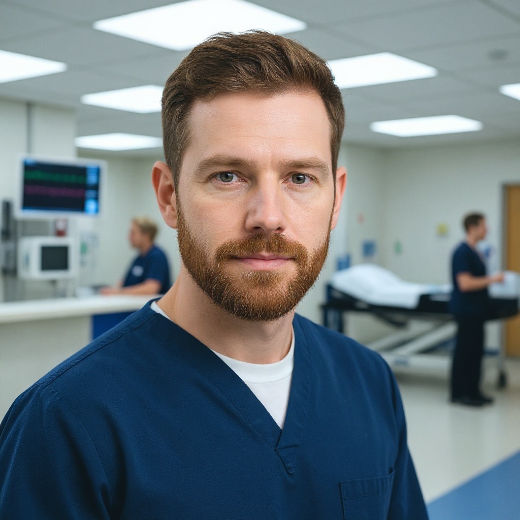
About the author
Michael O'Connell, DO, is a board-certified emergency medicine physician working as an attending physician at a busy Level I Trauma Center in Philadelphia, Pennsylvania. He also serves as a clinical instructor for medical residents and is active in wilderness medicine.