Understanding Mild Levoscoliosis: Causes, Diagnosis, and Evidence-Based Management

When a routine physical examination or imaging scan reveals an unexpected spinal curvature, it is natural to feel concerned. Mild levoscoliosis is one of the most frequently identified spinal variations in both adolescent and adult populations. Unlike the more commonly referenced right-sided curvature, a leftward deviation presents unique biomechanical considerations, diagnostic pathways, and management strategies. Fortunately, the overwhelming majority of cases fall into the mild category, meaning they rarely require invasive intervention or cause significant functional limitations. Instead, with informed monitoring, targeted physical conditioning, and lifestyle optimization, individuals can maintain optimal mobility, posture, and quality of life well into adulthood. Understanding the clinical parameters of this condition empowers patients and caregivers to separate evidence-based facts from widespread myths. This comprehensive guide explores the underlying anatomy, recognized risk factors, diagnostic standards, and scientifically supported treatment protocols for mild levoscoliosis. By integrating current research with practical clinical guidance, we will outline how to navigate this condition confidently and effectively.
Understanding the Anatomy and Classification of Spinal Curvature
To fully grasp the clinical significance of any spinal deviation, it is essential to first understand how specialists categorize curvature direction, severity, and location. The human spine is naturally designed with gentle anterior and posterior curves that distribute mechanical load efficiently during standing, walking, and dynamic movement. However, when an abnormal lateral deviation develops in the coronal plane, it is classified as scoliosis (Mayo Clinic). When this deviation bends toward the left side of the body, forming a characteristic "C" or "S" pattern, it is medically termed levoscoliosis. While right-sided curves (dextroscoliosis) are statistically more prevalent, particularly in the thoracic region during adolescence, leftward deviations frequently appear in the lumbar spine. This anatomical distinction matters because lumbar curvature can influence pelvic alignment, lower back musculature symmetry, and gait mechanics.
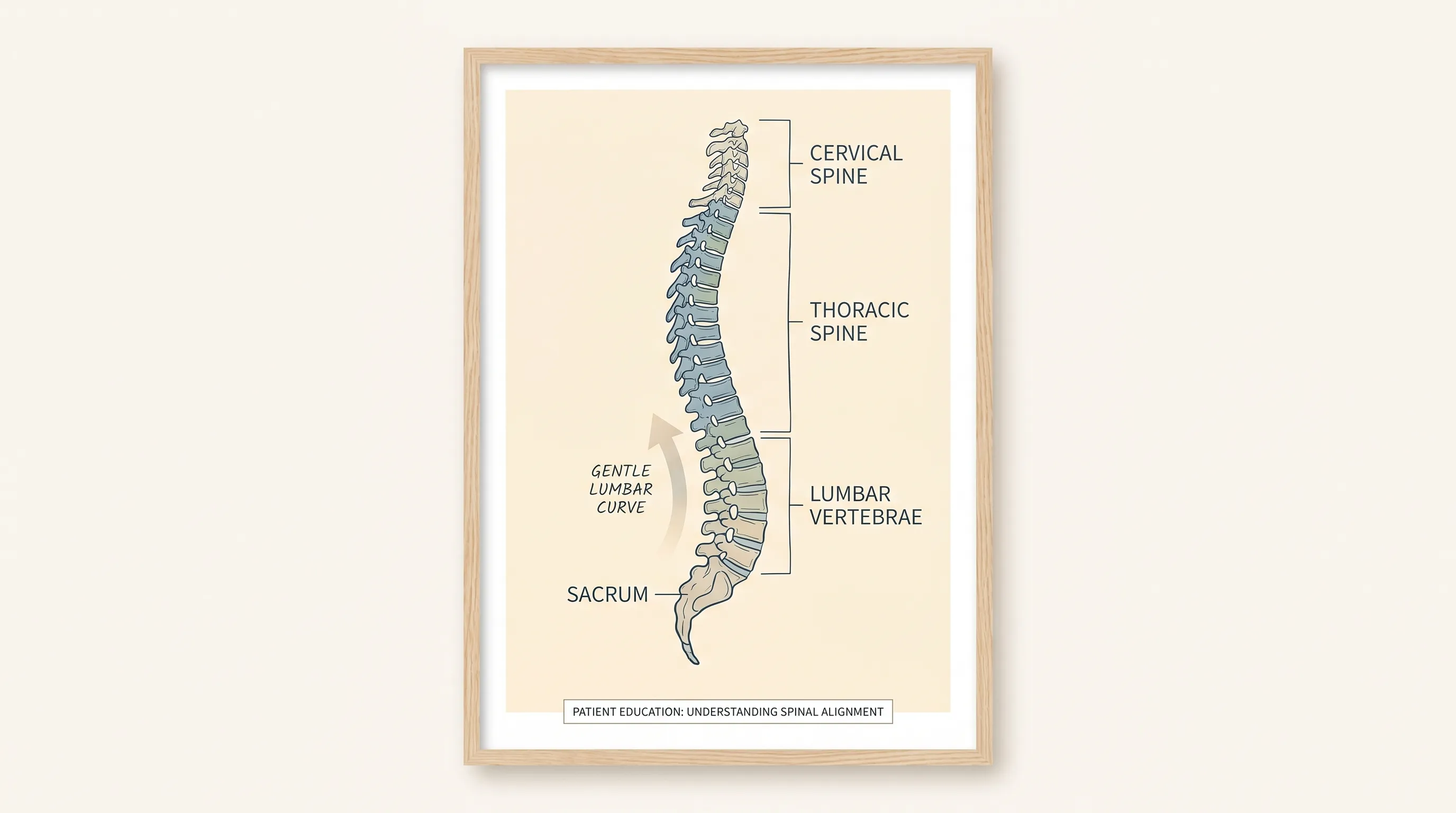
Clinical severity is universally measured using the Cobb angle, a standardized radiographic technique that calculates the degree of spinal deviation. A curve measuring between 10 and 24 degrees falls into the mild levoscoliosis range (Cleveland Clinic). This threshold is critical because it represents a structural variation that is clinically observable but typically does not compromise organ function, respiratory mechanics, or neurological integrity. Moderate scoliosis ranges from 25 to 40 degrees, often requiring more aggressive intervention such as specialized bracing during skeletal growth. Curves exceeding 40 to 45 degrees are classified as severe and may warrant surgical evaluation if progression threatens cardiopulmonary function or causes debilitating pain. Understanding where a specific diagnosis falls on this spectrum dictates the appropriate clinical pathway. Patients diagnosed with a mild curve generally enter an observation and conservative management phase, emphasizing muscular balance, flexibility, and proactive spinal health maintenance rather than aggressive correction protocols.
What Causes a Left-Sided Spinal Curve?
The etiology of spinal curvature has been extensively studied, with clinical consensus indicating that approximately 80 percent of all cases are idiopathic (National Institutes of Health), meaning they arise without a single identifiable structural cause. Idiopathic mild levoscoliosis typically emerges during periods of rapid skeletal growth, particularly between the ages of 10 and 15. While the exact biological trigger remains unclear, robust evidence points to a strong hereditary and genetic component. Family history significantly increases susceptibility, with research indicating that roughly 30 percent of diagnosed individuals have an immediate relative with some form of spinal curvature. Genetic markers related to connective tissue development, bone mineralization, and neuromuscular coordination are actively being investigated to better predict onset and progression patterns.
The remaining 20 percent of cases fall into secondary or acquired categories. Congenital scoliosis occurs when vertebral segments fail to form or segment properly during fetal development, sometimes resulting in a leftward deviation. Neuromuscular conditions such as cerebral palsy, muscular dystrophy, or spinal muscular atrophy can cause asymmetric muscle tone, gradually pulling the spine out of alignment. Degenerative scoliosis is increasingly common in adults over 50, resulting from age-related wear on intervertebral discs, facet joints, and vertebral bodies. As disc height decreases asymmetrically, the spine can gradually lean toward the left or right. Importantly, medical literature consistently emphasizes that lifestyle habits such as poor posture, heavy backpack use, sedentary behavior, or nutritional deficiencies do not cause structural scoliosis. However, these factors can exacerbate muscular imbalances, increase discomfort, and accelerate symptomatic progression in individuals who already possess an underlying curve.
| Classification | Cobb Angle Range | Typical Clinical Approach | Progression Risk |
|---|---|---|---|
| Mild | 10° to 24° | Observation, targeted exercise, postural training | Low; typically stabilizes after skeletal maturity |
| Moderate | 25° to 40° | Full-time bracing (if growing), intensive physical therapy | Moderate; requires active intervention during growth spurts |
| Severe | >40° to 45° | Surgical evaluation, spinal fusion, multidisciplinary care | High; may impact cardiopulmonary function if untreated |
Recognizing Symptoms and When to Seek Evaluation
One of the most defining characteristics of mild levoscoliosis is its subtle presentation. In many instances, the condition is entirely asymptomatic, meaning individuals experience no pain, neurological deficits, or functional restrictions. Instead, it is frequently identified incidentally during school screenings, pediatric wellness visits, or diagnostic imaging for unrelated concerns. When physical indicators do become noticeable, they usually reflect asymmetry rather than severe structural compromise. Common visual signs include uneven shoulder height, with one shoulder blade appearing more prominent than the other, or an apparent discrepancy in hip height when standing. The head may appear slightly offset from the midline of the torso, and one arm may hang marginally lower than its counterpart. Individuals may also notice clothing fitting unevenly, shirt hems hanging asymmetrically, or a subtle visible "C" shaped contour along the lower back.
While mild cases rarely produce systemic complications, understanding the warning signs of progression is essential. If a leftward lumbar curve begins to extend beyond the mild threshold, patients may experience localized lower back stiffness, muscular fatigue after prolonged standing, or intermittent nerve irritation manifesting as mild radiating sensations down the posterior thigh. It is crucial to differentiate these musculoskeletal symptoms from the severe manifestations associated with advanced curvature, which can include chronic ribcage compression, restrictive breathing patterns, reduced spinal mobility, or in rare advanced cases, neurological involvement affecting bowel or bladder function. Anyone experiencing persistent back pain, noticeable postural shifts, or progressive asymmetry should seek a professional orthopedic or spine specialist evaluation. Early assessment ensures accurate baseline measurements and establishes a personalized monitoring schedule tailored to individual growth velocity, skeletal maturity, and symptom profile.
Clinical Diagnosis and Measurement Standards
The diagnostic process for suspected spinal curvature follows a systematic, evidence-based pathway designed to quantify deviation accurately while ruling out underlying pathological conditions. Initial evaluation typically begins with a thorough clinical history and comprehensive physical examination. One of the most widely utilized screening tools is the Adam’s forward bend test, during which the individual stands with feet together and bends forward at the waist with arms extended downward. This position accentuates rotational asymmetry and rib hump formation, allowing clinicians to visually assess structural deviation and measure trunk rotation using a scoliometer (Mayo Clinic). A scoliometer reading of 5 to 7 degrees or higher generally warrants confirmatory imaging.
Standing full-spine radiographs serve as the diagnostic gold standard. These images capture the entire spinal column from the cervical base to the pelvis, enabling precise Cobb angle calculation. The measurement is derived by identifying the most tilted vertebrae above and below the curve’s apex, drawing perpendicular lines from their superior and inferior endplates, and measuring the intersecting angle. Modern digital imaging has significantly improved measurement accuracy, reducing inter-observer variability and allowing clinicians to track subtle changes over time. In certain clinical scenarios, particularly when neurological symptoms are present or when degenerative changes are suspected, magnetic resonance imaging (MRI) or computed tomography (CT) scans may be ordered. MRI provides exceptional visualization of soft tissue structures, including intervertebral discs, spinal cord integrity, and nerve root positioning, which is critical for ruling out intraspinal abnormalities. According to recent radiological studies, mild curves can occasionally be overlooked on focused lumbar MRI scans if the entire spine is not captured in the coronal plane, underscoring the importance of comprehensive imaging protocols when clinical suspicion exists.
Evidence-Based Management and Treatment Protocols
Once mild levoscoliosis is confirmed, the treatment philosophy shifts from correction to conservation and optimization. The primary clinical recommendation for individuals with a curve under 25 degrees is active observation rather than immediate intervention. This does not mean neglecting the condition; rather, it involves structured, periodic monitoring to ensure the curve remains stable. During periods of rapid adolescent growth, clinicians typically schedule physical examinations every four to six months, accompanied by follow-up standing X-rays if measurable changes occur. Most healthcare providers agree that the majority of mild cases will naturally plateau once skeletal maturity is reached, at which point the growth plates in the vertebrae fuse and the structural framework stabilizes. This predictable biological timeline allows families to focus on proactive health maintenance rather than invasive procedures.
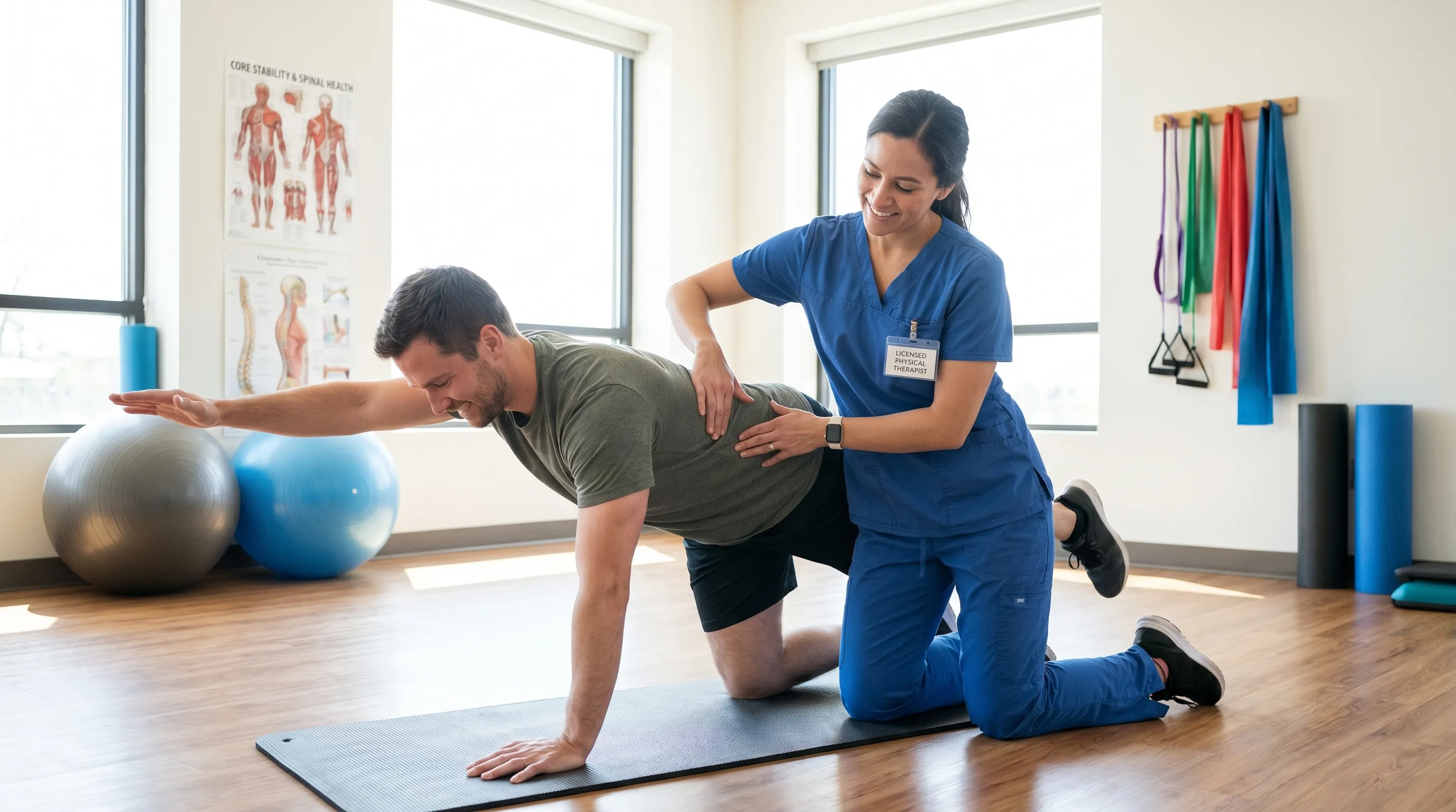
Physical therapy and targeted exercise represent the most impactful conservative management strategies (Cleveland Clinic). The therapeutic goal is not to force the spine into perfect alignment through manual manipulation, but rather to strengthen the deep core musculature, improve neuromuscular control, and correct compensatory postural habits. A well-designed rehabilitation program typically incorporates diaphragmatic breathing techniques to expand ribcage symmetry, isometric core stabilization to support the anterior spinal column, and targeted stretching to release hypertonic muscles along the convex side of the curve. Low-impact aerobic activities such as swimming, cycling, and brisk walking promote cardiovascular health while minimizing compressive spinal loading. Structured modalities like clinical Pilates and therapeutic yoga have gained substantial clinical support for their ability to enhance proprioception, improve flexibility, and foster body awareness. Patients are encouraged to engage in consistent, moderate-intensity movement routines three to five times weekly, prioritizing form and controlled breathing over speed or resistance.
Bracing is a highly effective intervention for moderate curves in growing adolescents, but it is not clinically indicated for mild levoscoliosis. Rigid orthoses are typically reserved for measurements between 25 and 40 degrees to mechanically restrict progression during critical growth windows (National Institutes of Health). In adults, once skeletal growth has ceased, bracing offers minimal structural benefit and may instead lead to muscle deconditioning if used improperly. Surgical intervention, involving spinal instrumentation and fusion, remains strictly reserved for severe, progressive curves exceeding 45 to 50 degrees that compromise pulmonary function, cause refractory pain, or threaten neurological integrity. For individuals managing mild levoscoliosis, surgery is neither necessary nor recommended. If episodic discomfort arises, conservative pain management strategies include over-the-counter nonsteroidal anti-inflammatory medications, alternating heat and ice therapy, myofascial release, and occasional chiropractic or osteopathic manipulation for symptomatic relief. These approaches address muscular tension and joint stiffness without attempting to alter the underlying spinal architecture.
Practical Lifestyle Strategies for Long-Term Spinal Health
Living well with a mild leftward spinal curve requires a holistic approach that integrates daily ergonomics, mindful movement, and supportive environmental adjustments. While posture alone does not cause structural deviation, maintaining optimal alignment reduces compensatory muscle fatigue, minimizes joint compression, and enhances overall biomechanical efficiency. Individuals should focus on distributing weight evenly through both feet when standing, avoiding prolonged single-side leaning, and utilizing lumbar support during extended sitting. Ergonomic workstations should feature adjustable chairs with adequate lower back contouring, monitors positioned at eye level to prevent forward head posture, and regular micro-breaks every 30 to 45 minutes to stand, stretch, and reset spinal positioning.
Sleep architecture plays a surprisingly influential role in spinal recovery and alignment maintenance. During sleep, the paraspinal muscles undergo critical repair, and intervertebral discs rehydrate. For mild levoscoliosis management, sleeping flat on the back is generally optimal, as it allows gravity to distribute evenly along the vertebral column. A medium-firm mattress that contours to the body’s natural curves without excessive sagging is recommended. Side sleepers should place a supportive pillow between the knees to prevent pelvic rotation and maintain hip neutrality. Stomach sleeping is typically discouraged due to the excessive cervical and lumbar rotation it forces. Additionally, maintaining a healthy body weight reduces chronic mechanical stress on the lumbar vertebrae and intervertebral discs, which can indirectly slow age-related degenerative changes that might otherwise exacerbate curvature-related discomfort.
Nutritional factors also contribute to long-term spinal resilience. Adequate calcium and vitamin D intake supports optimal bone mineral density, reducing the risk of osteoporotic compression that can accelerate degenerative curve progression. Anti-inflammatory dietary patterns rich in omega-3 fatty acids, leafy greens, and lean proteins help manage low-grade muscular inflammation and support connective tissue repair. Hydration remains equally vital, as intervertebral discs consist largely of water and rely on consistent fluid exchange to maintain shock-absorbing properties. When combined with regular therapeutic exercise, these lifestyle modifications create a comprehensive, sustainable framework for preserving spinal function and mobility.
Prognosis and Long-Term Outlook
The long-term prognosis for individuals diagnosed with mild levoscoliosis is overwhelmingly positive. Extensive longitudinal studies demonstrate that curves measuring less than 25 degrees rarely progress significantly after skeletal maturity (Mayo Clinic). Research published in peer-reviewed orthopedic journals indicates that adult progression typically averages less than 1 degree annually, if any, unless accompanied by advanced degenerative disc disease or significant osteoporotic changes. This means the vast majority of patients will never experience functional impairment, respiratory restriction, or neurological compromise directly attributable to their initial mild curve. Instead, they transition into a maintenance phase where periodic monitoring, consistent movement, and lifestyle optimization remain the primary focus.
Psychological well-being is another important consideration in long-term management. Receiving a spinal curvature diagnosis, particularly during adolescence, can sometimes trigger anxiety regarding appearance, athletic participation, or future health limitations. Open communication with healthcare providers, involvement in supportive patient communities, and education on realistic outcomes significantly reduce unnecessary distress. Most individuals with mild levoscoliosis participate fully in competitive sports, physically demanding professions, and active recreational hobbies without modification. When managed with a proactive, evidence-based approach, this condition becomes a manageable anatomical variation rather than a defining health limitation.
Frequently Asked Questions
Is mild levoscoliosis considered a serious medical condition?
Mild levoscoliosis, defined as a leftward spinal curvature measuring less than 25 degrees on the Cobb scale, is generally not considered serious. Most individuals experience minimal to no symptoms and can maintain fully active, unrestricted lives with routine monitoring and conservative lifestyle adjustments.
Can exercise or physical therapy correct a left-sided spinal curve?
While targeted physical therapy, core strengthening, and posture training cannot permanently reverse the structural curvature, they are highly effective for symptom management, muscular balance, pain relief, and preventing further progression. Exercise remains a cornerstone of conservative management and functional spinal health (Cleveland Clinic).
How often should mild levoscoliosis be monitored with imaging?
During periods of active skeletal growth, clinical guidelines recommend physical examinations every four to six months, with periodic standing X-rays to measure the Cobb angle. Once growth plates close, monitoring typically shifts to annual check-ups or symptom-driven evaluations as needed.
What sleeping positions are best for left-sided spinal curves?
Sleeping flat on the back with a supportive, medium-firm mattress is ideal for maintaining neutral spinal alignment. If side-sleeping is necessary, placing a firm pillow between the knees helps reduce rotational stress on the pelvis and lumbar spine, while a thin head pillow prevents cervical strain.
Does mild levoscoliosis progress into severe curvature in adulthood?
Significant progression after skeletal maturity is uncommon in mild cases. Research indicates that curves under 25 degrees rarely advance more than 10 to 15 degrees over a lifetime, provided there is no accelerated degenerative disc disease, traumatic injury, or underlying neuromuscular condition (National Institutes of Health).
Conclusion
Mild levoscoliosis represents a common, highly manageable spinal variation that requires understanding rather than alarm. By recognizing the clinical parameters of leftward curvature, embracing evidence-based monitoring protocols, and committing to consistent physical conditioning, individuals can effectively preserve mobility, minimize discomfort, and maintain optimal quality of life. The medical consensus is clear: most mild cases stabilize after skeletal maturity and rarely progress to severe stages. Through proactive posture management, targeted core strengthening, ergonomic optimization, and regular clinical follow-ups, patients transform a potential concern into a well-managed aspect of their overall health profile. Consulting qualified spine specialists, physical therapists, and orthopedic professionals ensures that every management strategy is tailored to individual anatomical needs and long-term wellness goals. With accurate information, structured monitoring, and a commitment to active living, mild levoscoliosis becomes a condition that is navigated with confidence, resilience, and informed clinical care.

About the author
Samuel Jones, MD, is a board-certified orthopedic surgeon specializing in joint replacement and orthopedic trauma. He is a team physician for a professional sports team and practices at a renowned orthopedic institute in Georgia.