Understanding and Resolving QL Muscle Pain: A Comprehensive Guide to Relief

Deep within the posterior trunk lies a frequently overlooked powerhouse: the quadratus lumborum. When this muscle becomes overworked or irritated, it can trigger a specific type of ql muscle pain that radiates through the lower back, hips, and even the groin. Millions of people mistakenly attribute this persistent, aching discomfort to a slipped disc, sacroiliac joint dysfunction, or kidney issues, only to find that standard treatments fall short. The truth is that this condition is often the result of modern sedentary habits, compensatory movement patterns, and unaddressed biomechanical weaknesses. Because the quadratus lumborum operates as both a stabilizer and a dynamic mover, it bears an extraordinary amount of daily stress. When postural imbalances accumulate or acute loads overwhelm the tissue, trigger points form, leading to chronic tension and restricted mobility. Fortunately, this condition responds exceptionally well to targeted rehabilitation, myofascial release, and evidence-based movement re-education. By understanding the underlying mechanics and implementing strategic lifestyle adjustments, you can achieve lasting relief and restore optimal spinal function.
Understanding the Quadratus Lumborum: Anatomy and Function
The quadratus lumborum is a deep, paired abdominal muscle that sits bilaterally along the posterior abdominal wall, directly adjacent to the lumbar vertebrae. Unlike the more superficial erector spinae or the expansive latissimus dorsi, the QL is often hidden from visual inspection, yet it plays a foundational role in spinal mechanics and load distribution. Anatomically, it originates from the posterior aspect of the iliac crest and the thick, robust iliolumbar ligament, extending superiorly to insert onto the transverse processes of the first through fourth lumbar vertebrae and the inferior border of the twelfth rib. This strategic attachment architecture allows it to bridge the critical gap between the pelvis, the lower spine, and the lower rib cage, functioning as a vital force-transfer hub during both static postures and dynamic movement, as detailed in anatomical references from the National Institutes of Health.
Free calculatorBMI CalculatorCalculate Body Mass IndexOpen the calculatorWhere the QL is Located and How It Connects to Surrounding Structures
The anatomical positioning of the quadratus lumborum places it in direct contact with several vital structures, which partially explains why its dysfunction can produce such confusing symptomatology. The muscle lies deep to the aponeurosis of the transversus abdominis and sits just lateral to the psoas major. Superiorly, its fibers anchor to the medial half of the lower border of the twelfth rib, integrating with the diaphragm and intercostal musculature. Inferiorly, it attaches to the posterior iliac crest and iliolumbar ligament, creating a continuous myofascial chain that links the thoracic cavity to the pelvic girdle. This intricate connectivity means that restrictions or tension in the QL can directly influence rib mobility, diaphragmatic function, hip positioning, and pelvic tilt. Furthermore, its proximity to the kidneys, ureters, and the dorsal rami of the lumbar nerves explains why clinical presentation can sometimes mimic visceral or neuropathic conditions. A thorough grasp of this anatomical landscape is essential for clinicians and patients alike when distinguishing muscular referral patterns from systemic pathology.
Primary Biomechanical Roles
The primary biomechanical functions of this muscle are highly specialized and context-dependent. It is fundamentally responsible for unilateral lateral flexion, which is the controlled motion of bending sideways at the waist. When only one side contracts, it pulls the pelvis upward or the rib cage downward, facilitating lateral trunk movement. When both sides contract simultaneously, they assist in extending the lumbar spine and stabilizing the torso against anterior shear forces during lifting or bending. Additionally, the quadratus lumborum plays a vital role in forced expiration, such as coughing, sneezing, or heavy vocalization, by actively depressing the twelfth rib to compress the thoracic cavity and assist the diaphragm in expelling air efficiently. During normal gait cycles, the muscle facilitates pelvic hiking on the swing side, ensuring proper foot clearance and maintaining a smooth, efficient walking pattern. Because of these diverse and constant responsibilities, the tissue is continuously engaged during standing, walking, lifting, and transitional movements. Research utilizing surface and needle electromyography (EMG) confirms that the muscle acts as a primary lumbar stabilizer during unilateral loading, making it highly susceptible to fatigue, microtrauma, and protective spasm when primary stabilizers like the transversus abdominis, multifidus, or gluteal complex become underactive or inhibited. When this intricate neuromuscular system breaks down, ql muscle pain becomes an inevitable clinical consequence.
Why QL Muscle Pain Develops: Causes and Pathophysiology
The development of chronic tension, fibrotic changes, and myofascial trigger points within the quadratus lumborum rarely stems from a single traumatic event. Instead, it is typically the cumulative result of prolonged mechanical stress, neuromuscular inhibition, tissue hypoxia, and lifestyle-driven postural adaptations. Understanding the pathophysiological cascade is essential for breaking the cycle of pain and implementing effective, long-lasting rehabilitation strategies.
Postural Asymmetries and Modern Lifestyles
Contemporary work and leisure habits place extraordinary, unrelenting demands on the posterior trunk. Prolonged sitting, particularly with asymmetrical postures such as crossing the legs, shifting body weight predominantly to one buttock, or leaning heavily to one side of a desk or steering wheel, forces one side of the quadratus lumborum into a chronically shortened, contracted state. Simultaneously, the contralateral muscle remains in an overstretched, eccentrically loaded position. Over time, this static loading disrupts normal tissue elasticity, restricts local capillary blood flow, and promotes the accumulation of metabolic waste products like lactic acid and inflammatory cytokines. The resulting tissue ischemia lowers the local pH environment, sensitizing nociceptors and creating a self-perpetuating cycle of protective muscle guarding, hypertonicity, and central pain processing. This physiological phenomenon clearly explains why office workers, long-distance drivers, and individuals engaged in prolonged screen time frequently report a dull, aching sensation along the lateral flank and lower lumbar region. Breaking this static cycle requires conscious postural resets and movement variability throughout the day, a practice strongly aligned with occupational health guidelines from the CDC.
Compensatory Overload and Muscle Imbalances
The human musculoskeletal system operates as a highly interconnected kinetic chain. When primary stabilizers like the gluteus medius, transversus abdominis, oblique muscles, or deep lumbar multifidus fail to activate properly due to inhibition, fatigue, or neurological dysfunction, secondary muscles are forced to assume roles they are not optimally designed to sustain. In the specific case of weak or inhibited hip abductors, the pelvis drops on the unsupported side during single-leg stance or walking. To counteract this pelvic drop and maintain horizontal alignment, the contralateral quadratus lumborum must work overtime to hike the hip and prevent excessive lumbar collapse. This compensatory overload rapidly depletes muscular endurance, alters normal firing sequences, and leads to the formation of hyperirritable myofascial trigger points. Clinical biomechanical studies consistently demonstrate that a significant percentage of non-specific low back pain cases are directly attributable to these compensatory movement patterns. When the core and glutes fail to share the load, the lateral trunk muscles absorb the excessive mechanical stress, resulting in tissue breakdown and chronic discomfort.
Acute Trauma and Repetitive Microtrauma
While chronic postural strain is the most common culprit behind this condition, acute injuries and repetitive microtrauma can also trigger significant ql muscle pain. Heavy lifting with compromised biomechanical form, particularly when combined with sudden lateral bending, twisting under load, or catching falling objects, can cause micro-tears in the muscle fibers, associated fascial layers, and the iliolumbar ligament attachment. Similarly, athletes and recreational participants engaged in asymmetric sports such as tennis, golf, baseball pitching, rock climbing, or unilateral carrying (e.g., heavy camera bags, toolboxes, or single-strap backpacks) subject the tissue to repetitive rotational and lateral shear forces. Without adequate recovery protocols, eccentric strengthening, and neuromuscular coordination training, these microtraumas accumulate over weeks and months. The result is chronic fibrosis, restricted range of motion, localized inflammation, and persistent discomfort that often mimics more serious spinal pathologies like disc herniations or facet joint syndromes. Early intervention and load management are critical to preventing acute strains from evolving into chronic, treatment-resistant syndromes.
Recognizing the Symptoms and Referral Patterns of QL Pain
Identifying quadratus lumborum involvement requires a nuanced understanding of its clinical presentation, as the symptoms frequently overlap with other lumbar, pelvic, and visceral conditions. Patients rarely describe the discomfort as a sharp, electric, or shooting nerve pain. Instead, they consistently report a deep, relentless, toothache-like sensation that worsens predictably with prolonged static postures, transitional movements, and specific mechanical positions.
Localized Discomfort and Trigger Points
The hallmark of this condition is a highly irritable trigger point located approximately midway between the iliac crest and the bottom of the lower rib cage, typically positioned one to two inches lateral to the lumbar spinous processes. When compressed during clinical palpation, this nodule can elicit a characteristic local twitch response, reproduce familiar pain patterns, and sometimes trigger referred symptoms in distant anatomical regions. The pain intensity often fluctuates predictably throughout the day, flaring up dramatically in the morning after sleeping on an unsupportive mattress, intensifying during long car rides, or worsening after prolonged periods of standing without shifting weight. Stretching the affected side, such as reaching overhead or bending laterally away from the affected side, frequently reproduces a sharp, cramping, or pulling sensation that limits functional mobility. Many individuals also report difficulty turning over in bed or transitioning from sitting to standing due to the sudden demand placed on the compromised tissue. Recognizing these predictable aggravating and easing factors is a crucial diagnostic clue.
Overlapping Referral Zones and Misdiagnosis Risks
According to authoritative clinical references like Simons, Travell & Simons’ Myofascial Pain and Dysfunction: The Trigger Point Manual, the referral map of the quadratus lumborum extends widely into the sacroiliac region, lateral hip, upper buttock, groin, and even along the lower ribs and lower abdominal quadrant. This widespread and highly variable referral pattern frequently leads to diagnostic confusion and unnecessary imaging or interventions. This clinical presentation closely mirrors documented patterns of myofascial pain syndrome recognized by the Mayo Clinic. Patients are commonly misdiagnosed with true SI joint dysfunction, lumbar disc herniations, hip osteoarthritis, or even renal colic due to the anatomical proximity of the kidney to the upper pole of the muscle. Skilled clinicians must perform a thorough differential evaluation to rule out visceral referral, discogenic pathology, radiculopathy, and joint degeneration before attributing symptoms solely to myofascial sources. Recognizing the distinct clinical features, such as pain that worsens with side-lying on the affected side, discomfort during forced coughing, and tenderness localized to the lateral lumbar gutter, helps differentiate ql muscle pain from other complex musculoskeletal syndromes.
Clinical Diagnosis and Differential Considerations
Proper diagnosis of quadratus lumborum syndrome relies on a comprehensive clinical examination that integrates detailed patient history, functional movement screening, manual tissue assessment, and targeted neurological testing. While advanced imaging like MRI or CT scans are rarely necessary for isolated muscular pain, they become invaluable when red flags are present or when conservative management fails.
Physical Examination and Palpation Techniques
Healthcare providers begin by observing static posture and dynamic movement patterns during gait, squatting, bending, and transitional activities. Patients with chronic involvement often present with a noticeable lateral pelvic tilt, with the affected side appearing functionally elevated or shifted. During functional range of motion testing, clinicians assess lateral bending mobility, carefully noting asymmetry, early end-feel restriction, and pain reproduction. Manual palpation is performed using deep, sustained, and progressive pressure along the muscle belly, the lateral border of the lumbar spine, and the superior and inferior fascial attachments. The presence of palpable taut bands, localized tenderness, and predictable referred pain upon trigger point compression strongly supports a myofascial diagnosis. Additional functional provocative tests, such as the prone lumbar extension test, where the patient attempts to lift their torso and thighs off a treatment table, often reproduces the characteristic deep aching sensation when the QL is actively contracting under body weight load.
Ruling Out Serious Underlying Conditions
Before initiating conservative management protocols, clinicians must systematically exclude pathologies that require urgent medical or surgical intervention. Red flags include unexplained weight loss, persistent fever, night pain completely unrelieved by positional changes, progressive neurological deficits in the lower extremities, bowel or bladder dysfunction, and a history of malignancy or recent high-impact trauma. If these systemic or neurological warning signs are absent, the likelihood of a benign muscular etiology increases substantially. A systematic, stepwise diagnostic approach ensures that patients receive highly targeted, effective interventions rather than generic, symptom-masking treatments that fail to address the specific tissue dysfunction driving the pain, consistent with global low back pain management standards published by the WHO. Differential diagnosis may also involve specialized orthopedic tests for sacroiliac joint irritation, such as the Gaenslen’s or Patrick’s test, and lumbar nerve root provocation maneuvers like the straight leg raise or slump test.
Evidence-Based Management and Relief Strategies
Addressing quadratus lumborum dysfunction requires a structured, multimodal approach that progresses strategically from acute symptom reduction to functional rehabilitation and long-term resilience building. Peer-reviewed research published in the Journal of Bodywork and Movement Therapies, Pain Medicine, and Archives of Physical Medicine and Rehabilitation consistently highlights the superior efficacy of combining manual therapy, targeted corrective exercise, dry needling, and patient education to achieve optimal clinical outcomes.
Acute Phase Interventions
During the initial flare-up phase, the primary therapeutic goal is to reduce nociceptive signaling, decrease protective muscle guarding, improve local microcirculation, and prevent central pain sensitization. Relative rest is strongly recommended, which involves actively avoiding movements that consistently reproduce sharp or cramping pain while intentionally maintaining light, comfortable, and non-provocative mobility. Applying moist heat for fifteen to twenty minutes, three to four times daily, promotes vasodilation, increases tissue pliability, facilitates metabolic waste clearance, and reduces the subjective perception of aching. Nonsteroidal anti-inflammatory drugs (NSAIDs) or clinically proven topical analgesics may be utilized under direct medical supervision to manage acute inflammatory responses. Importantly, complete bed rest or prolonged immobilization is actively discouraged, as extended inactivity leads to rapid muscular deconditioning, fascial adhesions, and joint stiffness that significantly prolong recovery timelines.
Targeted Stretching and Mobility Work
Once acute tissue sensitivity begins to diminish, controlled and progressive stretching becomes a cornerstone of rehabilitation. Effective and clinically validated techniques include the side-lying lateral flexion stretch, where the patient lies on their unaffected side, extends the top leg slightly backward to stabilize the pelvis, and reaches the top arm overhead while maintaining neutral spinal alignment. This position selectively elongates the shortened muscle fibers without excessively compressing the posterior facet joints. Another highly effective progression is the kneeling hip flexor combination stretch with a lateral overhead reach, which simultaneously addresses tight anterior chain structures and the lateral lumbar muscles while actively promoting pelvic neutrality and thoracic mobility. Holding these static stretches for thirty to forty-five seconds, repeated three to four times with controlled breathing, encourages sarcomere remodeling, restores optimal resting muscle length, and downregulates sympathetic nervous system arousal. Avoiding ballistic, bouncing, or aggressively forced movements prevents reflexive protective spasms and ensures safe, sustainable tissue adaptation.
Progressive Strengthening and Motor Control Integration
Long-term resolution and prevention of recurrence hinge entirely on restoring optimal neuromuscular control, rebuilding tissue tolerance, and building functional resilience in the posterior kinetic chain. Gluteus medius activation exercises, such as side-lying clamshells with light resistance bands, progressive banded hip abduction walks, and unilateral single-leg balance drills, systematically reduce the excessive compensatory burden placed on the lateral trunk muscles. Core stabilization protocols, including the dead bug, bird-dog, modified side plank progressions, and anti-rotation Pallof press drills, train the deep abdominal wall and multifidus to share spinal loading responsibilities and create stable, protected intra-abdominal pressure. Hip hinge mechanics training reinforces proper posterior chain dominance, ensuring that lifting, bending, and transitional movements originate efficiently from the hips rather than straining the lumbar spine. A 2018 systematic review confirmed that integrating precise myofascial trigger point release with carefully graded loading protocols significantly improves validated pain scores and functional mobility metrics within two to four clinical treatment cycles, supporting broader recommendations for low back pain rehabilitation.
| Management Phase | Primary Clinical Focus | Key Interventions & Protocols | Expected Functional Outcomes |
|---|---|---|---|
| Acute (Days 1–5) | Pain Reduction & Inflammation Control | Relative rest, consistent heat therapy, topical/NSAID support, gentle non-provocative mobility | Decreased protective guarding, improved tissue perfusion, reduced baseline pain perception |
| Subacute (Days 6–14) | Mobility Restoration & Trigger Point Resolution | Targeted static stretching, self-myofascial release with therapy balls, professional dry needling, manual soft tissue mobilization | Increased lateral flexion range of motion, reduced trigger point irritability, normalized pain-free gait |
| Rehabilitation (Weeks 2–6) | Strength & Neuromuscular Control | Gluteus medius activation, core anti-movement stabilization, hip hinge re-education, graded eccentric loading | Improved dynamic spinal stability, corrected movement compensation patterns, restored functional strength |
| Maintenance (Ongoing) | Prevention & Long-Term Performance | Ergonomic workspace optimization, daily mobility routines, progressive resistance training, movement variability practice | Significantly reduced recurrence risk, enhanced movement efficiency, sustained musculoskeletal resilience |

Ergonomic Modifications and Long-Term Prevention
Sustainable recovery extends far beyond clinical exercise sessions; it requires fundamental, intentional changes to daily movement habits, environmental setups, and activity management. Because the quadratus lumborum responds directly and predictably to mechanical loading, gravitational stress, and postural alignment, optimizing your workspace, sleep environment, and recreational routines is absolutely non-negotiable for preventing painful recurrence.
Workspace Optimization and Movement Breaks
Modern sedentary routines and prolonged static positioning rank among the strongest clinical predictors of chronic lateral back pain. To effectively mitigate cumulative static loading, home and office workstations must be consciously configured to promote a neutral pelvic alignment and balanced spinal curvature. Chairs with independently adjustable lumbar support and seat pan tilt help maintain the natural lordotic curve, preventing the anterior pelvic tilt and posterior trunk flexion that chronically lengthen and strain the lateral musculature. Implementing a strict thirty-minute movement rule forces the user to stand, walk, or perform gentle thoracic rotations regularly, interrupting tissue ischemia and resetting neuromuscular tone. Alternating the carrying shoulder for messenger bags, briefcases, or single-strap backpacks distributes external load symmetrically. When utilizing a standing desk, consciously shifting body weight evenly through both heels, engaging the glutes, and avoiding excessive hip hiking or leg crossing prevents chronic muscular shortening. These subtle micro-adjustments accumulate significantly over weeks and months, fundamentally altering the cumulative mechanical stress profile applied to the lower back throughout the workday.
Lifting Mechanics and Load Management
Improper lifting technique remains a frequent trigger for acute exacerbations, structural compromise, and chronic degenerative cycles. Safe and efficient load transfer begins with establishing a wide, stable base of support and actively engaging the anterior and lateral abdominal wall to create protective intra-abdominal pressure. The movement should be driven primarily by hip extension and posterior chain activation, keeping the torso relatively rigid, braced, and the external load positioned as close to the body’s center of mass as physically possible. Twisting or rotating the torso while holding a load places extreme torsional shear forces on the lumbar spine, disproportionately overloading one side of the muscle while the other resists. Training with progressive resistance, learning to execute the Valsalva maneuver correctly, and strictly avoiding sudden, uncontrolled ballistic movements protect the tissue from repetitive microtrauma. For athletes, manual laborers, and active individuals, incorporating unilateral loading drills and dedicated anti-rotation core exercises builds profound tissue tolerance and reinforces symmetrical motor patterns under dynamic stress conditions.
When to Seek Professional Medical Care
While many mild to moderate cases of muscular back tension respond favorably to disciplined self-management and home care protocols, certain clinical scenarios undeniably warrant expert evaluation and supervised intervention. Persistent symptoms that consistently interfere with restorative sleep, occupational performance, athletic participation, or basic activities of daily living clearly indicate the necessity of a structured, professionally guided rehabilitation program.
Red Flags and Warning Signs
Immediate and comprehensive medical evaluation becomes strictly necessary if back pain is accompanied by progressive lower extremity weakness, distinct numbness, or electrical tingling that follows a precise anatomical dermatomal pattern. Complete loss of bowel or bladder control, saddle anesthesia, severe unrelenting night pain that remains unresponsive to any positional changes, or unexplained constitutional symptoms are potential neurological or systemic emergencies that require urgent diagnostic imaging and specialist consultation. Additionally, if conservative home measures fail to produce meaningful functional improvement after a consistent four to six-week trial, professional assessment becomes essential to rule out hidden biomechanical deficits, developing central pain sensitization, or non-musculoskeletal pathologies.
Role of Physical Therapy and Advanced Manual Techniques
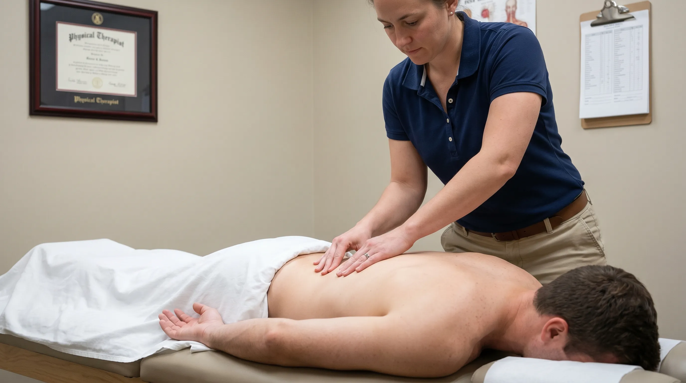
Licensed physical therapists, chiropractors, and sports medicine physicians play a critical, evidence-based role in resolving complex or chronic presentations. Published clinical trials consistently demonstrate that targeted manual therapy, instrument-assisted soft tissue mobilization (IASTM), and precise dry needling focusing on specific hyperirritable trigger points can reduce baseline pain intensity by more than fifty percent within just a few treatment sessions. Physical therapy extends far beyond passive symptom relief by providing graded exposure to previously painful movements, systematically correcting faulty motor patterns, and implementing scientifically backed progressive overload protocols tailored to individual capacity. A comprehensive biomechanical movement assessment identifies global kinetic chain dysfunctions, such as restricted ankle dorsiflexion, limited hip internal rotation, or profound thoracic spine stiffness, that chronically force the lumbar musculature to overcompensate. By diligently addressing the true root cause rather than temporarily masking localized symptoms, professional care delivers durable, functional recovery and thoroughly empowers patients with sustainable, long-term self-management strategies.
Frequently Asked Questions
How do I know if my pain is coming from my quadratus lumborum or my kidneys?
Kidney pain typically presents as a constant, deep, unrelenting ache located higher in the flank, often directly beneath the lower ribs, and is frequently accompanied by urinary changes, fever, chills, or nausea. It generally does not improve or worsen predictably with changes in body position or specific movements. In direct contrast, ql muscle pain intensifies sharply with lateral bending, twisting, prolonged sitting, or side-lying on the affected side, and frequently eases with gentle stretching, heat application, and postural correction. Firm palpation of the lateral muscle belly will reproduce the familiar localized aching in muscular cases, whereas true visceral or renal pain remains entirely unaffected by direct external tissue compression, a key distinction highlighted in renal vs musculoskeletal pain assessments.
Can dry needling effectively relieve chronic trigger points in the lower back?
Yes, robust clinical research strongly supports dry needling as a highly effective, minimally invasive intervention for rapidly deactivating stubborn, treatment-resistant myofascial trigger points. By carefully inserting a sterile monofilament needle directly into the hyperirritable sarcomere knot, the technique reliably elicits a localized twitch response that mechanically resets contracted fibers, restores localized microcirculation, flushes out inflammatory metabolites, and rapidly downregulates nociceptive signaling. Peer-reviewed studies consistently indicate significant reductions in baseline pain intensity and measurable improvements in functional range of motion within two to four clinical sessions when the procedure is appropriately combined with targeted corrective exercise and comprehensive movement education.
Why does my lower back hurt after sitting at my desk all day?
Prolonged sitting in a statically flexed, frequently asymmetrical position places the posterior trunk and lateral stabilizers in a chronically shortened, statically loaded state. This sustained posture severely restricts capillary blood flow to the deep stabilizing musculature and forces the lateral trunk muscles to maintain constant, low-level isometric contractions simply to keep the torso upright and prevent slumping. Over consecutive hours and days, this sustained muscular effort leads to progressive tissue ischemia, localized metabolic waste accumulation, and the gradual development of painful, protective trigger points. Implementing frequent scheduled movement breaks, consciously optimizing ergonomic workstation alignment, and performing gentle lateral thoracic and lumbar stretches throughout the workday effectively interrupts this harmful physiological cycle.
What are the best stretches for relieving tightness in the lateral lumbar region?
The most consistently effective and clinically safe stretches include the side-lying lateral flexion stretch, the kneeling anterior hip flexor release combined with an overhead lateral reach, and an extended side-lying child’s pose variation. These specific positions gently and progressively elongate the chronically shortened fibers without excessively compressing the posterior spinal facet joints or triggering protective neuromuscular reflexes. Maintaining each stretch statically for thirty to forty-five seconds while practicing slow, diaphragmatic breathing significantly enhances parasympathetic nervous system activation and promotes deep tissue relaxation. It is absolutely critical to avoid ballistic bouncing, aggressive overstretching, or forcing the end range of motion, as these counterproductive actions consistently trigger reflexive muscle tightening and delay the healing process.
How long does it typically take to recover from a quadratus lumborum strain?
Most uncomplicated, acute flare-ups resolve completely within two to six weeks when patients consistently adhere to relative rest principles, perform gentle daily mobility work, and progressively integrate foundational strengthening exercises. However, chronic cases persisting longer than three months frequently involve deeply ingrained movement dysfunctions, central nervous system pain sensitization, and significant global compensatory patterns throughout the kinetic chain. Successfully resolving these chronic presentations typically requires a highly structured, professionally supervised physical therapy program spanning eight to twelve continuous weeks, focusing on specialized manual therapy, precise neuromuscular re-education, and graded, systematic return to functional loading activities to ensure long-term prevention and structural resilience.
Conclusion
Navigating the complex clinical landscape of ql muscle pain requires a decisive paradigm shift away from passive, short-term symptom management and toward active, evidence-based, movement-focused rehabilitation. By thoroughly recognizing the intricate biomechanical role of the quadratus lumborum in dynamic spinal stabilization, deeply understanding how modern sedentary postures, repetitive asymmetries, and primary muscle inhibitions drive pathological tissue overload, and systematically implementing targeted stretching, progressive strengthening, and comprehensive ergonomic adjustments, individuals can achieve profound, durable, and completely natural relief from chronic discomfort. The strategic integration of skilled manual therapies, precise dry needling protocols, and carefully graded motor control training simultaneously addresses the acute pain generators and the underlying structural imbalances that perpetuate the dysfunction. Consistent daily adherence to sound movement principles, regular mobility maintenance, and timely professional guidance when necessary form the absolute foundation of a resilient, pain-free, and highly functional posterior kinetic chain. Embracing a proactive, scientifically grounded approach to spinal health transforms temporary recovery into a permanent opportunity for enhanced movement quality, optimized postural alignment, and sustained, high-level physical performance across the lifespan.
How we work
Researched and drafted with AI assistance, then reviewed by a human editor against the cited sources.
This article is general health information, not medical advice. Consult a qualified healthcare professional about your own situation.